. 2017 Oct 13;292(41):17011-17024.
doi: 10.1074/jbc.M117.788588. Epub 2017 Aug 18.

Proteolytic cleavage by the inner membrane peptidase (IMP) complex or Oct1 peptidase controls the localization of the yeast peroxiredoxin Prx1 to distinct mitochondrial compartments

Affiliations

Proteolytic cleavage by the inner membrane peptidase (IMP) complex or Oct1 peptidase controls the localization of the yeast peroxiredoxin Prx1 to distinct mitochondrial compartments

Fernando Gomes et al. J Biol Chem. .

Abstract

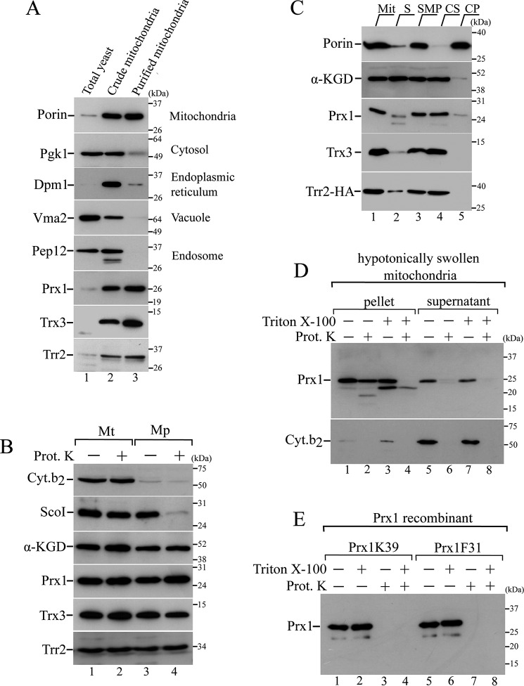
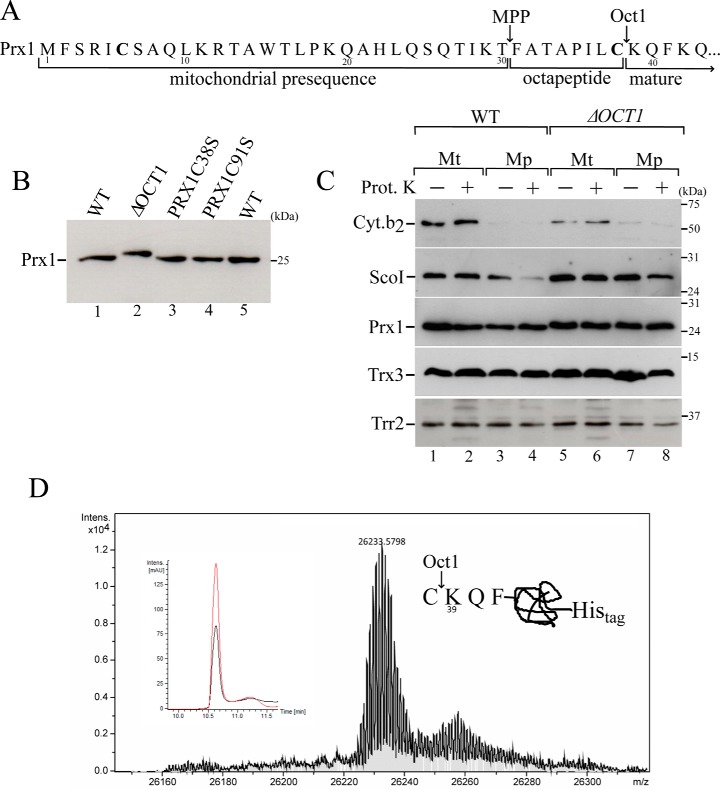
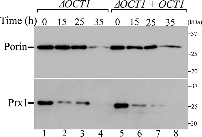
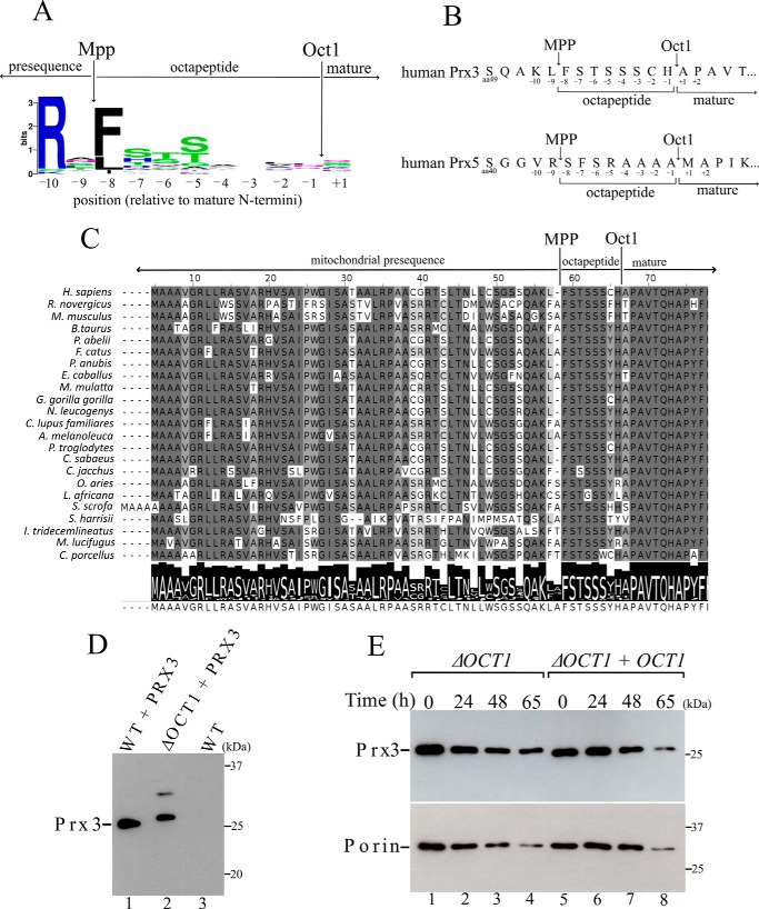
Yeast Prx1 is a mitochondrial 1-Cys peroxiredoxin that catalyzes the reduction of endogenously generated H2O2 Prx1 is synthesized on cytosolic ribosomes as a preprotein with a cleavable N-terminal presequence that is the mitochondrial targeting signal, but the mechanisms underlying Prx1 distribution to distinct mitochondrial subcompartments are unknown. Here, we provide direct evidence of the following dual mitochondrial localization of Prx1: a soluble form in the intermembrane space and a form in the matrix weakly associated with the inner mitochondrial membrane. We show that Prx1 sorting into the intermembrane space likely involves the release of the protein precursor within the lipid bilayer of the inner membrane, followed by cleavage by the inner membrane peptidase. We also found that during its import into the matrix compartment, Prx1 is sequentially cleaved by mitochondrial processing peptidase and then by octapeptidyl aminopeptidase 1 (Oct1). Oct1 cleaved eight amino acid residues from the N-terminal region of Prx1 inside the matrix, without interfering with its peroxidase activity in vitro Remarkably, the processing of peroxiredoxin (Prx) proteins by Oct1 appears to be an evolutionarily conserved process because yeast Oct1 could cleave the human mitochondrial peroxiredoxin Prx3 when expressed in Saccharomyces cerevisiae Altogether, the processing of peroxiredoxins by Imp2 or Oct1 likely represents systems that control the localization of Prxs into distinct compartments and thereby contribute to various mitochondrial redox processes.

Keywords: IMP complex; hydrogen peroxide; inner membrane peptidase complex; mitochondria; mitochondrial processing peptidase (MPP); octapeptidylpeptidase 1, Oct1; oxidative stress; peroxiredoxin; protein import; protein sorting; yeast.


Conflict of interest statement

The authors declare that they have no conflicts of interest with the contents of this article

Figures

Figure 1.
Figure 1.
Intramitochondrial localization of Trr2, Trx3, and Prx1. A, total yeast extract fractions (lane 1), crude mitochondrial fractions (lane 2), and highly pure mitochondrial fractions (lane 3) from BY4741 strain were analyzed by Western blotting using antibodies against markers for distinct cellular compartments as described on the right side of the gel. B, mitochondria were converted into mitoplasts by hypotonic shock (swelling). Equivalent amounts of mitochondria (Mt) and mitoplasts (Mp) were incubated in the presence or absence of 0.1 mg/ml proteinase K (Prot. K). Specific antibody markers are as follows: α-ketoglutarate dehydrogenase (α-KGD, a soluble matrix protein); Sco1 (an inner membrane protein that faces the intermembrane space); and cytochrome b2 (a soluble intermembrane protein). C, highly pure mitochondria (Mit, lane 1) were sonicated and separated into the soluble protein fraction (S, lane 2) and submitochondrial membranes fraction (SMP, lane 3). SMP fractions were then treated with 100 mm Na2CO3 and centrifuged to obtain the soluble (carbonate supernatant, CS, lane 4) and the insoluble (carbonate precipitate, CP, lane 5) fractions. Proteins from these different fractions were analyzed by Western blotting. D, mitochondria were initially converted into mitoplasts (hypotonic shock), followed by a centrifugation to separate the mitoplasts (pellet) from the supernatant (corresponding to the intermembrane space proteins). Both fractions were treated for 1 h with proteinase K (0.2 mg/ml) in the absence or presence of nonionic detergent Triton X-100 (0.5% v/v). E, recombinant Prx1 proteins (Prx1K39 and Prx1F31, representing the enzymes that were processed and unprocessed by Oct1, respectively) were incubated with proteinase K (0.2 mg/ml) in the absence or presence of Triton X-100 (0.5% v/v). These results are representative of at least two independent biological replicates, each one in two technical replicates.
Figure 2.
Figure 2.
Mitochondrial processing of Prx1 by Oct1 protease. A, scheme of the Prx1 N-terminal amino acid sequence. Arrows indicate the MPP and Oct1p cleavage sites based on a global mass spectrometric analysis (27). Cysteines present in the Prx1 presequence are highlighted in bold. B, total cell-free extracts from the wild type (WT, BY4741), OCT1 null mutant (ΔOCT1), and PRX1 null mutant expressing cysteine mutant versions of PRX1 (PRX1C38S and PRX1C91S) were analyzed by Western blotting. C, intramitochondrial localization of Prx1 in the wild-type and ΔOCT1 mitochondria (Mt). Mp, mitoplasts. Mitochondria isolated from cells grown on YPGal were processed according to legend of Fig. 1B. D, mass spectrometry analysis of C-terminal His-tagged Prx1 present in the matrix fraction. C-terminal His-tagged Prx1 expressed in the ΔPRX1 strain was purified by nickel affinity chromatography. The obtained mass was 26,233.57, which was consistent with the incorporation of two oxygen atoms to the Prx1 polypeptide. The inset depicts the corresponding reverse phase chromatogram. The red and black lines correspond to UV chromatogram at 280 and 254 nm, respectively. The scheme at the right side of the mass spectrum represents the cleavage site. These results are representative of at least two independent biological replicates, each one in two technical replicates. See Table 2 for strains and genotypes.
Figure 3.
Figure 3.
Mitochondrial processing of Prx1 by the IMP complex. A, Prx1 N-terminal amino acid sequence highlighting the likely Imp2 cleavage site. B, intramitochondrial localization of Prx1 in the ΔIMP2 mitochondria. Fractionation was performed as described in the legend of supplemental Fig. S1A. See Table 2 for strains and genotypes. C, mass spectrometry analysis of Prx1 sorted into the intermembrane space. C-terminal His-tagged Prx1 expressed in the ΔPRX1 strain was purified by nickel affinity chromatography. The obtained mass was 26918.92, which was consistent with the incorporation of three oxygen atoms to the Prx1 polypeptide. The insets depict the corresponding reverse phase chromatogram. The red and black lines correspond to UV chromatogram at 280 and 254 nm, respectively. The scheme at the right side of the mass spectrum represents the cleavage site. These results are representative of at least two independent biological replicates, each one in two technical replicates. See Table 2 for strains and genotypes.
Figure 4.
Figure 4.
Analysis of half-life of Prx1 in the presence or absence of Oct1 protease in organello. Mitochondria from ΔOCT1 and ΔOCT1 + OCT1 (re-expressing the OCT1 gene) yeast cells were incubated at 37 °C for the indicated time. Mitochondria were reisolated and analyzed by SDS-PAGE and immunoblotting. These results are representative of at least two independent biological replicates, each one in two technical replicates. See Table 2 for strains and genotypes.
Figure 5.
Figure 5.
Human mitochondrial Prx3 is cleaved by yeast Oct1. A, consensus analysis of the amino acid presequences. Arrows depict the cleavage sites of MPP and Oct1 (23). Amino acid frequency blots were generated using the WebLogo program. B, schematic representation of the amino acids in the N terminus of the human mitochondrial peroxiredoxins Prx5 and Prx3. The possible octapeptides that could be cleaved by Oct1 are highlighted. C, alignment of the N-terminal amino acid sequence of Prx3 from mammals. Arrows indicate the likely cleavage sites by MPP and Oct1 proteases. D, total cell-free extracts from the wild-type (WT) and OCT1 null mutant (ΔOCT1) expressing the non-tagged human Prx3. An upper band with lower intensity was observed only in the ΔOCT1 strain, whose meaning is unknown. Possibly an artifactual intermolecular disulfide Prx3 was generated in the denaturing conditions of the SDS-PAGE. E, stability of Prx3 in mitochondria isolated from ΔOCT1 and ΔOCT1 + OCT1 (re-expressing the OCT1 gene) yeast cells. These results are representative of at least two independent biological replicates, each one in two technical replicates. See Table 2 for strains and genotypes.

Similar articles

Cited by

References

    1. Herrmann J. M., and Riemer J. (2010) The intermembrane space of mitochondria. Antioxid. Redox Signal. 13, 1341–1358 - PubMed
    1. Ryan M. T., and Hoogenraad N. J. (2007) Mitochondrial-nuclear communications. Annu. Rev. Biochem. 76, 701–722 - PubMed
    1. Harbauer A. B., Zahedi R. P., Sickmann A., Pfanner N., and Meisinger C. (2014) The protein import machinery of mitochondria–A regulatory hub in metabolism, stress, and disease. Cell Metab. 19, 357–372 - PubMed
    1. Murphy M. P. (2009) How mitochondria produce reactive oxygen species. Biochem. J. 417, 1–13 - PMC - PubMed
    1. Kowaltowski A. J., de Souza-Pinto N. C., Castilho R. F., and Vercesi A. E. (2009) Mitochondria and reactive oxygen species. Free Radic. Biol. Med. 47, 333–343 - PubMed

MeSH terms