. 2017 Sep 6;37(36):8830-8844.
doi: 10.1523/JNEUROSCI.0730-17.2017. Epub 2017 Aug 16.

Calpain-Dependent Degradation of Nucleoporins Contributes to Motor Neuron Death in a Mouse Model of Chronic Excitotoxicity

Affiliations

Calpain-Dependent Degradation of Nucleoporins Contributes to Motor Neuron Death in a Mouse Model of Chronic Excitotoxicity

Kaori Sugiyama et al. J Neurosci. .

Abstract

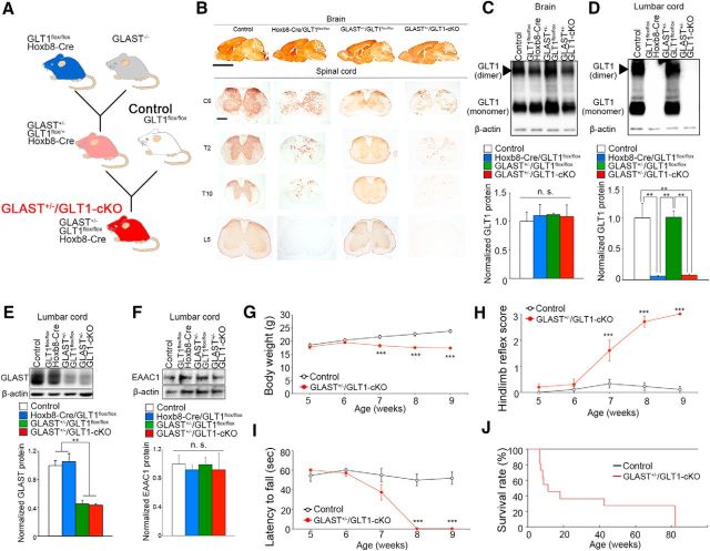
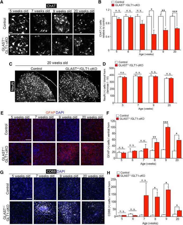
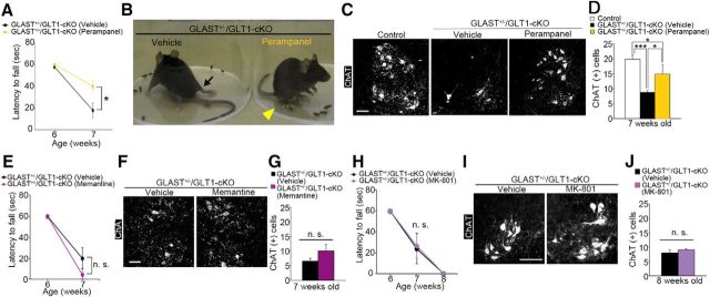
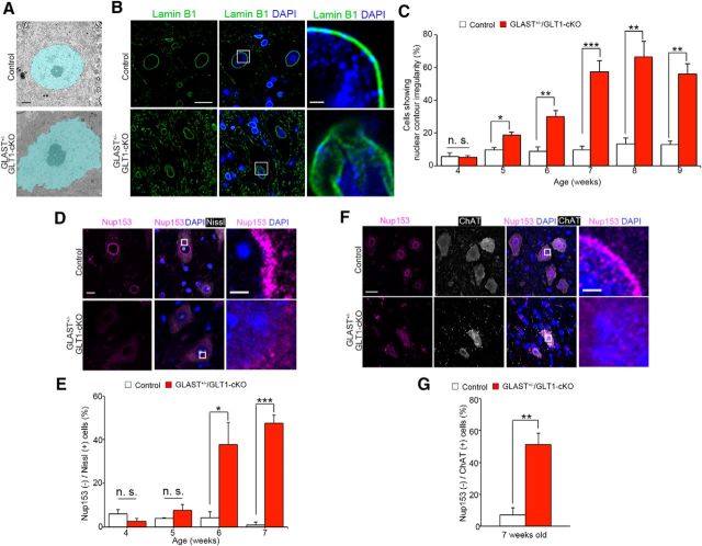
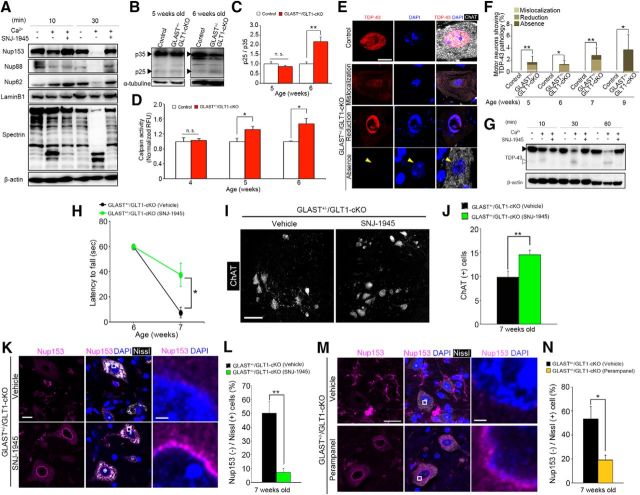
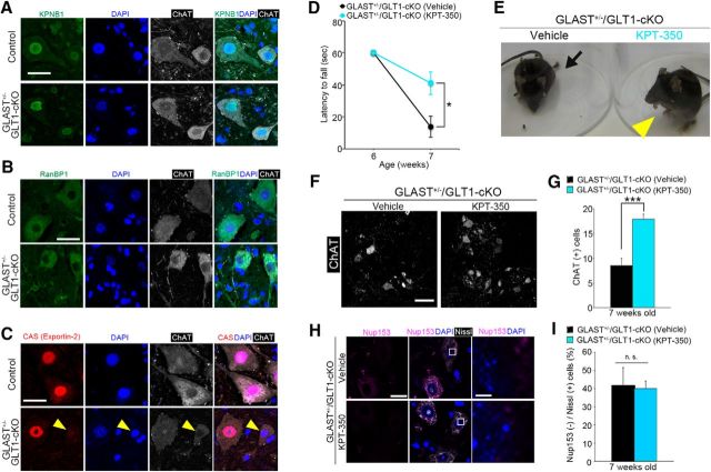
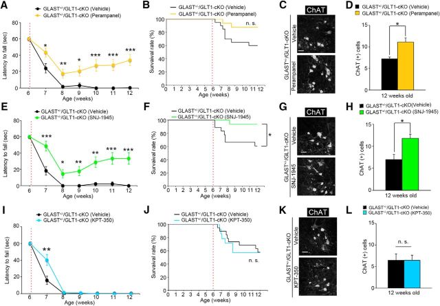
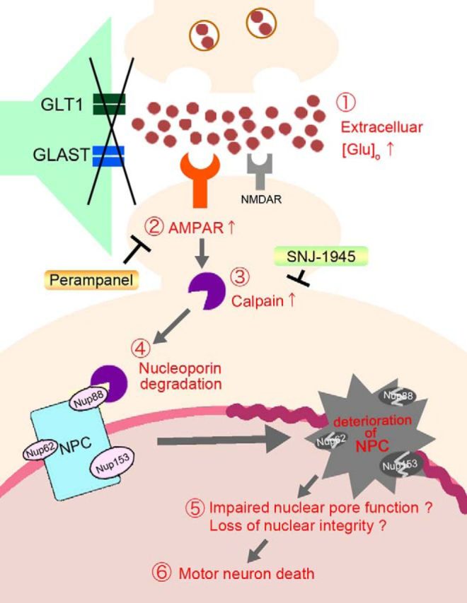
Glutamate-mediated excitotoxicity induces neuronal death by altering various intracellular signaling pathways and is implicated as a common pathogenic pathway in many neurodegenerative diseases. In the case of motor neuron disease, there is significant evidence to suggest that the overactivation of AMPA receptors due to deficiencies in the expression and function of glial glutamate transporters GLT1 and GLAST plays an important role in the mechanisms of neuronal death. However, a causal role for glial glutamate transporter dysfunction in motor neuron death remains unknown. Here, we developed a new animal model of excitotoxicity by conditionally deleting astroglial glutamate transporters GLT1 and GLAST in the spinal cords of mice (GLAST+/-/GLT1-cKO). GLAST+/-/GLT1-cKO mice (both sexes) exhibited nuclear irregularity and calpain-mediated degradation of nuclear pore complexes (NPCs), which are responsible for nucleocytoplasmic transport. These abnormalities were associated with progressive motor neuron loss, severe paralysis, and shortened lifespan. The nuclear export inhibitor KPT-350 slowed but did not prevent motor neuron death, whereas long-term treatment of the AMPA receptor antagonist perampanel and the calpain inhibitor SNJ-1945 had more persistent beneficial effects. Thus, NPC degradation contributes to AMPA receptor-mediated excitotoxic motor neuronal death, and preventing NPC degradation has robust protective effects. Normalization of NPC function could be a novel therapeutic strategy for neurodegenerative disorders in which AMPA receptor-mediated excitotoxicity is a contributory factor.SIGNIFICANCE STATEMENT Despite glial glutamate transporter dysfunction leading to excitotoxicity has been documented in many neurological diseases, it remains unclear whether its dysfunction is a primary cause or secondary outcome of neuronal death at disease state. Here we show the combined loss of glial glutamate transporters GLT1 and GLAST in spinal cord caused motor neuronal death and hindlimb paralysis. Further, our novel mutant exhibits the nuclear irregularities and calpain-mediated progressive nuclear pore complex degradation. Our study reveals that glial glutamate transporter dysfunction is sufficient to cause motor neuronal death in vivo.

Keywords: animal model; excitotoxicity; glutamate; motor neuron; transporter.


Figures

Figure 1.
Figure 1.
Conditional knockout of astroglial glutamate transporters in the spinal cord causes paralysis and motor neuron death. A, Schematic diagram showing the generation of GLAST+/−/GLT1-cKO mice. B, GLT1 immunohistochemistry in brain, cervival (C6), thoracic (T2 and T10), and lumbar (L5) spinal cord tissue at 3 months of age. Scale bars: top, 5 mm; bottom, 500 μm. C, D, Western blot analysis of GLT1 in the brains (C) and lumbar spinal cords (D) of control (GLT1flox/flox), Hoxb8-Cre/GLT1flox/flox, GLAST+/−/GLT1flox/flox, and Hoxb8-Cre/GLT1flox/flox /GLAST+/− (GLAST+/−/GLT1-cKO) mice at 4 weeks of age (n = 3 for each groups). GLT1 monomer band intensities were normalized to β-actin. **p < 0.01. n.s., not significant (post hoc Tukey-HSD test after one-way ANOVA). E, Western blot analysis of GLAST in the lumbar spinal cord of control, Hoxb8-Cre/GLT1flox/flox, GLAST+/−/GLT1flox/flox, and GLAST+/−/GLT1-cKO mice at 4 weeks of age (n = 3 for each groups). GLAST band intensities were normalized to β-actin. **p < 0.01 (post hoc Tukey HSD test after one-way ANOVA). F, Western blot analysis of EAAC1 in the lumbar spinal cord of control, Hoxb8-Cre/GLT1flox/flox, GLAST+/−/GLT1flox/flox, and GLAST+/−/GLT1-cKO mice at 4 weeks of age (n = 3 for each groups). EAAC1 band intensities were normalized to β-actin. n.s., not significant (one-way ANOVA). G–I, Body weight changes (G), hindlimb reflex score (H), and hanging wire test (I) of control (GLT1flox/flox, n = 15) and GLAST+/−/GLT1-cKO animals (Hoxb8-Cre/GLT1flox/flox /GLAST+/−, n = 16). ***p < 0.001 (post hoc unpaired two-tailed t test at corresponding time-point after two-way repeated-measures ANOVA). J, Percentage survival of control (n = 11) and GLAST+/−/GLT1-cKO (n = 14) animals calculated using the Kaplan–Meier method (p = 0.000016, log rank test). All data are expressed as the mean ± SEM.
Figure 2.
Figure 2.
Loss of motor neurons and gliosis in GLAST+/−/GLT1-cKO mice. A, ChAT immunofluorescence of the lumbar ventral horn in control and GLAST+/−/GLT1-cKO mice at 5, 7, 9, and 20 weeks of age. Scale bar, 50 μm. B, Quantification of ChAT-positive motor neurons from 5 to 9, and 20 weeks of age of control mice (P5W, n = 4; P6W, n = 4; P7W, n = 3; P8W, n = 3; P9W, n = 4; P20W, n = 3) and GLAST+/−/GLT1-cKO mice (P5W, n = 5; P6W, n = 4; P7W, n = 4; P8W, n = 3; P9W, n = 5; P20W, n = 4). *p < 0.05, **p < 0.05, ***p < 0.001. n.s., not significant (unpaired two-tailed t test). C, NeuN immunofluorescence in the lumbar dorsal horn from control and GLAST+/−/GLT1-cKO mice at 20 weeks of age. Scale bar, 200 μm. D, Quantification of NeuN-positive neurons in the superficial dorsal horn (laminae I–IV) of the spinal cord at 5, 7, 9, and 20 weeks of control mice (P5W, n = 3; P7W, n = 4; P9W, n = 3; P20W, n = 3) and GLAST+/−/GLT1-cKO mice (P5W, n = 3; P7W, n = 4; P9W, n = 4; P20W, n = 3) mice. E, GFAP immunofluorescence in the lumbar ventral horn from control and GLAST+/−/GLT1-cKO mice at 5, 7, 9, and 20 weeks of age. Scale bar, 100 μm. F, Quantification of GFAP-positive cells in the lumbar ventral horn from 5 to 9, and 20 weeks of age of control mice (P5W, n = 3; P6W, n = 3; P7W, n = 4; P8W, n = 3; P9W, n = 3; P20W, n = 3) and GLAST+/−/GLT1-cKO mice (P5W, n = 3; P6W, n = 3; P7W, n = 4; P8W, n = 3; P9W, n = 3; P20W, n = 3) mice. *p < 0.05, **p < 0.01, ***p < 0.001. G, CD68 immunofluorescence in the lumbar ventral horns from control and GLAST+/−/GLT1-cKO mice at 5, 7, 9, and 20 weeks of age. Scale bar, 100 μm. H, Quantification of CD68-positive cells in the lumbar ventral horn from 5 to 9, and 20 weeks of age of control mice (P5W, n = 3; P6W, n = 3; P7W, n = 4; P8W, n = 3; P9W, n = 3; P20W, n = 3) and GLAST+/−/GLT1-cKO mice (P5W, n = 3; P6W, n = 3; P7W, n = 4; P8W, n = 3; P9W, n = 3; P20W, n = 3). *p < 0.05. All data are expressed as the mean ± SEM. n.s., Not significant (unpaired two-tailed t test).
Figure 3.
Figure 3.
Overactivation of AMPA receptor but not NMDA receptor contributes to motor deficits and motor neuron loss in GLAST+/−/GLT1-cKO mice. A, Perampanel treatment delayed motor deficits in the hanging wire test (n = 15 for vehicle-treated and n = 17 for perampanel-treated GLAST+/−/GLT1-cKO mice). *p < 0.05 (post hoc unpaired two-tailed t test at corresponding time point after two-way repeated-measures ANOVA). B, Amelioration of hindlimb paralysis in GLAST+/−/GLT1-cKO mice after perampanel treatment. Arrow indicates the fully paralyzed hindlimb of vehicle-treated GLAST+/−/GLT1-cKO mice. Arrowhead indicates the hindlimb upon which the perampanel-treated GLAST+/−/GLT1-cKO mice could stand. C, ChAT immunofluorescence of the lumbar ventral horn from control, vehicle-treated, and perampanel-treated GLAST+/−/GLT1-cKO mice at 7 weeks of age. Scale bar, 100 μm. D, Quantification of ChAT-positive motor neurons at 7 weeks of age (n = 3 for control mice, n = 4 for vehicle-treated mice, and n = 5 for perampanel-treated GLAST+/−/GLT1-cKO mice). *p < 0.05, ***p < 0.001 (post hoc Tukey HSD test after one-way ANOVA). E, Memantine treatment could not ameliorate motor deficits in the hanging wire test (n = 9 for vehicle-treated and n = 7 for memantine-treated GLAST+/−/GLT1-cKO mice). n.s., Not significant (two-way repeated-measures ANOVA). F, ChAT immunofluorescence of the lumbar ventral horn from vehicle-treated and memantine-treated GLAST+/−/GLT1-cKO mice at 7 weeks of age. Scale bar, 100 μm. G, Quantification of ChAT-positive motor neurons at 7 weeks of age (n = 3 for vehicle-treated and 4 for memantine-treated GLAST+/−/GLT1-cKO mice). n.s., Not significant (unpaired two-tailed t test). H, MK-801 treatment could not ameliorate motor deficits in the hanging wire test (n = 5 for vehicle-treated and 6 for MK-801-treated GLAST+/−/GLT1-cKO mice). n.s., Not significant (two-way repeated-measures ANOVA). I ChAT immunofluorescence of the lumbar ventral horn from vehicle-treated and MK-801-treated GLAST+/−/GLT1-cKO mice at 8 weeks of age. Scale bar, 100 μm. J, Quantification of ChAT-positive motor neurons at 8 weeks of age (n = 3 for each groups). n.s., Not significant (unpaired two-tailed t test). All data are expressed as the mean ± SEM.
Figure 4.
Figure 4.
Nuclear contour irregularity and the loss of nuclear pore complex proteins are observed in spinal motor neurons of GLAST+/−/GLT1-cKO mice. A, Pseudocolored electron microscopic images of motor neurons in lumbar ventral horns from control and GLAST+/−/GLT1-cKO mice at 6 weeks of age. The nucleus of the motor neuron is pseudocolored in light blue. Scale bar, 2 μm. B, Lamin B1 immunofluorescence of lumbar ventral horns from control and GLAST+/−/GLT1-cKO mice at 7 weeks of age. Sections were double labeled with anti-lamin B1 (green) and DAPI (blue). Rectangles in middle panel are enlarged in the right panel. Scale bars: left, 10 μm; right, 5 μm. C, Quantitative analysis of Lamin B1-immunostained sections. The percentage of large ventral horn neurons showing nuclear contour irregularity is higher in GLAST+/−/GLT1-cKO mice from 5 weeks of age. Large ventral horn neurons showing nuclear contour irregularity were counted from control mice (P4W, n = 3; P5W, n = 3; P6W, n = 4; P7W, n = 5; P8W, n = 3; P9W, n = 3) and GLAST+/−/GLT1-cKO mice (P4W, n = 3; P5W, n = 4; P6W, n = 6; P7W, n = 4; P8W, n = 3; P9W, n = 3). *p < 0.05, **p < 0.01, ***p < 0.001. n.s., Not significant (unpaired two-tailed t test). D, Nup153 immunofluorescence of lumbar ventral horns from control and GLAST+/−/GLT1-cKO mice at 6 weeks of age. Sections were triple labeled with anti-Nup153 (magenta), DAPI (blue), and Neurotrace (fluorescent Nissl stain, white). Rectangles in the middle panel are enlarged in right panel. Scale bars: left, 10 μm; right, 5 μm. E, Quantitative analysis of Nup153-immunostained sections. The percentage of Nup153-negative large ventral horn neurons is higher in GLAST+/−/GLT1-cKO mice from 6 weeks of age. Nup153-negative large ventral horn neurons were counted from control mice (n = 3 at each age), and GLAST+/−/GLT1-cKO mice (P4W, n = 3; P5W, n = 4; P6W, n = 3; P7W, n = 4) mice. *p < 0.05, ***p < 0.001. n.s., Not significant (unpaired two-tailed t test). All data are expressed as the mean ± SEM. F, Nup153 immunofluorescence of lumbar ChAT-positive motor neurons from control and GLAST+/−/GLT1-cKO mice at 7 weeks of age. Sections were triple-labeled with anti-Nup153 (magenta), DAPI (blue), and ChAT (white). Rectangles in middle panel are enlarged in right panel. Scale bars: left, 10 μm; right, 5 μm. G, Quantitative analysis of Nup153(−)/ChAT(+) motor neurons. The percentage of Nup153 (−)/ ChAT(+) motor neurons is similar to that of Nup153(−) large ventral horn neurons in GLAST+/−/GLT1-cKO mice at 7 weeks of age (n = 4 for control and n = 5 for GLAST+/−/GLT1-cKO). **p < 0.01 (unpaired two-tailed t test). All data are expressed as the mean ± SEM.
Figure 5.
Figure 5.
Calpain overactivation contributes to motor deficits and motor neuron loss in GLAST+/−/GLT1-cKO mice. A, Representative immunoblot that shows proteolysis of Nup153, Nup88, Nup62, laminB1, and α-spectrin in mouse brain homogenate that was incubated with exogenous calcium and/or a calpain inhibitor, SNJ-1945 (100 μm). B, Representative immunoblot showing proteolysis of p35 in lumbar ventral horn from control and GLAST+/−/GLT1-cKO mice at 5 and 6 weeks of age. C, Quantification of the p25/p35 ratio from control mice (P5W, n = 3; P6W, n = 4) and GLAST+/−/GLT1-cKO mice (n = 3 at each age). Band intensities of p35 and p25 were normalized to α-tubulin. **p < 0.01. n.s., Not significant (unpaired two-tailed t test). D, Measurement of calpain activity of lumbar ventral horn form control mice (P4W, n = 5; P5W, n = 6; P6W, n = 5) and GLAST+/−/GLT1-cKO mice (P4W, n = 6; P5W, n = 5; P6W, n = 6). Relative fluorescence units (RFUs) were compared by calculating the fold difference in enzyme activity. *p < 0.05; n.s., Not significant (unpaired two-tailed t test). E, TDP-43 immunofluorescence in the lumbar ventral horns from control at 7 weeks of age (the top row of a panel) and GLAST+/−/GLT1-cKO mice at 7 (the second and third rows of a panel) and 9 (the bottom row of a panel) weeks of age. Sections were double labeled with TDP-43 (red) and ChAT (white). Nuclei were visualized with DAPI. The second row of a panel shows mislocalization of TDP-43 to the cytoplasm in motor neurons of GLAST+/−/GLT1-cKO mice. The third row of a panel shows reduction of nuclear TDP-43 without cytoplasmic TDP-43 immunoreactivity in motor neurons of GLAST+/−/GLT1-cKO mice. The bottom row of a panel shows loss of TDP-43 from both the nucleus (yellow arrowhead) and the cytoplasm in motor neurons of GLAST+/−/GLT1-cKO mice. Scale bar, 10 μm. F, The percentage of motor neurons showing aberrant staining pattern of TDP-43 from 5, 6, 7, and 9 weeks of age of control mice (P5W, n = 7; P6W, n = 6; P7W, n = 5; P9W, n = 6) and GLAST+/−/GLT1-cKO mice (P5W, n = 7; P6W, n = 5; P7W, n = 6; P9W, n = 5). *p < 0.05, **p < 0.01 (unpaired two-tailed t test). G, Representative immunoblot that shows proteolysis of TDP-43 and β-actin in mouse brain homogenate that was incubated with exogenous calcium and/or a calpain inhibitor, SNJ-1945 (100 μm). Black and white arrowheads indicate a full-length and cleaved fragment of TDP-43, respectively. H, SNJ-1945 treatment delayed motor deficits in the hanging wire test (n = 8 for each groups). *p < 0.05 (post hoc unpaired two-tailed t test at corresponding time point after two-way repeated-measures ANOVA). I, ChAT immunofluorescence of the lumbar ventral horns from vehicle-treated and SNJ-1945-treated GLAST+/−/GLT1-cKO mice at 7 weeks of age. Scale bar, 100 μm. J, Quantification of ChAT-positive motor neurons at 7 weeks of age (n = 5 for vehicle-treated and n = 6 for SNJ-1945-treated GLAST+/−/GLT1-cKO mice). **p < 0.01 (unpaired two-tailed t test). K, Nup153 immunofluorescence of lumbar ventral horns from vehicle-treated and SNJ-1945-treated GLAST+/−/GLT1-cKO mice at 7 weeks of age. Sections were triple labeled with anti-Nup153 (magenta), DAPI (blue), and Neurotrace (fluorescent Nissl stain, white). Rectangles in the middle panel are enlarged in the right panel. Scale bars: left, 10 μm; right, 5 μm. L, Quantitative analysis of Nup153-immunostained sections. The percentage of Nup153-negative large ventral horn neurons decreased in GLAST+/−/GLT1-cKO mice treated with SNJ-1945 (n = 4 for each groups). **p < 0.01 (unpaired two-tailed t test). M, Nup153 immunofluorescence of lumbar ventral horns from vehicle-treated and perampanel-treated GLAST+/−/GLT1-cKO mice at 7 weeks of age. Sections were triple labeled with anti-Nup153 (magenta), DAPI (blue), and Neurotrace (fluorescent Nissl stain, white). Rectangles in the middle panel are enlarged in the right panel. Scale bars: left, 20 μm; right, 5 μm. N, Quantitative analysis of Nup153-immunostained sections. The percentage of Nup153-negative large ventral horn neurons decreased in GLAST+/−/GLT1-cKO mice treated with perampanel (n = 4 for vehicle-treated and n = 5 for perampanel-treated GLAST+/−/GLT1-cKO mice). *p < 0.05 (unpaired two-tailed t test). All data are expressed as the mean ± SEM.
Figure 6.
Figure 6.
Nuclear export inhibitor KPT-350 transiently ameliorates motor deficits, motor neuron loss in GLAST+/−/GLT1-cKO mice. A, KPNB1 immunofluorescence of motor neurons in the lumbar ventral horn from control and GLAST+/−/GLT1-cKO mice at 7 weeks of age. Sections were triple labeled with anti-KPNB1 (green), DAPI (blue), and anti-ChAT (white). Scale bar, 10 μm. B, RanBP-1 immunofluorescence of motor neurons in the lumbar ventral horn from control and GLAST+/−/GLT1-cKO mice at 7 weeks of age. Sections were triple labeled with anti-RanBP-1 (green), DAPI (blue), and anti-ChAT (white). Scale bar, 10 μm. C, CAS immunofluorescence of motor neurons in the lumbar ventral horn from control and GLAST+/−/GLT1-cKO mice at 7 weeks of age. Sections were triple labeled with anti-CAS (red), DAPI (blue), and anti-ChAT (white). Arrowheads indicate the reduction or absence of nuclear CAS immunoreactivity in GLAST+/−/GLT1-cKO mice. Scale bar, 10 μm. D, KPT-350 treatment delayed motor deficits in the hanging wire test (n = 10 for vehicle-treated and n = 9 for KPT-350-treated GLAST+/−/GLT1-cKO mice). *p < 0.05 (post hoc unpaired two-tailed t test at corresponding time point after two-way repeated-measures ANOVA). E, Amelioration of hindlimb paralysis in GLAST+/−/GLT1-cKO mice after KPT-350 treatment. Arrow indicates the fully paralyzed hindlimb of the vehicle-treated GLAST+/−/GLT1-cKO mice. Arrowhead indicates the hindlimb upon which the KPT-350-treated GLAST+/−/GLT1-cKO mice could stand. F, ChAT immunofluorescence of the lumbar ventral horn from vehicle-treated and KPT-350-treated GLAST+/−/GLT1-cKO mice at 7 weeks of age. Scale bar, 100 μm. G, Quantification of ChAT-positive motor neurons at 7 weeks of age (n = 7 for vehicle-treated and n = 5 for KPT-350-treated GLAST+/−/GLT1-cKO mice). ***p < 0.001 (unpaired two-tailed t test). H, Nup153 immunofluorescence of lumbar ventral horns from vehicle-treated and KPT-350-treated GLAST+/−/GLT1-cKO mice at 7 weeks of age. Sections were triple labeled with anti-Nup153 (magenta), DAPI (blue), and Neurotrace (fluorescent Nissl stain, white). Rectangles in the middle panel are enlarged in the right panel. Scale bars: left, 20 μm; right, 5 μm. (I) Quantitative analysis of Nup153-immunostained sections. The percentage of Nup153-negative large ventral horn neurons is not changed in GLAST+/−/GLT1-cKO mice treated with KPT-350 (n = 3 for each groups). n.s., Not significant (unpaired two-tailed t test). All data are expressed as the mean ± SEM.
Figure 7.
Figure 7.
Effects of long-term treatment with perampanel, SNJ-1945, and KPT-350 in GLAST+/−/GLT1-cKO mice. A, Perampanel treatment for 6 weeks delayed motor deficits in the hanging wire test (n = 20 for vehicle-treated and n = 16 for perampanel-treated GLAST+/−/GLT1-cKO mice). B, Percentage survival of vehicle-treated (n = 20) and perampanel-treated (n = 16) GLAST+/−/GLT1-cKO mice calculated using the Kaplan–Meier method (p = 0.076, log rank test). n.s., Not significant. C, ChAT immunofluorescence of the lumbar ventral horn from vehicle-treated and perampanel-treated GLAST+/−/GLT1-cKO mice at 12 weeks of age. D, Quantification of ChAT-positive motor neurons at 12 weeks of age (n = 4 for vehicle-treated and n = 6 for perampanel-treated GLAST+/−/GLT1-cKO mice). E, SNJ-1945 treatment for 6 weeks delayed motor deficits in the hanging wire test (n = 18 for vehicle-treated and n = 16 for SNJ-1945-treated GLAST+/−/GLT1-cKO mice). F, Percentage survival of vehicle-treated (n = 18) and SNJ-1945-treated (n = 16) GLAST+/−/GLT1-cKO mice calculated using the Kaplan–Meier method (p = 0.026, log rank test). *p < 0.05. G, ChAT immunofluorescence of the lumbar ventral horn from vehicle-treated and perampanel-treated GLAST+/−/GLT1-cKO mice at 12 weeks of age. H, Quantification of ChAT-positive motor neurons at 12 weeks of age (n = 4 for vehicle-treated and n = 6 for SNJ-1945-treated GLAST+/−/GLT1-cKO mice). I, Amelioration of motor dysfunction by KPT-350 treatment was observed at 7 weeks of age but not at later (n = 19 for vehicle-treated and n = 14 for KPT-350-treated GLAST+/−/GLT1-cKO mice). J, Percentage survival of vehicle-treated (n = 19) and KPT-350-treated (n = 14) GLAST+/−/GLT1-cKO mice calculated using the Kaplan-Meier method (p = 0.86, logrank test). n.s., Not significant. K, ChAT immunofluorescence of the lumbar ventral horn from vehicle-treated and KPT-350-treated GLAST+/−/GLT1-cKO mice at 12 weeks of age. L, Quantification of ChAT-positive motor neurons at 12 weeks of age (n = 4 for vehicle-treated and n = 6 for KPT-350-treated GLAST+/−/GLT1-cKO mice). Scale bars: C, G, K, 100 μm. A, E, I, *p < 0.05, **p < 0.01, ***p < 0.001 (post hoc unpaired two-tailed t test at corresponding time point after two-way repeated-measures ANOVA). D, H, L, *p < 0.05. n.s., Not significant (unpaired two-tailed t test). Red broken lines represent the first day of administration. All data are expressed as the mean ± SEM.
Figure 8.
Figure 8.
Model of AMPA receptor-induced spinal motor neuron death. In spinal cord-specific GLT1 and GLAST double-knock-out mice, the reduction of astroglial glutamate transporters (GLAST and GLT1) induces spinal motor neuron death as follows: (1) an increase in extracellular glutamate concentration; (2) an increase in glutamate-induced AMPA receptor activation, thereby leading to an increase in intracellular Ca2+ levels; (3) the Ca2+ overload-induced activation of calpain; (4) hyperactivated calpain degradation of NPCs; (5) impairment of nuclear pore function may progress to nuclear “leakiness”; and (6) induces motor neuron death. Glutamate-induced spinal motor neuron death can be reversed following long-term treatment with the antiepileptic AMPA receptor antagonist perampanel and the calpain inhibitor SNJ-1945. AMPAR, AMPA receptor; NMDAR, NMDA receptor; Nup62, Nucleoporin 62; Nup88, Nucleoporin 88; Nup153, Nucleoporin 153.

Similar articles

Cited by

References

    1. Aida T, Yoshida J, Nomura M, Tanimura A, Iino Y, Soma M, Bai N, Ito Y, Cui W, Aizawa H, Yanagisawa M, Nagai T, Takata N, Tanaka KF, Takayanagi R, Kano M, Götz M, Hirase H, Tanaka K (2015) Astroglial glutamate transporter deficiency increases synaptic excitability and leads to pathological repetitive behaviors in mice. Neuropsycopharmacology 40:1569–1579. 10.1038/npp.2015.26 - DOI - PMC - PubMed
    1. Akamatsu M, Yamashita T, Hirose N, Teramoto S, Kwak S (2016) The AMPA receptor antagonist perampanel robustly rescues amyotrophic lateral sclerosis (ALS) pathology in sporadic ALS model mice. Sci Rep 6:28649. 10.1038/srep28649 - DOI - PMC - PubMed
    1. Bano D, Young KW, Guerin CJ, Lefeuvre R, Rothwell NJ, Naldini L, Rizzuto R, Carafoli E, Nicotera P (2005) Cleavage of the plasma membrane Na+/Ca2+ exchanger in excitotoxicity. Cell 120:275–285. 10.1016/j.cell.2004.11.049 - DOI - PubMed
    1. Bano D, Dinsdale D, Cabrera-Socorro A, Maida S, Lambacher N, McColl B, Ferrando-May E, Hengartner MO, Nicotera P (2010) Alteration of the nuclear pore complex in Ca(2+)-mediated cell death. Cell Death Differ 17:119–133. 10.1038/cdd.2009.112 - DOI - PubMed
    1. Carriedo SG, Yin HZ, Weiss JH (1996) Motor neurons are selectively vulnerable to AMPA/kainate receptor-mediated injury in vitro. J Neurosci 16:4069–4079. - PMC - PubMed

Publication types

LinkOut - more resources