. 2017 Sep 6;37(36):8678-8687.
doi: 10.1523/JNEUROSCI.0798-17.2017. Epub 2017 Aug 7.

AgRP Neurons Can Increase Food Intake during Conditions of Appetite Suppression and Inhibit Anorexigenic Parabrachial Neurons

Affiliations

AgRP Neurons Can Increase Food Intake during Conditions of Appetite Suppression and Inhibit Anorexigenic Parabrachial Neurons

Rachel A Essner et al. J Neurosci. .

Abstract

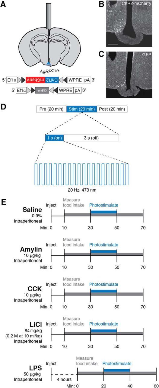
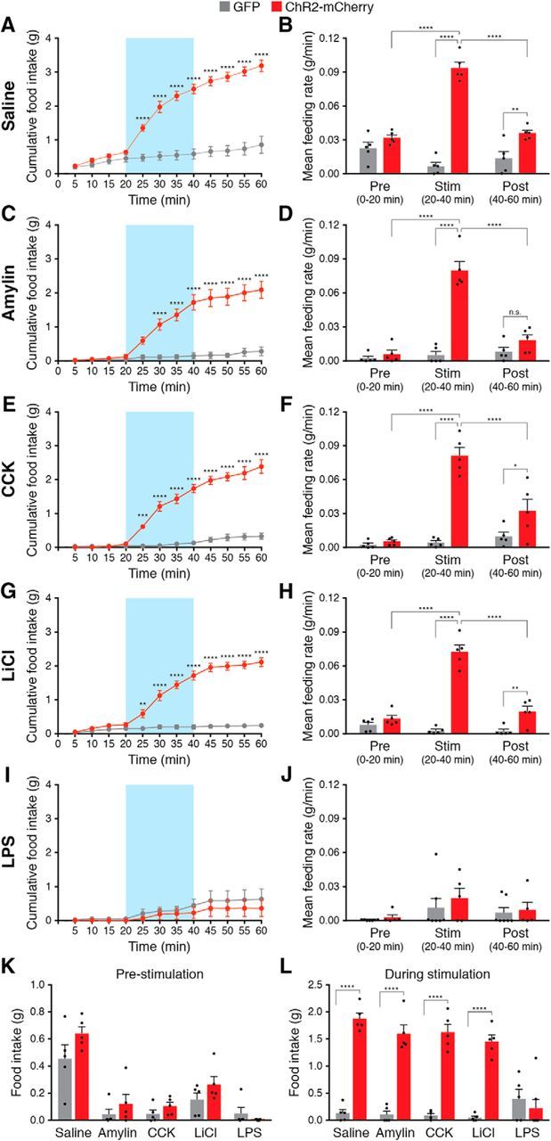
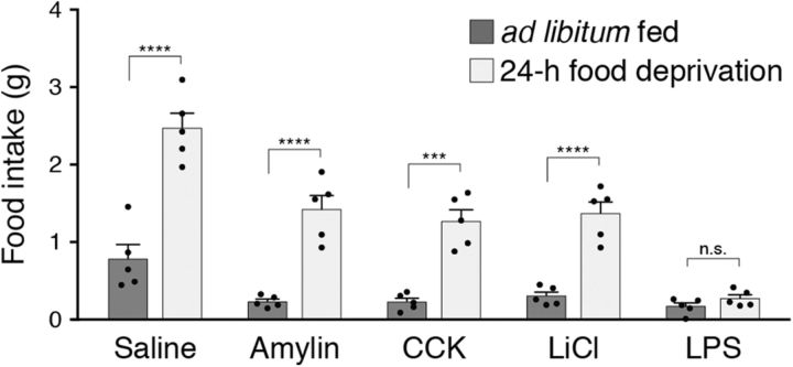
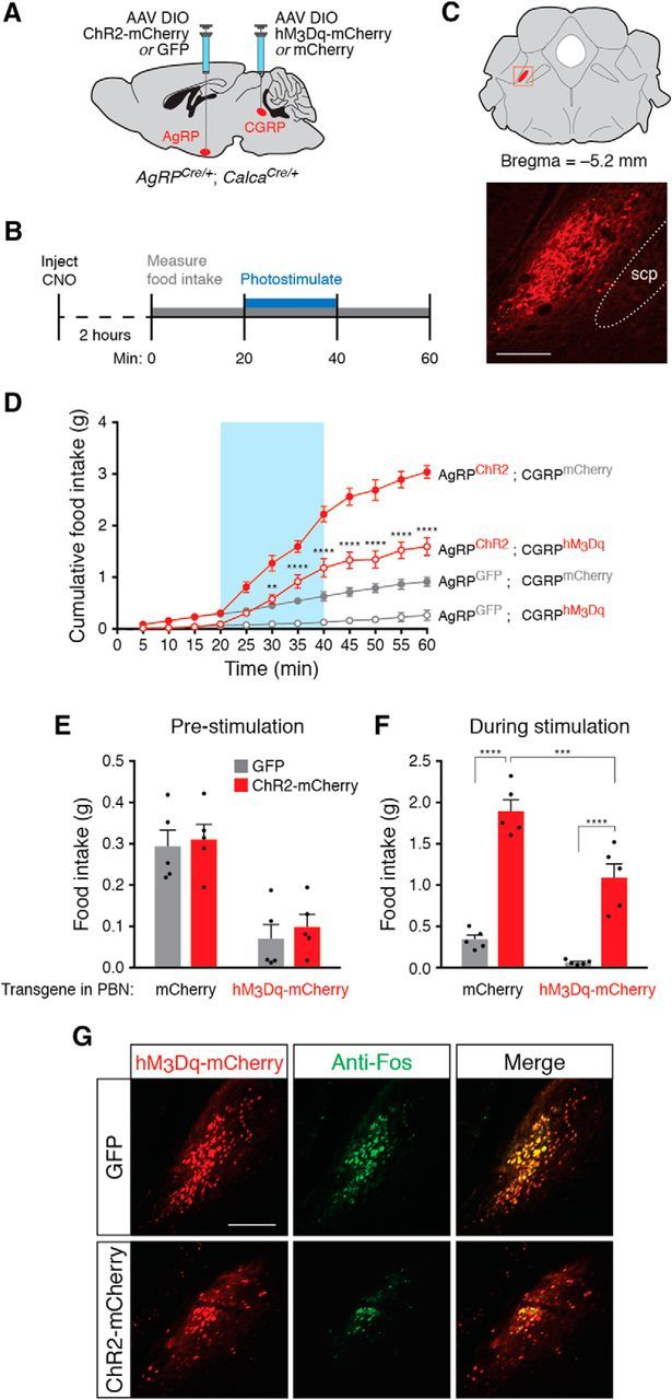
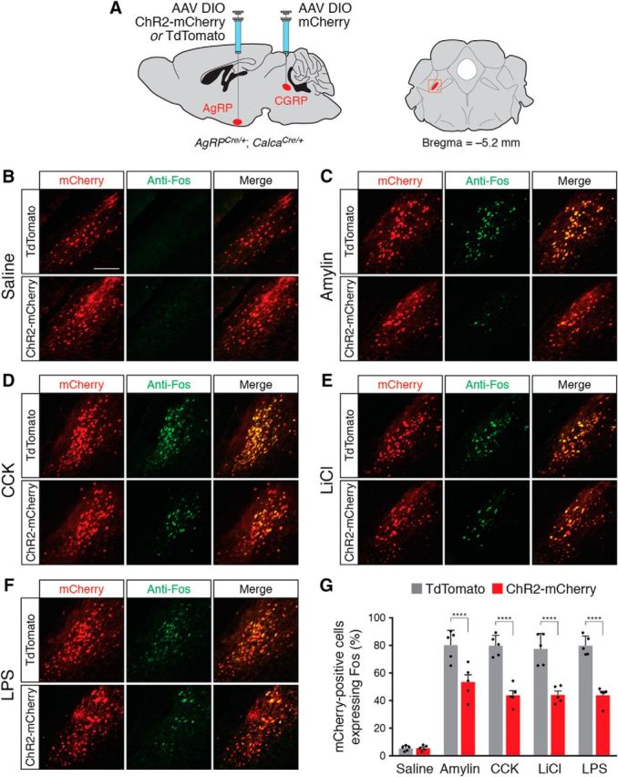
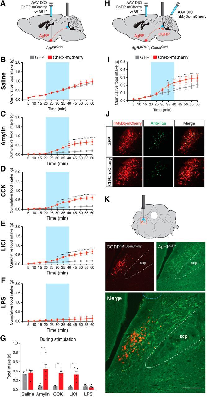
To maintain energy homeostasis, orexigenic (appetite-inducing) and anorexigenic (appetite suppressing) brain systems functionally interact to regulate food intake. Within the hypothalamus, neurons that express agouti-related protein (AgRP) sense orexigenic factors and orchestrate an increase in food-seeking behavior. In contrast, calcitonin gene-related peptide (CGRP)-expressing neurons in the parabrachial nucleus (PBN) suppress feeding. PBN CGRP neurons become active in response to anorexigenic hormones released following a meal, including amylin, secreted by the pancreas, and cholecystokinin (CCK), secreted by the small intestine. Additionally, exogenous compounds, such as lithium chloride (LiCl), a salt that creates gastric discomfort, and lipopolysaccharide (LPS), a bacterial cell wall component that induces inflammation, exert appetite-suppressing effects and activate PBN CGRP neurons. The effects of increasing the homeostatic drive to eat on feeding behavior during appetite suppressing conditions are unknown. Here, we show in mice that food deprivation or optogenetic activation of AgRP neurons induces feeding to overcome the appetite suppressing effects of amylin, CCK, and LiCl, but not LPS. AgRP neuron photostimulation can also increase feeding during chemogenetic-mediated stimulation of PBN CGRP neurons. AgRP neuron stimulation reduces Fos expression in PBN CGRP neurons across all conditions. Finally, stimulation of projections from AgRP neurons to the PBN increases feeding following administration of amylin, CCK, and LiCl, but not LPS. These results demonstrate that AgRP neurons are sufficient to increase feeding during noninflammatory-based appetite suppression and to decrease activity in anorexigenic PBN CGRP neurons, thereby increasing food intake during homeostatic need.SIGNIFICANCE STATEMENT The motivation to eat depends on the relative balance of activity in distinct brain regions that induce or suppress appetite. An abnormal amount of activity in neurons that induce appetite can cause obesity, whereas an abnormal amount of activity in neurons that suppress appetite can cause malnutrition and a severe reduction in body weight. The purpose of this study was to determine whether a population of neurons known to induce appetite ("AgRP neurons") could induce food intake to overcome appetite-suppression following administration of various appetite-suppressing compounds. We found that stimulating AgRP neurons could overcome various forms of appetite suppression and decrease neural activity in a separate population of appetite-suppressing neurons, providing new insights into how the brain regulates food intake.

Keywords: AgRP; CGRP; ChR2; appetite; food intake; parabrachial nucleus.


Figures

Figure 1.
Figure 1.
Optogenetic and pharmacological paradigms used to manipulate feeding behaviors. A, Diagram showing placement of a mono fiber-optic cannula above the arcuate nucleus and AAV constructs used to transduce AgRP neurons. Gray and black triangles represent loxP and lox2722 sites, respectively. B, C, Representative images showing unilateral expression of ChR2-mCherry (B) or GFP (C) in the arcuate nucleus. Dashed line shows approximate location of tips of fiber-optic implants. Scale bar, 250 μm. D, Photostimulation protocol. During the 20 min photostimulation period, 10 ms light pulses were delivered at 20 Hz for 1 s every 4 s. E, Injection timeline for food intake experiments.
Figure 2.
Figure 2.
AgRP neuron stimulation is sufficient to increase feeding following administration of noninflammatory anorexigenic compounds. Stimulation of AgRP neurons rapidly and reversibly increased cumulative food intake (left) and mean feeding rate (right) following administration of saline (A, B), amylin (C, D), CCK (E, F), and LiCl (G, H), but not following administration of LPS (I, J). Blue background represents the 20 min photostimulation period. All values represent the mean ± SEM, black dots represent individual animals (n = 5 animals per group). K, Total food consumed before stimulation of AgRP neurons across conditions. L, Total food consumed during stimulation of AgRP neurons across conditions. *p < 0.05, **p < 0.01, ***p < 0.001, ****p < 0.0001, Bonferroni post hoc tests between groups; n.s., not significant.
Figure 3.
Figure 3.
Food intake over 1 h following administration of anorexigenic compounds in ad libitum fed and 24 h food deprived animals. All values represent the mean ± SEM, black dots represent individual animals (n = 5 animals per group). ***p < 0.001, ****p < 0.0001, Bonferroni post hoc tests between feeding conditions; n.s., not significant.
Figure 4.
Figure 4.
AgRP neuron stimulation is sufficient to increase feeding during chemogenetic stimulation of PBN CGRP neurons. A, Viral injection strategy to transduce arcuate AgRP neurons and parabrachial CGRP neurons. B, Timeline of CNO administration and AgRP neuron photostimulation for each experimental trial. C, Top, Coronal diagram representing the location of PBN CGRP neurons; red square represents parabrachial region depicted in photomicrograph. Bottom, Representative image showing mCherry fluorescence in parabrachial CGRP neurons. scp, Superior cerebellar peduncle. Scale bar, 250 μm. D, Optogenetic stimulation of AgRP neurons increases food intake during CGRP neuron-mediated appetite suppression. All values represent the mean ± SEM (n = 5 animals per group). Blue background represents the 20 min photostimulation period. **p < 0.01, ****p < 0.0001, Bonferroni post hoc tests between AgRPGFP;CGRPhM3Dq and AgRPChR2;CGRPhM3Dq experimental groups at each time point. E, Total food consumed before stimulation of AgRP neurons across conditions. F, Total food consumed during stimulation of AgRP neurons across conditions. **p < 0.01, ****p < 0.0001, Bonferroni post hoc tests between groups. G, Representative histological images showing coincident hM3Dq-mCherry (red) and Fos (green) expression in CGRP neurons following injection of CNO in ChR2-mCherry- or GFP-transduced animals and photostimulation of AgRP neurons. Scale bar, 250 μm.
Figure 5.
Figure 5.
AgRP neuron stimulation reduces Fos expression in PBN CGRP neurons following administration of anorexigenic compounds. A, Injection strategy in AgRPCre/+; CalcaCre/+ double knock-in mice. Right, Coronal diagram representing the location of CGRP neurons; red square represents parabrachial region depicted in photomicrographs. BF, Representative histological images showing coincident mCherry (red) and Fos (green) expression in CGRP neurons following injection of saline (B), amylin (C), CCK (D), LiCl (E), and LPS (F), in ChR2-mCherry- or TdTomato-transduced animals, following photostimulation of AgRP neurons. Scale bar, 250 μm. G, Quantification of coexpression of mCherry and Fos in the parabrachial nucleus. All values represent the mean ± SD, black dots represent individual animals (n = 5 animals per group). ****p < 0.0001, Bonferroni post hoc tests between TdTomato- and ChR2-mCherry-transduced animals.
Figure 6.
Figure 6.
Stimulation of projections from AgRP neurons to parabrachial neurons increases feeding following administration of noninflammatory anorexigenic compounds. A, Injection strategy in AgRPCre/+ mice for AgRP-to-PBN projection experiments. B, Stimulation of AgRP neuron projections has no effect on baseline food intake following administration of saline. Blue background represents the 20 min photostimulation period. CF, Stimulation of AgRP neuron projections increases food intake following administration of amylin (C), CCK (D), and LiCl (E), but not following administration of LPS (F). *p < 0.05, **p < 0.01, ***p < 0.001, ****p < 0.0001, Bonferroni post hoc tests between groups. G, Total food consumed during stimulation of AgRP neurons across conditions. All values represent the mean ± SEM, black dots represent individual animals (n = 5 animals per group). **p < 0.01, ***p < 0.001, Bonferroni post hoc tests between groups. H, Injection strategy in AgRPCre/+; CalcaCre/+ double knock-in mice for AgRP-to-PBN projection experiments with hM3Dq-mediated stimulation of CGRP neurons. I, Stimulation of AgRP neuron projections increases food intake following injection of CNO. **p < 0.01, ***p < 0.001, Bonferroni post hoc tests between groups. J, Representative histological images showing coincident hM3Dq-mCherry (red) and Fos (green) expression in CGRP neurons following injection of CNO in ChR2-mCherry- or GFP-transduced animals and photostimulation of AgRP neurons. Scale bar, 250 μm. K, AgRP neuron projections throughout the lateral parabrachial nucleus. Top, Coronal diagram representing the location of CGRP neurons; red square represents parabrachial region depicted in photomicrographs. Bottom, Representative images showing hM3Dq-mCherry transduced CGRP neurons and GFP-transduced AgRP fiber projections in the lateral parabrachial nucleus. scp, Superior cerebellar peduncle. Light blue dashed line at top of bottom image shows approximate location of tips of fiber-optic implants. Scale bar, 250 μm.
Figure 7.
Figure 7.
Model of functional anatomy of AgRP neuron projections. AgRP neurons increase food intake by projecting to downstream populations (green connections) that initiate feeding including the bed nucleus of the stria terminalis (BNST), lateral hypothalamus (LH), paraventricular hypothalamic nucleus (PVH), and paraventricular thalamic nucleus (PVT). AgRP neurons also inhibit other nonfeeding behavioral/physiological states by projecting to downstream regions (red connections) such as the MeA to inhibit fear and aggression or arcuate kisspeptin neurons (Kiss) to inhibit reproduction. Our results demonstrate that AgRP neurons disengage noninflammatory appetite-suppressing states by projecting to the PBN (dark red connections).

Similar articles

Cited by

References

    1. Aponte Y, Atasoy D, Sternson SM (2011) AGRP neurons are sufficient to orchestrate feeding behavior rapidly and without training. Nat Neurosci 14:351–355. 10.1038/nn.2739 - DOI - PMC - PubMed
    1. Atasoy D, Betley JN, Su HH, Sternson SM (2012) Deconstruction of a neural circuit for hunger. Nature 488:172–177. 10.1038/nature11270 - DOI - PMC - PubMed
    1. Becskei C, Grabler V, Edwards GL, Riediger T, Lutz TA (2007) Lesion of the lateral parabrachial nucleus attenuates the anorectic effect of peripheral amylin and CCK. Brain Res 1162:76–84. 10.1016/j.brainres.2007.06.016 - DOI - PubMed
    1. Betley JN, Cao ZF, Ritola KD, Sternson SM (2013) Parallel, redundant circuit organization for homeostatic control of feeding behavior. Cell 155:1337–1350. 10.1016/j.cell.2013.11.002 - DOI - PMC - PubMed
    1. Betley JN, Xu S, Cao ZF, Gong R, Magnus CJ, Yu Y, Sternson SM (2015) Neurons for hunger and thirst transmit a negative-valence teaching signal. Nature 521:180–185. 10.1038/nature14416 - DOI - PMC - PubMed

Publication types