. 2017 Sep 6;37(36):8816-8829.
doi: 10.1523/JNEUROSCI.2125-16.2017. Epub 2017 Aug 8.

GABAergic Interneuron Differentiation in the Basal Forebrain Is Mediated through Direct Regulation of Glutamic Acid Decarboxylase Isoforms by Dlx Homeobox Transcription Factors

Affiliations

GABAergic Interneuron Differentiation in the Basal Forebrain Is Mediated through Direct Regulation of Glutamic Acid Decarboxylase Isoforms by Dlx Homeobox Transcription Factors

Trung N Le et al. J Neurosci. .

Abstract

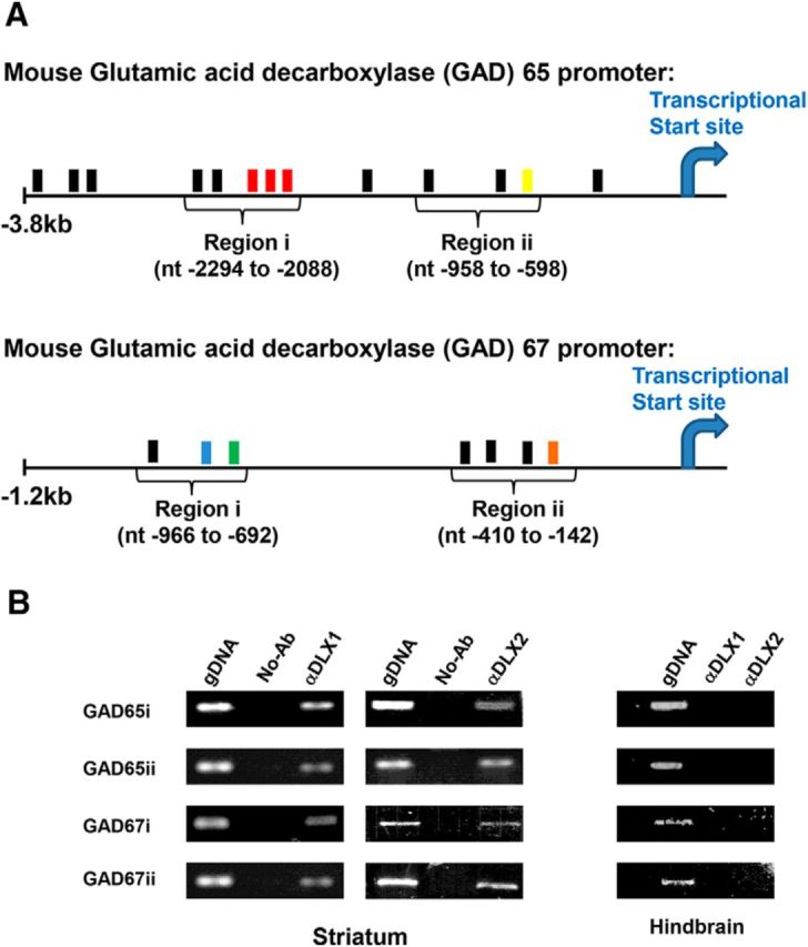
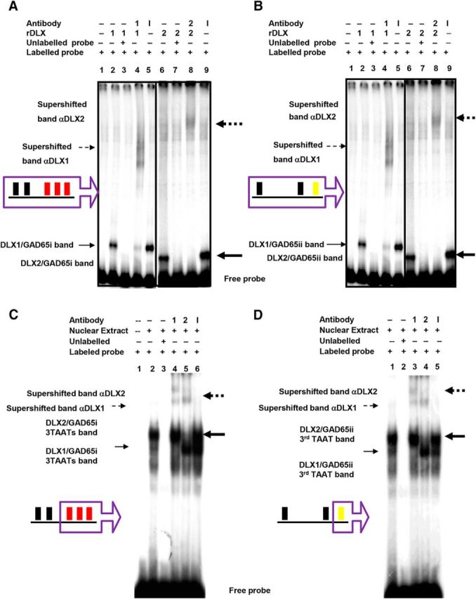
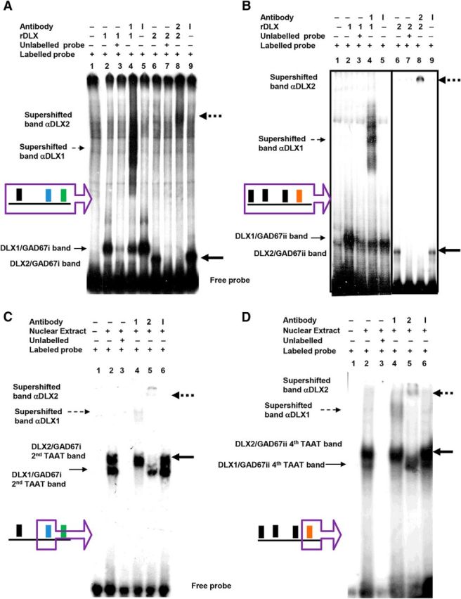
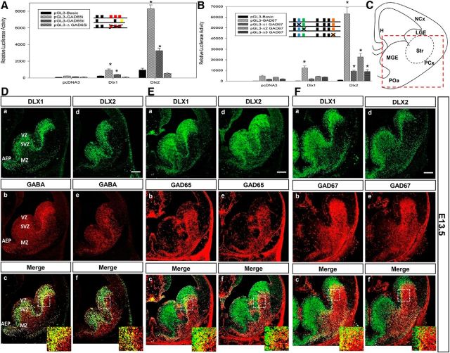
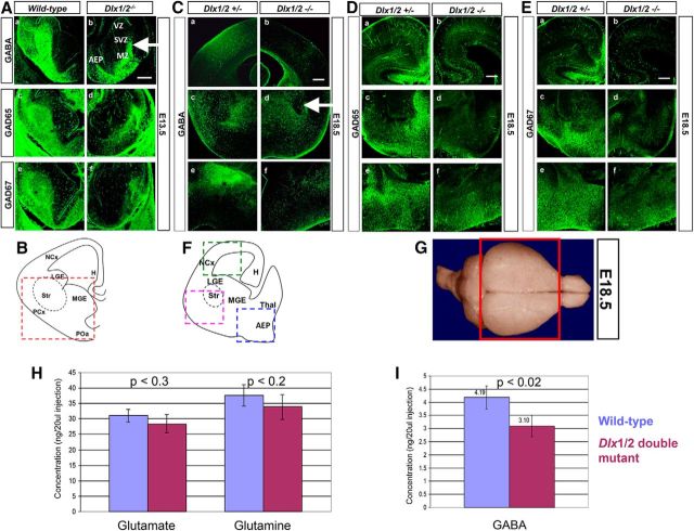
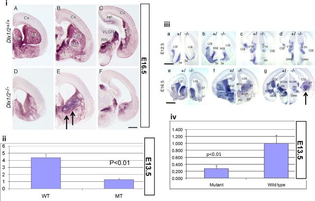
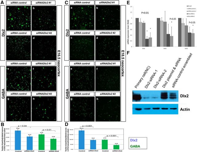
GABA is the key inhibitory neurotransmitter in the cortex but regulation of its synthesis during forebrain development is poorly understood. In the telencephalon, members of the distal-less (Dlx) homeobox gene family are expressed in, and regulate the development of, the basal ganglia primodia from which many GABAergic neurons originate and migrate to other forebrain regions. The Dlx1/Dlx2 double knock-out mice die at birth with abnormal cortical development, including loss of tangential migration of GABAergic inhibitory interneurons to the neocortex (Anderson et al., 1997a). We have discovered that specific promoter regulatory elements of glutamic acid decarboxylase isoforms (Gad1 and Gad2), which regulate GABA synthesis from the excitatory neurotransmitter glutamate, are direct transcriptional targets of both DLX1 and DLX2 homeoproteins in vivo Further gain- and loss-of-function studies in vitro and in vivo demonstrated that both DLX1 and DLX2 are necessary and sufficient for Gad gene expression. DLX1 and/or DLX2 activated the transcription of both Gad genes, and defects in Dlx function disrupted the differentiation of GABAergic interneurons with global reduction in GABA levels in the forebrains of the Dlx1/Dlx2 double knock-out mouse in vivo Identification of Gad genes as direct Dlx transcriptional targets is significant; it extends our understanding of Dlx gene function in the developing forebrain beyond the regulation of tangential interneuron migration to the differentiation of GABAergic interneurons arising from the basal telencephalon, and may help to unravel the pathogenesis of several developmental brain disorders.SIGNIFICANCE STATEMENT GABA is the major inhibitory neurotransmitter in the brain. We show that Dlx1/Dlx2 homeobox genes regulate GABA synthesis during forebrain development through direct activation of glutamic acid decarboxylase enzyme isoforms that convert glutamate to GABA. This discovery helps explain how Dlx mutations result in abnormal forebrain development, due to defective differentiation, in addition to the loss of tangential migration of GABAergic inhibitory interneurons to the neocortex. Reduced numbers or function of cortical GABAergic neurons may lead to hyperactivity states such as seizures (Cobos et al., 2005) or contribute to the pathogenesis of some autism spectrum disorders. GABAergic dysfunction in the basal ganglia could disrupt the learning and development of complex motor and cognitive behaviors (Rubenstein and Merzenich, 2003).

Keywords: GABA; forebrain development; glutamic acid decarboxylase; homeobox; interneuron.


Figures

Figure 1.
Figure 1.
Candidate DLX target sequences within Gad1 (GAD67) and Gad2 (GAD65) promoter regions were identified through the presence of canonical homeodomain DNA binding motifs within the Gad promoters and confirmed by chromatin immunoprecipitation (ChIP). A, The specific regions within the regulatory elements of the mouse GAD65/Gad2 (GenBank AB032757; Gad2 NM_008078.2) and GAD67/Gad1 (GenBank Z49978; Gad1 NM_008077.5) promoters, designated GAD65i/ii and GAD67i/ii, contain putative homeodomain DNA-binding sites (TAAT/ATTA), shown in filled boxes. DLX proteins occupy colored boxes and do not occupy black boxes, respectively, based on subsequent experiments. Nucleotide positions are indicated with respect to transcriptional start sites. B, DLX1 and DLX2 occupy the Gad1 and Gad2 promoters in E13.5 GE tissues using ChIP. ChIP assays of GAD65/Gad2 and GAD67/Gad1 promoter regions showed that DLX1 and DLX2 occupy both regions (region i and region ii) of each Gad gene promoter using E13 GE tissues and specific DLX1 and DLX2 antibodies. Negative controls used no addition of antibodies and/or E13.5 hindbrain tissues. The positive controls used E13.5 genomic DNA. PCR bands were subcloned and sequenced for confirmation.
Figure 2.
Figure 2.
DLX1 and DLX2 proteins specifically bind to Gad65/Gad2 promoter regions i and ii in situ. EMSA showed recombinant DLX1 or DLX2 binding to α-P32-labeled (A) Gad65 promoter region i, and (B) Gad65 promoter region ii oligonucleotides containing homeodomain binding sites (Fig. 1A, regions in purple boxes). GE nuclear-extracted proteins also bound to α-P32-labeled oligonucleotides containing specific TAAT/ATTA binding motif for (C) Gad65 region i 3 TAAT motifs, and for (D) Gad65 region ii third TAAT motif (Fig. 1A, motifs in purple boxes). A, B, Radioactive oligonucleotide probes were incubated alone (lane 1), with DLX1 recombinant proteins (lanes 2–5), with DLX2 recombinant proteins (lanes 6–9), with unlabeled competitive probes (lane 3, 7), with specific DLX1 or 2 antibodies (lanes 4, 8), and with nonspecific antibodies (lanes 5, 9). C, D, Radioactive oligonucleotide probes were incubated alone (lane 1), with GE nuclear extract (lanes 2–6), with unlabeled competitive probes (lane 3), with specific DLX1 antibody (lane 4), with specific DLX2 antibody (lane 5), with nonspecific antibodies (lane 6). AD, Binding of DLX proteins to a specific oligonucleotide sequence results in a gel shifted band, indicated by solid arrows. Binding of DLX protein to a specific oligonucleotide and to a specific DLX antibody resulted in a gel supershifted band, indicated by broken arrows. (DLX1: unbold arrow, DLX2: bold arrow). r, Recombinant; 1, DLX1 protein or anti-DLX1 antibody; 2, DLX2 protein or anti-DLX2 antibody; I, irrelevant/nonspecific antibody.
Figure 3.
Figure 3.
DLX1 and DLX2 proteins specifically bind to Gad67/Gad1 promoter regions i and ii in situ. EMSA showed recombinant DLX1 or DLX2 binding to α-P32-labeled (A) Gad67 promoter region i, and (B) Gad67 promoter region ii oligonucleotides containing candidate homeodomain binding sites (Fig. 1A, regions in purple boxes). GE nuclear-extracted proteins also bound to α-P32-labeled oligonucleotides containing specific TAAT/ATTA binding motif for (C) the Gad67 region i second TAAT motif, and for (D) the Gad67 region ii fourth TAAT motif (Fig. 1A, motifs in purple boxes). A, B, Radioactive oligonucleotide probes were incubated alone (lane 1), with recombinant DLX1 proteins (lanes 2–5), with recombinant DLX2 proteins (lanes 6–9), with unlabeled competitive probes (lanes 3, 7), with specific DLX1 or 2 antibodies (lanes 4, 8), and with nonspecific antibodies (lanes 5, 9). C, D, radioactive oligonucleotide probes were incubated alone (lane 1), with GE nuclear extract (lanes 2–6), with unlabeled competitive probes (lane 3), with specific DLX1 antibody (lane 4), with specific DLX2 antibody (lane 5), with nonspecific antibodies (lane 6). AD, Binding of DLX proteins to a specific oligonucleotide sequence results in a gel shifted band, indicated by solid arrows. Binding of DLX protein to a specific oligonucleotide and to a specific DLX antibody results in a gel supershifted band, indicated by broken arrows. (DLX1: unbold arrow, DLX2: bold arrow). r, Recombinant; 1, DLX1 protein or anti-DLX1 antibody; 2, DLX2 protein or anti-DLX2 antibody; I, irrelevant/nonspecific antibody.
Figure 4.
Figure 4.
A, B, Dlx1 and Dlx2 activate transcription of Gad1 (Gad67) and Gad2 (Gad65) reporter constructs in vitro. Transient transfection assays in C6 glioma cells with (A) Gad65 promoter regions i or ii, and (B) Gad67 1.3 kb promoter constructs (contains regions i and ii; Fig. 1A), containing homeodomain binding sites cloned into a pGL3-luciferase reporter construct, were performed in the absence or presence of DLX1 or DLX2 coexpression. DLX1 and DLX2 activate transcription of the reporter genes using the Gad65 and Gad67 promoters, with DLX2 as a more robust activator. Mutations of specific TAAT/ATTA binding motifs within (A) Gad65 or (B) Gad67 promoter sequences lead to a significant reduction of transcriptional activation of these reporter gene constructs. All luciferase activities were relative and normalized to the activity level of the internal control β-gal. Average ± SEM of at least triplicate experiments. *p < 0.05. ΔGAD65i, Mutation of 3 TAATs of Gad65 region I; Δ1GAD67, mutation of the second TAAT of Gad67 region I; Δ2GAD67, mutation of the third TAAT of Gad67 region I; Δ3GAD67, mutation of the fourth TAAT of Gad67 region ii. CF, Coexpression of DLX homeodomain proteins and the GABA neurotransmitter in wild-type E13.5 basal telencephalon. C, Schematic diagram of coronal section of the E13.5 forebrain, showing basal telencephalon in red dashed box. Sections were double-labeled with specific antibodies against DLX1 (Da, Ea, Fa), DLX2 (Dd, Ed, Fd), GABA or GAD65 or GAD67 (Db, Eb, Fb, De, Ee, Fe) of E13.5 ganglionic eminences. Da, Ea, Fa, Dd, Ed, Fd, DLX1- or DLX2-positive cells (green) in the VZ and SVZ of the LGE, MGE, and AEP. Db, Eb, Fb, De, Ed, Fe, GABA or GAD65 or GAD67-labeled cells (red) in the same tissue sections throughout the basal telencephalon, predominantly in SVZ and MZ of the LGE and AEP. Bottom, The overlay of the two images with GABA/GAD65/GAD67 coexpressed with DLX proteins in most SVZ interneurons (yellow). Scale bar, 200 μm. Insets, Representative of a ∼10× enlargement of the dashed inset box to better demonstrate colabeled cells. H, Hippocampus; LGE, lateral ganglionic eminence; NCx, neocortex; PCx, paleocortex; POa, anterior preoptic area; Str, striatum.
Figure 5.
Figure 5.
AF, GABA expression in the developing forebrain of Dlx1/2 wild-type compared with Dlx1/2 DKO mice. A, In the E13.5 wild-type, GABA (Aa), GAD65 (Ac), and GAD67 (Ae) expression is predominantly localized to the SVZ and MZ of the AEP and LGE compared with that in the absence of Dlx1 and Dlx2 function (Ab, Ad, Af). B, Schematic diagram of coronal section of the E13.5 forebrain, showing basal telencephalon in red dashed box. In the E18.5 wild-type, (C) GABA, (D) GAD65, (E) GAD67 expression is shown in neocortex (Ba), striatum (Bc), and AEP (Be) compared with the Dlx1/2 double mutant (Bb, Bd, Bf). F, Schematic diagram of coronal section of the E18.5 forebrain, showing neocortex (green dashed box), striatum (pink dshed box), and AEP (blue dashed box). Scale bars, 200 μm. H, Hippocampus; LGE, lateral ganglionic eminence; NCx, neocortex; PCx, paleocortex; POa, anterior preoptic area; Str, striatum. GI, GABA levels are reduced in the Dlx1/2-null forebrains using HPLC. G, Forebrains of wild-type and Dlx1/2 double-mutant were dissected as depicted in the red box of the diagram. H, In the E18.5 forebrain, glutamate and glutamine (precursors of GABA) levels do not change when comparing wild-type and Dlx1/2 knock-out forebrain (excluding the olfactory bulbs). I, GABA neurotransmitter level decreases by ∼26% in the Dlx1/2 knock-out compared with wild-type forebrain. Homoserine was used as internal control. Sample concentrations were measured from standard curves and expressed as nanogram per 20 μl injection into the HPLC apparatus. Average ± SEM with associated p values.
Figure 6.
Figure 6.
Expression and level of Gad mRNA isoforms in the rostral and caudal forebrain in the wild-type and Dlx1/2 DKO demonstrated by digoxigenin in situ hybridization and quantitative real-time PCR. i, Gad65 isoform mRNA is expressed throughout the basal forebrain in a diffuse pattern in the wild-type (iAiC, from rostral to caudal) compared with the Dlx1/2 knock-out forebrain (iDiF, from rostral to caudal). Black arrows depicted ectopic accumulation of Gad65 mRNA in the basal ganglia. ii, Gad65 mRNA level was examined by real-time PCR in the basal ganglia and compared between wild-type (WT) and double-mutant (MT) littermates at E13.5. iii, Gad67 isoform mRNA is also expressed throughout the basal forebrain in a diffuse pattern in the wild-type (iiiaiiig, left, from rostral to caudal) and compared with Dlx1/2 DKO forebrain (iiiaiiig, right, from rostral to caudal). Black arrow depicted accumulation of Gad67 mRNA in the embryonic striatum at E16.5. iv, Gad67 mRNA levels were examined by quantitative real-time PCR in the basal ganglia and compared between wild-type and double mutant littermates at E13.5. The real-time PCR results show relative fold differences between wild-type and mutant littermates, and were normalized using the house keeping gene GAPDH as an internal control. Average ± SEM with associated p values. CGE, Caudal ganglionic eminence; LGE, lateral ganglionic eminence; MGE, medial ganglionic eminence; ST, striatum; VTH, ventral thalamus; AH, anterior hypothalamic nucleus; DMH, dorsal medial nucleus; OT, olfactory tract; SE, septum; VP, ventral pallidum; PO, preoptic area; VLGN, ventral lateral geniculate nucleus; ZI, zona incerta nucleus; RN, red nucleus. Scale bars: iAiF, iiiaiiid, 200 μm; iiie–iiig, 400 μm .
Figure 7.
Figure 7.
AD, Knockdown of Dlx2 expression by siRNA in E16.5 and E18.5 embryonic neocortical primary cultures. Primary dissociated forebrain cultures of neocortex (A, C) at E16.5 (A) and E18.5 (C) were transfected with a duplex control scrambled siRNA or two different duplex siRNAs targeting Dlx2 coding sequences. Transfection of duplex siRNA targeting Dlx2 reduced DLX2 expression (green; Ab, Ad, Cb, Cd) and concomitantly decreased GABA expression (green; Af, Ah, Cf, Ch) in embryonic neocortical primary cultures compared with control scrambled siRNA-transfected cells (Aa, Ac, Ae, Ag, Ca, Cc, Ce, Cg). Scale bars, 200 μm. E, Using quantitative RT-PCR, different versions of siRNA (siRNA constructs 1 and 2) knocked down Dlx2 expression in primary embryonic neocortical (NC) cultures, compared with a control scrambled siRNA control (p < 0.01). Controls were performed separately for each siRNA construct to Dlx2. Embryonic neocortical cells endogenously express DLX2, and were used as a positive control. Concomitantly, transfection with either Dlx2 siRNA resulted in statistically significant decreases in Gad1 and Gad2 expression (p < 0.05). F, For testing of siRNA transfection efficiency, cotransfection of both Dlx2 siRNAs and the siRNA control, and pooled Dlx2 siRNA with pcDNA3/Dlx2 plasmid was also performed in embryonic NC cells. Western analysis with anti-DLX2 antibody showed that different versions of siRNA (siRNA constructs 1 and 2) knocked down DLX2 expression compared with endogenous DLX2 levels. Coexpression of a Dlx2 expression plasmid with Dlx2 siRNA rescued DLX2 expression. An siRNA control (scrambled siRNA) and untransfected embryonic neocortical lysate were used as controls, respectively. β-Actin was used as loading control. B, D, Quantification of DLX2- and GABA-positive cells following reduction of DLX2 expression mediated by duplex siRNA in E16.5 (B) and E18.5 (D) neocortical primary cultures (n = 4). DLX2- or GABA-positive cells were counted and compared with the total of number of immunopositive cells transfected with scrambled siRNA control. All comparisons were performed in at least three trials. Average ± SEM with associated p values.

Similar articles

Cited by

References

    1. Al-Jaberi N, Lindsay S, Sarma S, Bayatti N, Clowry GJ (2015) The early fetal development of human neocortical GABAergic interneurons. Cereb Cortex 25:631–645. 10.1093/cercor/bht254 - DOI - PMC - PubMed
    1. Anderson SA, Eisenstat DD, Shi L, Rubenstein JL (1997a) Interneuron migration from basal forebrain to neocortex: dependence on Dlx genes. Science 278:474–476. 10.1126/science.278.5337.474 - DOI - PubMed
    1. Anderson SA, Qiu M, Bulfone A, Eisenstat DD, Meneses J, Pedersen R, Rubenstein JL (1997b) Mutations of the homeobox genes Dlx-1 and Dlx-2 disrupt the striatal subventricular zone and differentiation of late born striatal neurons. Neuron 19:27–37. 10.1016/S0896-6273(00)80345-1 - DOI - PubMed
    1. Anderson SA, Kaznowski CE, Horn C, Rubenstein JL, McConnell SK (2002) Distinct origins of neocortical projection neurons and interneurons in vivo. Cereb Cortex 12:702–709. 10.1093/cercor/12.7.702 - DOI - PubMed
    1. Anderson SA, Marín O, Horn C, Jennings K, Rubenstein JL (2001) Distinct cortical migrations from the medial and lateral ganglionic eminences. Development 128:353–363. - PubMed

Publication types

MeSH terms