. 2017 Aug 18;7(1):8753.
doi: 10.1038/s41598-017-09326-7.

Osteocyte regulation of orthodontic force-mediated tooth movement via RANKL expression

Affiliations

Osteocyte regulation of orthodontic force-mediated tooth movement via RANKL expression

Ayumi Shoji-Matsunaga et al. Sci Rep. .

Abstract

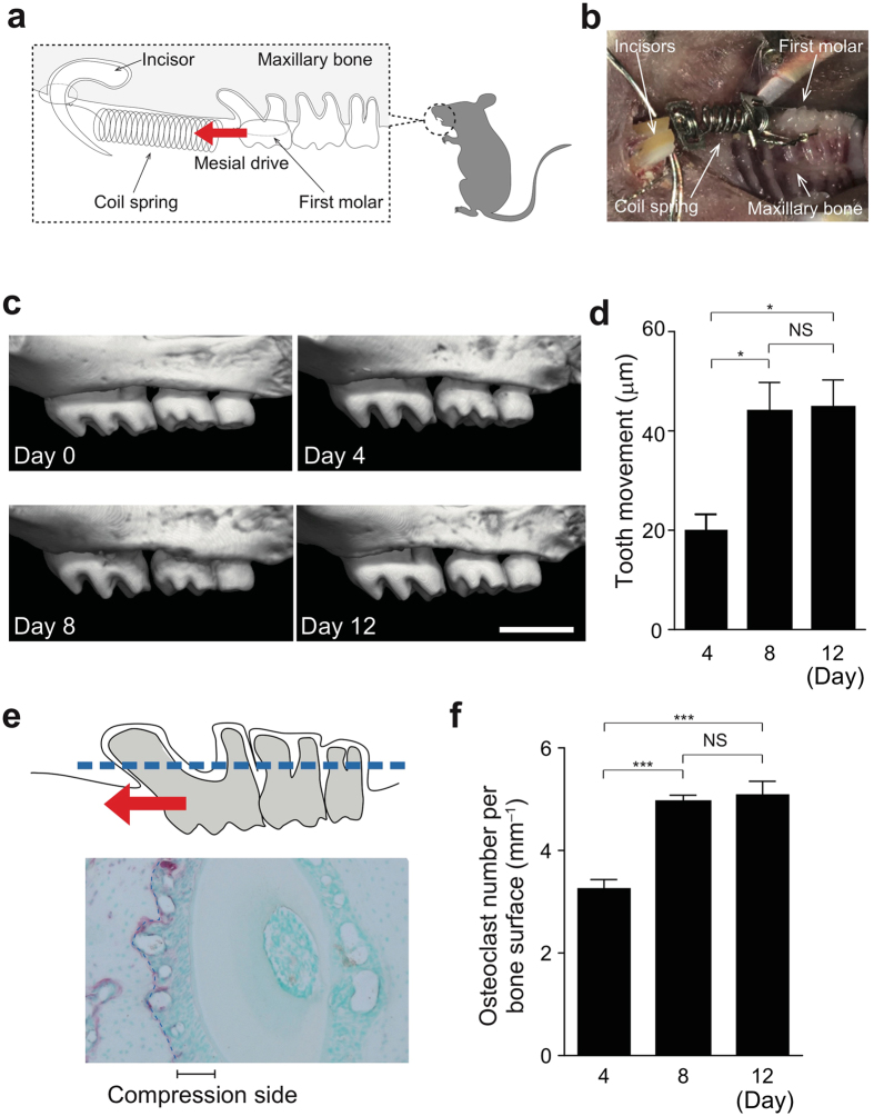
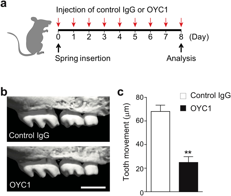
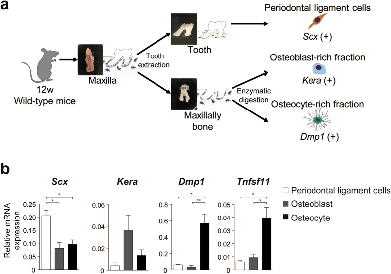
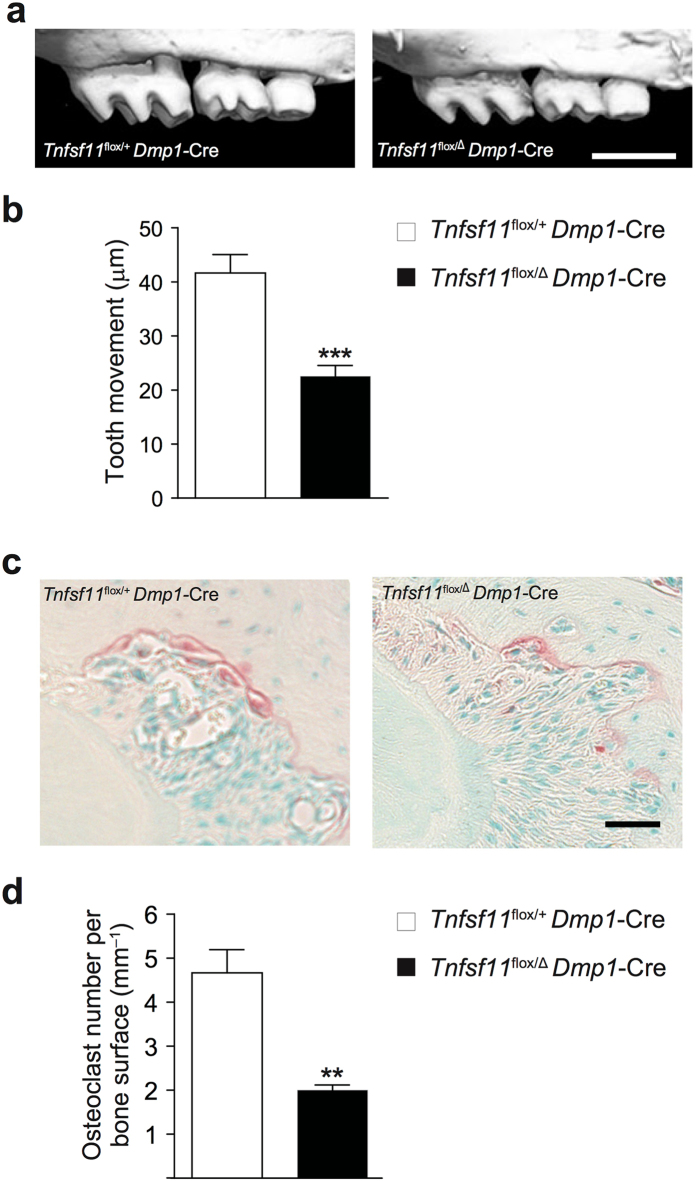
Orthodontic tooth movement is achieved by the remodeling of the alveolar bone surrounding roots of teeth. Upon the application of orthodontic force, osteoclastic bone resorption occurs on the compression side of alveolar bone, towards which the teeth are driven. However, the molecular basis for the regulatory mechanisms underlying alveolar bone remodeling has not been sufficiently elucidated. Osteoclastogenesis is regulated by receptor activator of nuclear factor-κB ligand (RANKL), which is postulated to be expressed by the cells surrounding the tooth roots. Here, we show that osteocytes are the critical source of RANKL in alveolar bone remodeling during orthodontic tooth movement. Using a newly established method for the isolation of periodontal tissue component cells from alveolar bone, we found that osteocytes expressed a much higher amount of RANKL than other cells did in periodontal tissue. The critical role of osteocyte-derived RANKL was confirmed by the reduction of orthodontic tooth movement in mice specifically lacking RANKL in osteocytes. Thus, we provide in vivo evidence for the key role of osteocyte-derived RANKL in alveolar bone remodeling, establishing a molecular basis for orthodontic force-mediated bone resorption.


Conflict of interest statement

The authors declare that they have no competing interests.

Figures

Figure 1
Figure 1
Experimental tooth movement model. (a) Schematic diagram of orthodontic tooth movement. The upper first molar was moved mesially by a closed–coil spring. The red arrow indicates the direction of orthodontic force. (b) Intra–oral photograph. (c) Tooth movement during the experiment time course. Representative micro-CT images in wild–type mice after spring insertion (0, 4, 8 and 12 days). (d) Quantification of tooth movement in each group (n = 3 per time point). (e) Measurement of osteoclasts on alveolar bone around the distal buccal root of the upper first molar (area enclosed by the dotted line). (f) The number of osteoclasts on the alveolar bone surface (n = 3 per time point). Scale bar: 1 mm. Error bars, means ± s.e.m.; *P < 0.05; ***P < 0.001; NS, not significant.
Figure 2
Figure 2
Effect of anti-mouse RANKL monoclonal antibody (OYC1) on tooth movement. (a) Schematic diagram of the injection time course. OYC1 or control IgG was injected locally into the buccal and palatal gingiva around the upper left first molar of wild-type mice. (b) Representative micro-CT images of a moved tooth after treatment of OYC1 or control IgG. (c) Quantification of tooth movement in each group (n = 3–4). Error bars, means ± s.e.m.; **P < 0.01.
Figure 3
Figure 3
RANKL expression in periodontal tissues. (a) Establishment of isolation of cells from periodontal tissue. Periodontal ligament cells, osteoblasts and osteocytes were collected according to the schematic flow chart (see methods). (b) Profiling of gene expression in periodontal ligament cells, osteoblasts and osteocytes (quantitative RT-PCR analysis) (n = 5 per each fraction). Statistical analysis of Dmp1 expression was carried out using Kruskal-Wallis test and Dunn’s multiple comparison test. Error bars, means ± s.e.m.; *P < 0.05; **P < 0.01.
Figure 4
Figure 4
Tooth movement in osteocyte-specific RANKL deficient mice. (a) Representative micro-CT images of a moved tooth. Tnfsf11 flox/+ Dmp1-Cre mice were used as a control. (b) Quantification of tooth movement in each group (n = 6–7). (c) TRAP staining of the compression side of the palatal roots. (d) The number of osteoclasts on the alveolar bone surface. Scale bars: (a), 1 mm; (c), 50 μm. Error bars, means ± s.e.m.; **P < 0.01; ***P < 0.001.

References

    1. Nakashima T, Hayashi M, Takayanagi H. New insights into osteoclastogenic signaling mechanisms. Trends Endocrinol. Metab. 2012;23:582–590. doi: 10.1016/j.tem.2012.05.005. - DOI - PubMed
    1. Takayanagi H. Osteoimmunology: shared mechanisms and crosstalk between the immune and bone systems. Nat. Rev. Immunol. 2007;7:292–304. doi: 10.1038/nri2062. - DOI - PubMed
    1. Baron R, Hesse E. Update on bone anabolics in osteoporosis treatment: Rationale, current status, and perspectives. J. Clin. Endocrinol. Metab. 2012;97:311–325. doi: 10.1210/jc.2011-2332. - DOI - PMC - PubMed
    1. Seeman E, Delmas PD. Bone quality–the material and structural basis of bone strength and fragility. N. Engl. J. Med. 2006;354:2250–2261. doi: 10.1056/NEJMra053077. - DOI - PubMed
    1. Dallas SL, Prideaux M, Bonewald LF. The osteocyte: An endocrine cell… and more. Endocr. Rev. 2013;34:658–690. doi: 10.1210/er.2012-1026. - DOI - PMC - PubMed

Publication types