. 2017 Aug 18;7(1):8843.
doi: 10.1038/s41598-017-09414-8.

Fluticasone/salmeterol reduces remodelling and neutrophilic inflammation in severe equine asthma

Affiliations

Fluticasone/salmeterol reduces remodelling and neutrophilic inflammation in severe equine asthma

Michela Bullone et al. Sci Rep. .

Abstract

Asthmatic airways are inflamed and undergo remodelling. Inhaled corticosteroids and long-acting β2-agonist combinations are more effective than inhaled corticosteroid monotherapy in controlling disease exacerbations, but their effect on airway remodelling and inflammation remains ill-defined. This study evaluates the contribution of inhaled fluticasone and salmeterol, alone or combined, to the reversal of bronchial remodelling and inflammation. Severely asthmatic horses (6 horses/group) were treated with fluticasone, salmeterol, fluticasone/salmeterol, or with antigen avoidance for 12 weeks. Lung function, central and peripheral airway remodelling, and bronchoalveolar inflammation were assessed. Fluticasone/salmeterol and fluticasone monotherapy decreased peripheral airway smooth muscle remodelling after 12 weeks (p = 0.007 and p = 0.02, respectively). On average, a 30% decrease was observed with both treatments. In central airways, fluticasone/salmeterol reversed extracellular matrix remodelling after 12 weeks, both within the lamina propria (decreased thickness, p = 0.005) and within the smooth muscle layer (p = 0.004). Only fluticasone/salmeterol decreased bronchoalveolar neutrophilia (p = 0.03) to the same extent as antigen avoidance already after 8 weeks. In conclusion, this study shows that fluticasone/salmeterol combination decreases extracellular matrix remodelling in central airways and intraluminal neutrophilia. Fluticasone/salmeterol and fluticasone monotherapy equally reverse peripheral airway smooth muscle remodelling.


Conflict of interest statement

The authors declare that they have no competing interests.

Figures

Figure 1
Figure 1
Pulmonary function and bronchoreversibility tests performed in study I (a,b) and II (c,d). Bronchodilator response was evaluated before and 30 minutes after the administration of inhaled albuterol 500 μg (b) or intravenous N-butyl-scopolamine 0.03 mg/kg (d) to detect residual airway obstruction. Values are expressed as means ± S.E.M. Dashed lines represent normal threshold in healthy horses. RL: pulmonary resistance; EL: pulmonary elastance; ΔPL: swing in transpulmonary pressure. ‡different from baseline of the same group for fluticasone/salmeterol (p < 0.0001), ¤different from baseline for antigen avoidance (p < 0.05), *different from baseline for fluticasone (p < 0.0001), #different from baseline for salmeterol (p < 0.0001), §difference between groups at the time point indicated (p < 0.05).
Figure 2
Figure 2
Bronchoalveolar lavage fluid cytology of study I (a) and II (b). Values are expressed as means ± S.E.M. *different from baseline of the same group (p < 0.05); ‡Difference between groups at the time point indicated (p < 0.05). BALF: bronchoalveolar lavage fluid cytology.
Figure 3
Figure 3
Peripheral airway smooth muscle remodelling. The corrected quantity of ASM, as well as ASM cell hypertrophy and hyperplasia were assessed by histology in study I (a,c, and e, respectively) and II (b,d, and f). Each symbol represents one horse (mean value of multiple measures performed). *Different from baseline of the same group (p < 0.05); §difference between groups at the time point indicated (p < 0.05). ASM: airway smooth muscle; Pi: internal perimeter of the airway. (g,h) Representative examples of the cumulative frequency distribution of peripheral ASM remodelling (ASM/Pi2) in one horse treated with fluticasone/salmeterol (g: n = 16 airways/time) and another one treated with antigen avoidance (h: n = 19 airways/time). Dashed lines identify median values.
Figure 4
Figure 4
Peripheral ECM remodelling. (a) Representative image of a small airway of an asthmatic horse in which smooth muscle stains dark pink, collagen stains yellow, and elastin stains black (Russell-Movat pentachrome staining). Scale bar: 50 μm. (b,c) Corrected area of total ECM within the lamina propria of small airways of the horses participating in study I (b) and II (b). Each symbol represents one horse (mean value of multiple measures performed). (d-e) ECM fraction within the peripheral ASM layer of horses participating in study I (d) and II (e). *Different from baseline of the same group (p < 0.05). ECM: extracellular matrix; EF: elastic fibers; Pi: internal perimeter.
Figure 5
Figure 5
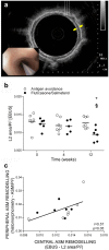
Assessment of central airway remodelling with EBUS. (a) Representative image of a central airway obtained with EBUS in a horse with asthma. Thin white arrows indicate the 3 hyperechoic layers of the bronchial wall representing, from the airway lumen going outwards, the bronchial epithelium, the inner, and the outer borders of the bronchial cartilage. The distance between the thick yellow arrows illustrates the thickness of L2. (b) EBUS-assessed central airway remodelling in study I. (c) Correlation between EBUS-assessed central airway remodelling and peripheral ASM remodelling assessed at histology at baseline (T0) in study I. Each symbol represents one horse (mean value of multiple measures performed). L2: second layer; EBUS: endobronchial ultrasound; ASM: airway smooth muscle; Pi: internal perimeter. *Different from baseline of the same group (p < 0.05); §difference between groups at the time point indicated (p < 0.05).
Figure 6
Figure 6
Central ASM and ECM remodelling. ASM area, myocyte size, ECM fraction of the ASM, and thickness of the ECM in the lamina propria were assessed in study I (ad) and II (eh). Each symbol represents one horse (mean value of multiple measures performed). Dashed line in panels d and h corresponds to the mean thickness of ECM reported in healthy horses in a previous study. *Different from baseline of the same group (p < 0.05); ♮p = 0.05 from baseline of the same group. ASM: airway smooth muscle; ECM: extracellular matrix.
Figure 7
Figure 7
Study design. Grey background indicates antigen exposure. BAL: bronchoalveolar lavage; EBB: endobronchial biopsies; EBUS: endobronchial ultrasound.

Similar articles

Cited by

References

    1. Bergeron C, Tulic MK, Hamid Q. Airway remodelling in asthma: from benchside to clinical practice. Can Respir J. 2010;17:e85–93. doi: 10.1155/2010/318029. - DOI - PMC - PubMed
    1. Grainge CL, et al. Effect of bronchoconstriction on airway remodeling in asthma. N Engl J Med. 2011;364:2006–2015. doi: 10.1056/NEJMoa1014350. - DOI - PubMed
    1. James AL, et al. Airway smooth muscle thickness in asthma is related to severity but not duration of asthma. Eur Respir J. 2009;34:1040–1045. doi: 10.1183/09031936.00181608. - DOI - PubMed
    1. James AL, et al. Airway smooth muscle hypertrophy and hyperplasia in asthma. Am J Respir Crit Care Med. 2012;185:1058–1064. doi: 10.1164/rccm.201110-1849OC. - DOI - PubMed
    1. Lambert RK, Wiggs BR, Kuwano K, Hogg JC, Pare PD. Functional significance of increased airway smooth muscle in asthma and COPD. J Appl Physiol. 1993;74:2771–2781. doi: 10.1063/1.354625. - DOI - PubMed

Publication types

MeSH terms

Grants and funding