. 2017 Oct:111:102-113.
doi: 10.1016/j.yjmcc.2017.08.008. Epub 2017 Aug 16.

Spontaneous activation of a MAVS-dependent antiviral signaling pathway determines high basal interferon-β expression in cardiac myocytes

Affiliations

Spontaneous activation of a MAVS-dependent antiviral signaling pathway determines high basal interferon-β expression in cardiac myocytes

Efraín E Rivera-Serrano et al. J Mol Cell Cardiol. 2017 Oct.

Abstract

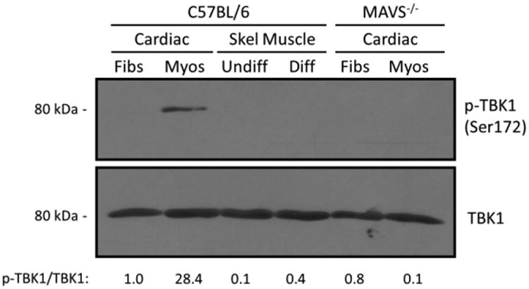
Viral myocarditis is a leading cause of sudden death in young adults as the limited turnover of cardiac myocytes renders the heart particularly vulnerable to viral damage. Viruses induce an antiviral type I interferon (IFN-α/β) response in essentially all cell types, providing an immediate innate protection. Cardiac myocytes express high basal levels of IFN-β to help pre-arm them against viral infections, however the mechanism underlying this expression remains unclear. Using primary cultures of murine cardiac and skeletal muscle cells, we demonstrate here that the mitochondrial antiviral signaling (MAVS) pathway is spontaneously activated in unstimulated cardiac myocytes but not cardiac fibroblasts or skeletal muscle cells. Results suggest that MAVS association with the mitochondrial-associated ER membranes (MAM) is a determinant of high basal IFN-β expression, and demonstrate that MAVS is essential for spontaneous high basal expression of IFN-β in cardiac myocytes and the heart. Together, results provide the first mechanism for spontaneous high expression of the antiviral cytokine IFN-β in a poorly replenished and essential cell type.

Keywords: Cardiac myocyte; Cardiomyocyte; Interferon; MAM; MAVS; Mitochondria.


Figures

Figure 1
Figure 1. Basal IFN-β expression is higher in cardiac myocytes than in cardiac fibroblasts and skeletal muscle cells
(A) Primary cultures of murine cardiac and undifferentiated (undiff.) and differentiated (diff.) skeletal muscle cells were generated from wild-type mice and were immunostained for the muscle-specific sarcomeric actin alpha Sr-1. Nuclei were counter-stained with DAPI. Scale bar = 50 μm. (B) RNA was harvested from the indicated cardiac cell or differentiated skeletal muscle cell primary cultures, analyzed by RT-qPCR, and copy number was normalized to GAPDH. Data are representative of means for two independent experiments from two independently generated primary cultures ± SD. *, Significantly different from all others (P < 0.05).
Figure 2
Figure 2. Mitochondrial proteins and MAVS are more abundant in muscle cells than cardiac fibroblasts
(A) Primary cardiac or skeletal muscle cultures were immunostained with antibodies against TOM20 or MAVS. Nuclei were counterstained with DAPI. Scale bar = 50 μm. (B) Quantification of mitochondrial or MAVS density in the cell measured as the fraction of the cell occupied by either TOM20 or MAVS (mean ± SD; n = 6-12). *, Significantly different from cardiac myocytes and skeletal muscle cells (P < 0.05). (C) Whole-cell protein extracts were harvested from the indicated primary cultures, resolved by SDS-PAGE, and immunoblotted using the indicated primary antibodies. Densitometry values for normalized TOM20 or MAVS band intensities are expressed relative to the band intensity of cardiac fibroblasts. (D) Primary cardiac cultures were incubated with MitoTracker®, fixed, and immunostained using an antibody against MAVS. Scale bar = 10 μm. Results are representative of at least two independent experiments.
Figure 3
Figure 3. Mitochondria are highly associated with the MAM in cardiac myocytes compared to cardiac fibroblasts and skeletal muscle cells
A) Primary cultures were immunostained with antibodies against TOM20 (mitochondria) and FACL4 (MAM); and nuclei were counterstained with DAPI. Images are representative of at least two independent experiments and > 10 cells analyzed per experiment. Scale bar = 10 μm. B) Pearson's co-localization coefficient (mean ± SEM; n = 8-12) of pixel overlap between TOM20 and FACL4. *, Significantly different from cardiac fibroblasts and skeletal muscle cells (P < 0.05). C) Whole-cell protein extracts from the indicated primary cultures were resolved by SDS-PAGE and immunoblotted using the indicated primary antibodies. Densitometry values for MFN2 band intensities normalized to actin are expressed relative to the band intensity of cardiac fibroblasts.
Figure 4
Figure 4. MAVS is highly associated with the MAM, but not with peroxisomes, in stimulated cardiac fibroblasts and unstimulated cardiac myocytes
A) Primary cultures were immunostained with antibodies against MAVS and FACL4 (MAM) or PMP70 (peroxisome); and nuclei were counterstained with DAPI. Images are representative of at least two independent experiments and > 10 cells analyzed per experiment. Scale bar = 10 μm. B) Pearson's co-localization coefficient (mean ± SEM; n = 8-12) of pixel overlap between MAVS and FACL4 or PMP70. *, Significantly different from cardiac fibroblasts and skeletal muscle cells (P < 0.05).
Figure 5
Figure 5. MAVS and TRAF3 are intimately associated in unstimulated cardiac myocytes independently of RIG-I and MDA5 expression
Primary cardiac cultures generated from either RIG-I-/-MDA5-/- or MAVS-/- mice were fixed and subjected to an in situ proximity ligation assay (PLA) using the indicated primary antibodies and the respective species-specific secondary PLA probes. Confocal microscopy was used to obtain z-stack images of PLA signals (red) and nuclei (blue) representative of the total volume of the cells. Scale bar = 10 μm. The number of PLA signals per cell was quantified for each condition and each plotted value represents a single scored cell. *, Significantly different from cardiac fibroblasts and skeletal muscle cells (P < 0.05).
Figure 6
Figure 6. TRAF3 is spontaneously activated in unstimulated cardiac myocytes and its activation is MAVS-dependent
A) Primary cultures were immunostained for TRAF3 and nuclei were counterstained with DAPI. Histograms display measured fluorescence intensity along the drawn line in the inset panels. B) Cardiac fibroblasts derived from either wild-type or MAVS-/- mice were infected with reovirus T3D and immunostained for TRAF3 and viral antigen. Arrows depict redistribution of activated TRAF3 to perinuclear punctate bodies upon virus infection. C,D) Cardiac myocytes generated from MAVS-/- (C) or RIG-I-/- MDA5-/- (D) mice were immunostained as in panel A. Scale bar = 10 μm. Results are representative of at least two independent experiments and > 20 cells per condition.
Figure 7
Figure 7. TBK1 is activated in unstimulated cardiac myocytes and its activation is MAVS-dependent
Whole-cell protein lysates obtained from the indicated primary cultures were resolved by SDS-PAGE and immunoblotted with antibodies against TBK1 (total) or phosphorylated (p-)TBK1. Densitometry values for normalized p-TBK1 band intensities are expressed relative to the band intensity of wild-type cardiac fibroblasts.
Figure 8
Figure 8. MAVS is a determinant of high basal levels of IFN-β expression in cardiac myocytes and the adult heart
A) Primary cardiac myocytes and fibroblasts were generated from either wild-type or MAVS-/- mice. RNA was harvested and analyzed by RT-qPCR, and IFN-β copy number was normalized to GAPDH. Data are representative of means for two independent experiments from two independently generated primary cultures ± SD. *, Significantly different from all others (P < 0.05). B) RNA was harvested from adult mouse hearts and analyzed by RT-qPCR. After background subtraction, IFN-β copy number was normalized to GAPDH. Data are means for 3-4 mice from two independent experiments ± SD. *, Significantly different from wild-type (P < 0.05). C) Model for basal expression of IFN-β in cardiac cells. Inactive components are indicated by dashed borders and grey lettering. Components that were not directly assessed are indicated in italics.

References

    1. Althof N, Harkins S, Kemball CC, Flynn CT, Alirezaei M, Whitton JL. In vivo ablation of type I interferon receptor from cardiomyocytes delays coxsackieviral clearance and accelerates myocardial disease. J Virol. 2014;88:5087–5099. - PMC - PubMed
    1. Arnoult D, Soares F, Tattoli I, Girardin SE. Mitochondria in innate immunity. EMBO reports. 2011;12:901–910. - PMC - PubMed
    1. Azuma K, Ichimura K, Mita T, Nakayama S, Jin WL, Hirose T, Fujitani Y, Sumiyoshi K, Shimada K, Daida H, Sakai T, Mitsumata M, Kawamori R, Watada H. Presence of alpha-smooth muscle actin-positive endothelial cells in the luminal surface of adult aorta. Biochem Biophys Res Commun. 2009;380:620–626. - PubMed
    1. Banerjee I, Fuseler JW, Price RL, Borg TK, Baudino TA. Determination of cell types and numbers during cardiac development in the neonatal and adult rat and mouse. American journal of physiology Heart and circulatory physiology. 2007;293:H1883–1891. - PubMed
    1. Baty CJ, Sherry B. Cytopathogenic effect in cardiac myocytes but not in cardiac fibroblasts is correlated with reovirus-induced acute myocarditis. Journal of virology. 1993;67:6295–6298. - PMC - PubMed

MeSH terms