Polyurethane/Gelatin Nanofibrils Neural Guidance Conduit Containing Platelet-Rich Plasma and Melatonin for Transplantation of Schwann Cells
- PMID: 28823058
- PMCID: PMC11481952
- DOI: 10.1007/s10571-017-0535-8
Polyurethane/Gelatin Nanofibrils Neural Guidance Conduit Containing Platelet-Rich Plasma and Melatonin for Transplantation of Schwann Cells
Abstract
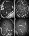
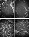
The current study aimed to enhance the efficacy of peripheral nerve regeneration using a biodegradable porous neural guidance conduit as a carrier to transplant allogeneic Schwann cells (SCs). The conduit was prepared from polyurethane (PU) and gelatin nanofibrils (GNFs) using thermally induced phase separation technique and filled with melatonin (MLT) and platelet-rich plasma (PRP). The prepared conduit had the porosity of 87.17 ± 1.89%, the contact angle of 78.17 ± 5.30° and the ultimate tensile strength and Young's modulus of 5.40 ± 0.98 MPa and 3.13 ± 0.65 GPa, respectively. The conduit lost about 14% of its weight after 60 days in distilled water. The produced conduit enhanced the proliferation of SCs demonstrated by a tetrazolium salt-based assay. For functional analysis, the conduit was seeded with 1.50 × 10⁴ SCs (PU/GNFs/PRP/MLT/SCs) and implanted into a 10-mm sciatic nerve defect of Wistar rat. Three control groups were used: (1) PU/GNFs/SCs, (2) PU/GNFs/PRP/SCs, and (3) Autograft. The results of sciatic functional index, hot plate latency, compound muscle action potential amplitude and latency, weight-loss percentage of wet gastrocnemius muscle and histopathological examination using hematoxylin-eosin and Luxol fast blue staining, demonstrated that using the PU/GNFs/PRP/MLT conduit to transplant SCs to the sciatic nerve defect resulted in a higher regenerative outcome than the PU/GNFs and PU/GNFs/PRP conduits.
Keywords: Gelatin; Melatonin; Neural guidance conduit; Platelet-rich plasma; Polyurethane; Schwann cells.
Conflict of interest statement
The authors declare that they have no conflicts of interest.
Figures


Similar articles
-
Sciatic nerve regeneration by transplantation of Schwann cells via erythropoietin controlled-releasing polylactic acid/multiwalled carbon nanotubes/gelatin nanofibrils neural guidance conduit.J Biomed Mater Res B Appl Biomater. 2018 May;106(4):1463-1476. doi: 10.1002/jbm.b.33952. Epub 2017 Jul 4. J Biomed Mater Res B Appl Biomater. 2018. PMID: 28675568
-
Enhanced sciatic nerve regeneration by poly-L-lactic acid/multi-wall carbon nanotube neural guidance conduit containing Schwann cells and curcumin encapsulated chitosan nanoparticles in rat.Mater Sci Eng C Mater Biol Appl. 2020 Apr;109:110564. doi: 10.1016/j.msec.2019.110564. Epub 2019 Dec 17. Mater Sci Eng C Mater Biol Appl. 2020. PMID: 32228906
-
A nanofibrous polycaprolactone/collagen neural guidance channel filled with sciatic allogeneic schwann cells and platelet-rich plasma for sciatic nerve repair.J Biomater Appl. 2025 Feb;39(7):797-806. doi: 10.1177/08853282241297446. Epub 2024 Nov 5. J Biomater Appl. 2025. PMID: 39498821
-
Sciatic Nerve Regeneration in Rat Model With PLGA-MWCNT Conduit Loaded by Fibrin Hydrogel Containing Nanolycopene and Schwann Cells.J Biomed Mater Res B Appl Biomater. 2025 Sep;113(9):e35643. doi: 10.1002/jbm.b.35643. J Biomed Mater Res B Appl Biomater. 2025. PMID: 40853029
-
Sophisticated polycaprolactone/gelatin nanofibrous nerve guided conduit containing platelet-rich plasma and citicoline for peripheral nerve regeneration: In vitro and in vivo study.Int J Biol Macromol. 2020 May 1;150:380-388. doi: 10.1016/j.ijbiomac.2020.02.102. Epub 2020 Feb 11. Int J Biol Macromol. 2020. PMID: 32057876
Cited by
-
Tissue engineering and stem cell-based therapeutic strategies for premature ovarian insufficiency.Regen Ther. 2023 Nov 25;25:10-23. doi: 10.1016/j.reth.2023.11.007. eCollection 2024 Mar. Regen Ther. 2023. PMID: 38108045 Free PMC article. Review.
-
Rational design of biodegradable thermoplastic polyurethanes for tissue repair.Bioact Mater. 2021 Dec 31;15:250-271. doi: 10.1016/j.bioactmat.2021.11.029. eCollection 2022 Sep. Bioact Mater. 2021. PMID: 35386346 Free PMC article. Review.
-
Platelet-Rich Plasma Therapy in the Treatment of Diseases Associated with Orthopedic Injuries.Tissue Eng Part B Rev. 2020 Dec;26(6):571-585. doi: 10.1089/ten.TEB.2019.0292. Epub 2020 Nov 3. Tissue Eng Part B Rev. 2020. PMID: 32380937 Free PMC article. Review.
-
Ascorbic Acid Facilitates Neural Regeneration After Sciatic Nerve Crush Injury.Front Cell Neurosci. 2019 Mar 21;13:108. doi: 10.3389/fncel.2019.00108. eCollection 2019. Front Cell Neurosci. 2019. PMID: 30949031 Free PMC article.
-
Recent Achievements in the Development of Biomaterials Improved with Platelet Concentrates for Soft and Hard Tissue Engineering Applications.Int J Mol Sci. 2024 Jan 26;25(3):1525. doi: 10.3390/ijms25031525. Int J Mol Sci. 2024. PMID: 38338805 Free PMC article. Review.
References
-
- Atik B, Erkutlu I, Tercan M, Buyukhatipoglu H, Bekerecioglu M, Pence S (2011) The effects of exogenous melatonin on peripheral nerve regeneration and collagen formation in rats. J Surg Res 166:330–336 - PubMed
-
- Chan-Chan L, Solis-Correa R, Vargas-Coronado R, Cervantes-Uc J, Cauich-Rodríguez J, Quintana P, Bartolo-Pérez P (2010) Degradation studies on segmented polyurethanes prepared with HMDI, PCL and different chain extenders. Acta Biomater 6:2035–2044 - PubMed
-
- Chang HM, Liu CH, Hsu WM, Chen LY, Wang HP, Wu TH, Chen KY, Ho WH, Liao WC (2014) Proliferative effects of melatonin on Schwann cells: implication for nerve regeneration following peripheral nerve injury. J Pineal Res 56:322–332 - PubMed
-
- Chen J, Dong R, Ge J, Guo B, Ma PX (2015) Biocompatible, biodegradable, and electroactive polyurethane-urea elastomers with tunable hydrophilicity for skeletal muscle tissue engineering. ACS Appl Mater Interfaces 7:28273–28285 - PubMed
MeSH terms
Substances
Grants and funding
LinkOut - more resources
Full Text Sources
Other Literature Sources
Research Materials

