. 2017 Dec;13(4):529-544.
doi: 10.1007/s11302-017-9579-y. Epub 2017 Aug 19.

Inhibition of P2X7 receptors improves outcomes after traumatic brain injury in rats

Affiliations

Inhibition of P2X7 receptors improves outcomes after traumatic brain injury in rats

Xiaofeng Liu et al. Purinergic Signal. 2017 Dec.

Abstract

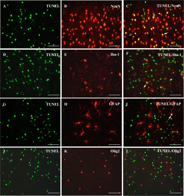
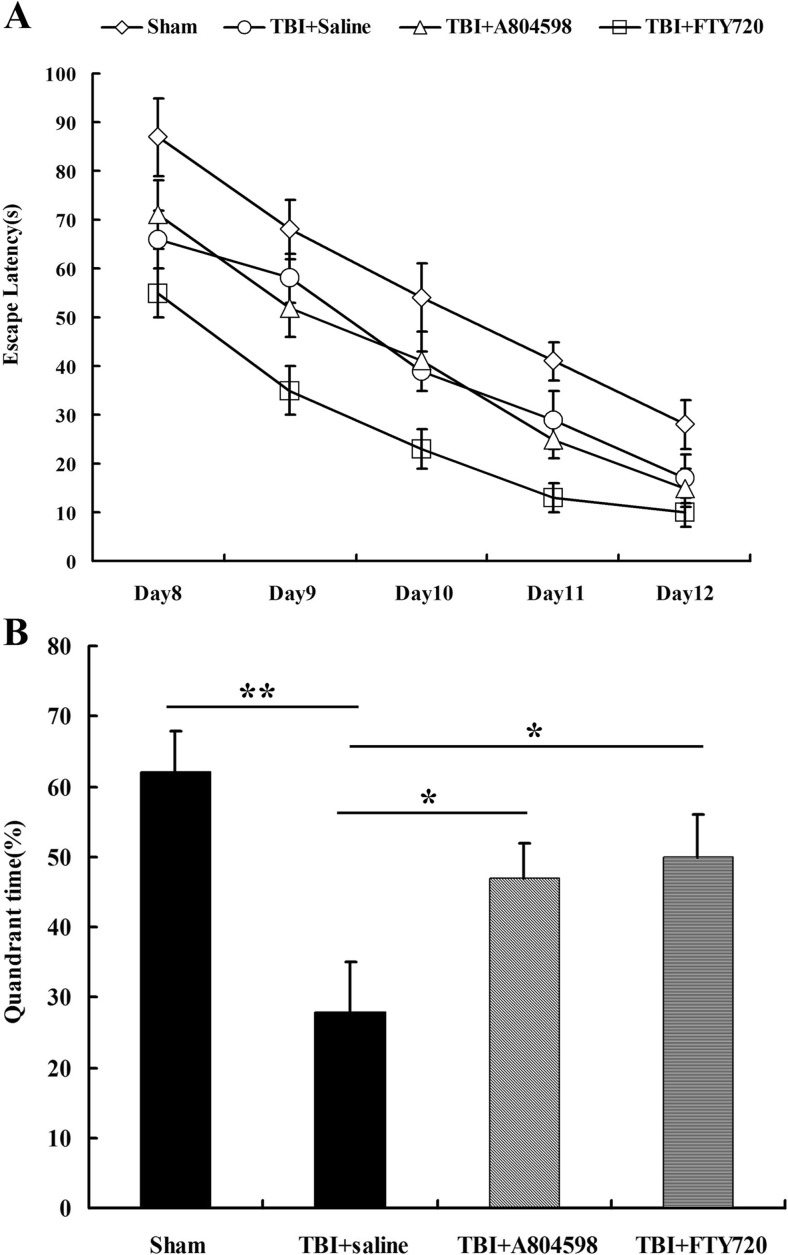
Traumatic brain injury (TBI) is the leading cause of death and disability for people under the age of 45 years worldwide. Neuropathology after TBI is the result of both the immediate impact injury and secondary injury mechanisms. Secondary injury is the result of cascade events, including glutamate excitotoxicity, calcium overloading, free radical generation, and neuroinflammation, ultimately leading to brain cell death. In this study, the P2X7 receptor (P2X7R) was detected predominately in microglia of the cerebral cortex and was up-regulated on microglial cells after TBI. The microglia transformed into amoeba-like and discharged many microvesicle (MV)-like particles in the injured and adjacent regions. A P2X7R antagonist (A804598) and an immune inhibitor (FTY720) reduced significantly the number of MV-like particles in the injured/adjacent regions and in cerebrospinal fluid, reduced the number of neurons undergoing apoptotic cell death, and increased the survival of neurons in the cerebral cortex injured and adjacent regions. Blockade of the P2X7R and FTY720 reduced interleukin-1βexpression, P38 phosphorylation, and glial activation in the cerebral cortex and improved neurobehavioral outcomes after TBI. These data indicate that MV-like particles discharged by microglia after TBI may be involved in the development of local inflammation and secondary nerve cell injury.

Keywords: Microglial cells; Microvesicles; Neuroinflammation; P2X7R; Traumatic brain injury.


Conflict of interest statement

Conflicts of interest

Xiaofeng Liu declares that she has no conflict of interest.

Zhengqing Zhao declares that she has no conflict of interest.

Ruihua Ji declares that she has no conflict of interest.

Jiao Zhu declares that she has no conflict of interest.

Qian-Qian Sui declares that she has no conflict of interest.

Gillian E. Knight declares that she has no conflict of interest.

Geoffrey Burnstock declares that he has no conflict of interest.

Cheng He declares that she has no conflict of interest.

Hongbin Yuan declares that she has no conflict of interest.

Zhenghua Xiang declares that she has no conflict of interest.

Ethical approval

All experimental procedures were approved by the Institutional Animal Care and Use Committee at Second Military Medical University and conformed to the UK Animals (Scientific Procedures) Act 1986 and associated guidelines on the ethical use of animals.

Figures

Fig. 1
Fig. 1
Expression of P2X7R-ir (red) in the cerebral cortex of control and TBI rats. Subpanel a is P2X7R-ir and subpanel d is an immerged image from A (red) and Iba-1-ir (green, a marker for microglial cell) from the control group. Note that almost all the P2X7-ir are also labeled with Iba-1. The immunostaining results show that P2X7R-ir is predominately expressed in microglial cells. b, c, g, h, i P2X7R-ir from the TBI groups at 6, 12, 24, 48, and 72 h, respectively. e, j, l, f, l P2X7R-ir from TBI+A804598 and TBI+FTY720 groups. Note that microvesicle (MV)-like particles were detected mainly at 6 and 12 h after TBI, and A804598 and FTY720 decreased significantly the number of vesicles. In the early stages from 6 to 24 h after TBI, the number of P2X7R-ir microglial cells did not change significantly but increased from 48 h after TBI. Both A804598 and FTY720 decreased the number of microglia cells significantly at 96 h after TBI. All scale bars = 60 μm
Fig. 2
Fig. 2
Quantitative analysis of MV-like particles (a) and microglial cells (b) with P2X7R-ir after TBI. The number of MV-like particles and microglial cells is expressed as mean ± SEM (n = 6). Note that MV-like particles were mainly detected at 6 and 12 h after TBI; both A804598 and FTY720 reduced significantly the number of MV-like particles at 6 and 12 h and the number of microglial cells at 96 h. **p < 0.01
Fig. 3
Fig. 3
Double immunofluorescence of P2X7R-ir (red), Iba-1 (green), GFAP (green), and MBP (green) in the cerebral cortex of rats 12 h after TBI. a, c, e P2X7R-ir. b, d, f Merged images of Iba-1-ir, GFAP-ir, and MBP-ir with a, c, and e, respectively. b Magnified image of the region of b indicated by an arrow. Note that almost all the MV-like particles and cells with P2X7-ir are also labeled with Iba-1-ir but not with GFAP-ir or MBP-ir. The scale bars in af = 60 μm, b = 10 μm
Fig. 4
Fig. 4
Flow cytometry and immunocytochemical analysis of MV-like particles in CSF after TBI. ad Results from control, TBI, TBI+A804598, and TBI+FTY720 groups at 12 h after TBI. e Summary of flow cytometry of CSF from different groups. Note that the number of MV-like particles reaches a peak at 12 h. Both A804598 and FTY720 significantly reduced the number of MV-like particles at 6 and 12 h. f Immunocytochemical analysis of MV-like particles with P2X7R (red) and Iba-1 immunostaining (green). Note that most of MV-like particles with P2X7R-ir are also labeled by Iba-1, although some of them are not. An arrow indicates an MV-like particle with Iba-1 not labeled by P2X7R-ir (shown at higher magnification in f)
Fig. 5
Fig. 5
Expression of P2X7R detected by Western blotting in the cerebral cortex from TBI (a), TBI+A804598 (b), and TBI+FTY720 (c) groups. d Summary of P2X7R/GAPDH ratio. Data are representative of five rats per group. The ratio of P2X7R/GAPDH was analyzed with one-way ANOVA followed by Dunnett’s post hoc test (**p < 0.01 vs. control sham-operated rats). The results show that expression of P2X7R protein increased significantly from 6 h to 7 days after TBI but was not affected by A804598 or FTY720
Fig. 6
Fig. 6
Effect of A804598 and FTY720 on cell apoptosis in the cerebral cortex after TBI. a, c, e, g, i, k TUNEL staining from the control, 6, 12, 24, 48, and 96 h groups, respectively. d, h, l, f, j TUNEL staining from TBI+A804598 and TBI+FTY720. M is the summary of the number of TUNEL-positive cells in the different groups. Note that A804598 or FTY720 treatment significantly reduced the number of apoptotic cells. Values are expressed as the mean ± SEM. **p < 0.01 TBI+A804598 groups or TBI+FTY720 groups versus saline-treated groups. All the scale bars = 60 μm
Fig. 7
Fig. 7
Cell type analysis of apoptotic cells in the cerebral cortex at 6 h after TBI. a, d, g, j TUNEL-positive cells. b, e, h, k NeuN-positive, GFAP-positive, Iba-1-positive, and Olig2-positive cells in the same fields of a, d, g, and j, respectively. c, f, i, l Immerged images from a and b, d and e, g and h, and j and k. Note that most of the apoptotic cells were NeuN-positive, occasionally Iba-1- or GFAP-positive, but never Olig2-positive. The results indicate that most of the apoptotic cells after TBI are neurons. All scale bars = 60 μm
Fig. 8
Fig. 8
Effect of A804598 and FTY720 on neuronal survival rate in the cerebral cortex after TBI. ad NeuN immunostaining from the control group, TBI group, TBI+A804598 group, and TBI+FTY720 group 14 days after TBI, respectively. e Summaries of the number of survival neurons. Note that both A804598 and FTY720 treatment increased significantly the number of surviving neurons. Values are expressed as mean ± SEM. *p < 0.05, **p < 0.01. 2w (2 weeks) = 14 days. All scale bars = 60 μm
Fig. 9
Fig. 9
Effect of A804598 and FTY720 on expression of interleukin (IL)-1β in the cerebral cortex after TBI. IL-1β protein was detected by ELISA. Both A804598 and FTY720 reduced significantly the levels of IL-1β. Data are expressed as mean ± SEM. *p < 0.05, **p < 0.01
Fig. 10
Fig. 10
Expression of phosphorylation P38 (pP38) detected by Western blotting in the cerebral cortex from TBI (a), TBI+A804598 (b), and TBI+FTY720 (c) groups. d Summary of pP38/actin ratio. Data are representative of five rats per group. The ratio of pP38/actin was analyzed with one-way ANOVA followed by Dunnett’s post hoc test (**p < 0.01 TBI vs. TBI+A804598 or TBI+FTY720 rats). Statistical analysis shows that pP38 protein increased significantly at 12 and 24 h after TBI, which was inhibited by A804598 or FTY720, significantly
Fig. 11
Fig. 11
Effect of A804598 and FTY720 on astrogliosis in the cerebral cortex after TBI. af Astrocytes with GFAP-ir at 4 days, 7 days (1w), and 14 days (2w) in the TBI+saline groups (ac), TBI+A804598 groups (d, f), and TBI+FTY720 group (e) respectively. g Quantitative analysis of average area optical density (AAOD) of astrocytes with GFAP-ir. Data are expressed as mean ± SEM (n = 6). **p < 0.01. Statistical analysis shows that density of GFAP-ir increased gradually after TBI. A804598 or FTY720 reduced the density of GFAP-ir significantly at 7 and 14 days after TBI
Fig. 12
Fig. 12
Effect of A804598 and FTY720 on spatial learning and memory in the water maze. a Escape latency to find the platform during the 5 days of the training trial from the 8th to the 12th day after TBI. b Time spent during the probe trial in the quadrant where the platform had been located. Values are expressed as mean ± SEM. **p < 0.01, *p < 0.05. Statistical analysis shows that spatial learning and memory were affected significantly by TBI. Both A804598 and FTY720 improved spatial learning and memory after TBI

References

    1. Jennett B. Epidemiology of head injury. J Neurol Neurosurg Psychiatry. 1996;60(4):362–369. doi: 10.1136/jnnp.60.4.362. - DOI - PMC - PubMed
    1. Langlois JA, Rutland-Brown W, Wald MM. The epidemiology and impact of traumatic brain injury: a brief overview. J Head Trauma Rehabil. 2006;21(5):375–378. doi: 10.1097/00001199-200609000-00001. - DOI - PubMed
    1. Davis AE. Mechanisms of traumatic brain injury: biomechanical, structural and cellular considerations. Crit Care Nurs Q. 2000;23(3):1–13. doi: 10.1097/00002727-200011000-00002. - DOI - PubMed
    1. Gaetz M. The neurophysiology of brain injury. Clin Neurophysiol. 2004;115(1):4–18. doi: 10.1016/S1388-2457(03)00258-X. - DOI - PubMed
    1. Cernak I. Animal models of head trauma. NeuroRx. 2005;2(3):410–422. doi: 10.1602/neurorx.2.3.410. - DOI - PMC - PubMed

Substances