. 2017 Sep 7;170(6):1234-1246.e14.
doi: 10.1016/j.cell.2017.07.045. Epub 2017 Aug 17.

Activation and Desensitization Mechanism of AMPA Receptor-TARP Complex by Cryo-EM

Affiliations

Activation and Desensitization Mechanism of AMPA Receptor-TARP Complex by Cryo-EM

Shanshuang Chen et al. Cell. .

Abstract

AMPA receptors mediate fast excitatory neurotransmission in the mammalian brain and transduce the binding of presynaptically released glutamate to the opening of a transmembrane cation channel. Within the postsynaptic density, however, AMPA receptors coassemble with transmembrane AMPA receptor regulatory proteins (TARPs), yielding a receptor complex with altered gating kinetics, pharmacology, and pore properties. Here, we elucidate structures of the GluA2-TARP γ2 complex in the presence of the partial agonist kainate or the full agonist quisqualate together with a positive allosteric modulator or with quisqualate alone. We show how TARPs sculpt the ligand-binding domain gating ring, enhancing kainate potency and diminishing the ensemble of desensitized states. TARPs encircle the receptor ion channel, stabilizing M2 helices and pore loops, illustrating how TARPs alter receptor pore properties. Structural and computational analysis suggests the full agonist and modulator complex harbors an ion-permeable channel gate, providing the first view of an activated AMPA receptor.

Keywords: chemical synapse; glutamate receptor; ion channel gating; ligand gated ion channel; membrane protein; neurotransmitter; structural biology; synapse.


Figures

Figure 1
Figure 1. Activated states of the GluA2-TARP γ2 complex
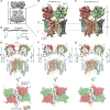
(A) Initial cryo-EM reconstruction of full-length GluA2-TARP γ2 complex bound with quisqualate and (R,R)-2b. Higher resolution reconstructions focused on the LBD/TMD layers. (B) and (C) Cryo-EM reconstructions of the GluA2-TARP γ2 complex bound with kainate and quisqualate in the presence of (R,R)-2b, respectively. (D) Whole-cell patch clamp recording from cells expressing the GluA2-TARP γ2 complex. The ratio between steady-state currents elicited by kainate and quisqualate is 0.80 ± 0.03 (mean ± standard deviation, n = 6). (E)–(G) Structures of the LBD/TMD layers in the previously determined MPQX complex (E), with kainate/(R,R)-2b (F) and with quisqualate/(R,R)-2b (G). The LBD D1 and D2 lobes are shown in darker and lighter shades, respectively. COMs of D1 and D2 are indicated by black dots, whose distances from a reference point, COM of Thr617 Cα atoms, are labeled in the “side views”; COM distances between proximal helices between opposing subunits, helix G (G) for A/C subunits and helix K (K) for B/D subunits, are indicated in the “top-down views”. Distances are in angstroms (Å). See also Figures S1, S2 and Table S1.
Figure 2
Figure 2. Conformational changes upon receptor activation
(A)–(C) Structural comparison of opposing subunit pairs, A/C and B/D, and adjacent subunits, A/D, showing the LBDs and M3 helices in the MPQX (A), kainate (B) and quisqualate (C) GluA2-TARP γ2 complexes. COMs of D1 and D2 lobes are indicated by black dots. Cα atoms of residues near M3 bundle crossing are shown as spheres. LBD clamshell closure is represented by schematic cartoons. Structures were superimposed using main-chain atoms of the M3 helices. In views showing LBD dimers (right panel), locations representing COMs of Gly-Thr linkers derived from isolated LBD structures are indicated by orange dots. Distances are in angstroms (Å). See also Figures S3, S4 and Movie S1.
Figure 3
Figure 3. Ion channel pore of quisqualate/(R,R)-2b complex
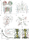
(A) View of LBD/TMD layers and, in inset, close-up of the TMD region showing receptor TMD, TM4 of TARP and solvent accessible pathway of the ion channel pore, along the central 2-fold axis (dashed line). In the inset only two subunits are shown. A/C and B/D subunits are colored in green and salmon, respectively, with the pore loops highlighted in yellow. Cα atoms of Arg586 (Q/R site) and Thr617 are shown as blue and grey spheres, respectively. Pore radii calculated without the side-chain model of Arg586 are depicted by purple, green and red dots representing pore radii of >3.3 Å, 1.8−3.3 Å, and <1.8 Å, respectively. (B) The M2 helices are stabilized through hydrophobic side-chain interactions with adjacent M1 and M3 helices. The M2 helix is represented as cartoon in transparent solvent accessible surface. Well-resolved side-chains are shown as sticks, and Cαs of key glycine residues in M3 are defined by grey spheres. (C) “Top-down” view of M2 helices and the pore loops. COMs of M2 helices (salmon and green spheres), distances and angles are shown. (D) Na+ ion permeation trajectory captured during MD simulation of the quisqualate/(R,R)-2b structure (with R586Q mutation), showing spontaneous entry of the ion. Inset shows the number of water molecules along the pore axis averaged over a 20 ns simulation of the quisqualate/(R,R)-2b and the MPQX structures (PDB code: 3KG2). (E) Orthogonal views revealing asymmetric water and ion distributions in the channel lumen of the quisqualate/(R,R)-2b structure. The 30% water occupancy isosurface is shown in semi-transparent surface. The trajectory of the permeating Na+ ion is shown as yellow dots. See also Figures S3, S4 and S5.
Figure 4
Figure 4. GluA2 LBD and TARP γ2 interactions
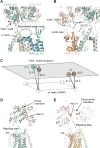
(A) and (B) GluA2 LBD-TARP γ2 interface viewed parallel to the membrane at A/C and B/D positions, respectively. Structures of GluA2-TARP γ2 complex bound with quisqualate/(R,R)-2b (green and salmon) and MPQX (grey) were superimposed using main-chain atoms of receptor TMD. The Cα atoms of the ‘KGK’ motif (697–699) are shown as spheres. In the quisqualate/(R,R)-2b bound structure, lysine and glycine Cα atoms are blue and yellow, respectively. (C) Schematic diagram illustrating displacement of the “KGK” motif and change in distances between the “KGK” motif and TARP α1 helix upon receptor activation. (D) and (E) Possible interactions between TARP and LBD dimer interface (D) and between TARP and LBD dimer-dimer interface (E) in GluA2-TARP γ2 complex bound with quisqualate/(R,R)-2b. Unstructured S2-M4 linkers are represented by dashes lines.
Figure 5
Figure 5. A TARP-bound desensitized state
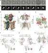
(A) and (B) Raw particle images (A) and representative 2D-class averages (B) illustrating the conformational heterogeneity of the GluA2-TARP γ2 quisqualate complex. (C) Density map of the most well defined 3D class where refinement was focused on the LBD/TMD layers. The view is along the LBD dimer interface showing the separation of the D1-D1 interface and formation of the D2–D2 interface. (D) and (E) “Side” and “top-down” views of the quisqualate-bound GluA2-TARP γ2 complex, showing the ruptured LBD dimer interface. COM distances are measured similarly as the non-desensitized structures (Figures 1E–1G). (F)–(H) Conformational changes associated with receptor desensitization revealed by superposition of LBD-M3 pairs from quisqualate bound GluA2-TARP γ2 complex structures in the absence (in color) or presence of (R,R)-2b (in grey), using main-chain atoms of the receptor TMD. Views are similar as in Figure 2. Angles formed between vectors connecting COMs of D1 and D2 lobes in complexes without and with (R,R)-2b are labeled at both A/C and B/D positions in (H). Distances are in angstroms (Å). See also Figures S1, S6, S7 and Movie S1.
Figure 6
Figure 6. TARPs act as LBD bouy
(A) Helix E, M3 helices and the M3-S2 linker highlighted in the quisqualate/(R,R)-2b bound GluA2-TARP γ2 complex. (B)–(E) The E and M3 helices, and the M3-S2 linkers of the GluA2-TARP γ2 complex bound with MPQX (B), kainate/(R,R)-2b (C), quisqualate/(R,R)-2b (D) and quisqualate alone (E) viewed parallel to the membrane. Distances between proximal and opposing helix E COM pairs are labeled; “elevation” is the distance perpendicular to the membrane between the helix E COM and Thr617. (F) “Elevation” and “separation” plot of opposing helix E COMs highlights the role of TARPs. Distances from the GluA2-TARP γ2 complex and intact GluA2 structures are indicated by solid and open spheres, and grouped separately in solid and dashed circles, respectively. Distances are in angstroms (Å).
Figure 7
Figure 7. Mechanisms of receptor activation and desensitization
Shown is the LBD/TMD layer of two receptor and two TARP subunits. TARPs function as a molecular buoy on which the LBD layer ‘floats’, ensuring tension exerted on the M3-S2 linkers is efficiently transmitted to open the channel gate rather than causing the LBD to approach the membrane.

References

    1. Adams PD, Afonine PV, Bunkóczi G, Chen VB, Davis IW, Echols N, Headd JJ, Hung L-W, Kapral GJ, Grosse-Kunstleve RW. PHENIX: a comprehensive Python-based system for macromolecular structure solution. Acta Crystallographica Section D: Biological Crystallography. 2010;66:213–221. - PMC - PubMed
    1. Ahmed AH, Ptak CP, Fenwick MK, Hsieh C-L, Weiland GA, Oswald RE. Dynamics of cleft closure of the GluA2 ligand-binding domain in the presence of full and partial agonists revealed by hydrogen-deuterium exchange. J Biol Chem. 2013;288:27658–27666. - PMC - PubMed
    1. Armstrong N, Gouaux E. Mechanisms for activation and antagonism of an AMPA-sensitive glutamate receptor: crystal structures of the GluR2 ligand binding core. Neuron. 2000;28:165–181. - PubMed
    1. Armstrong N, Jasti J, Beich-Frandsen M, Gouaux E. Measurement of conformational changes accompanying desensitization in an ionotropic glutamate receptor. Cell. 2006;127:85–97. - PubMed
    1. Bowie D, Mayer ML. Inward recification of both AMPA and kainate subtype glutamate receptors generated by polyamine-mediated ion channel block. Neuron. 1995;15:453–462. - PubMed

LinkOut - more resources