. 2017 Dec:108:73-82.
doi: 10.1016/j.nbd.2017.08.006. Epub 2017 Aug 18.

Intravenous immune-modifying nanoparticles as a therapy for spinal cord injury in mice

Affiliations

Intravenous immune-modifying nanoparticles as a therapy for spinal cord injury in mice

Su Ji Jeong et al. Neurobiol Dis. 2017 Dec.

Abstract

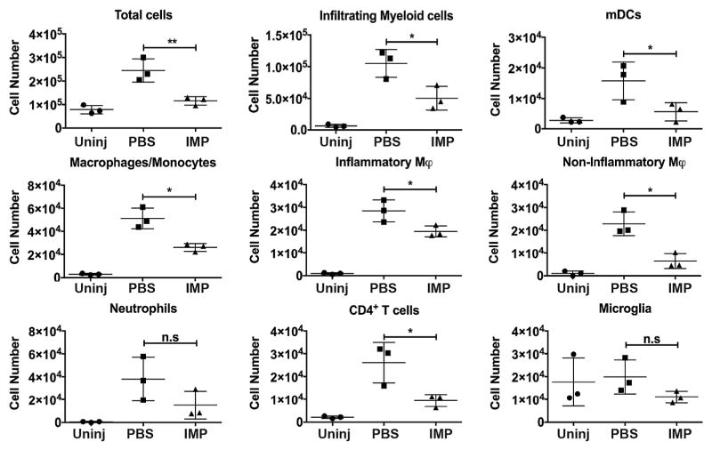
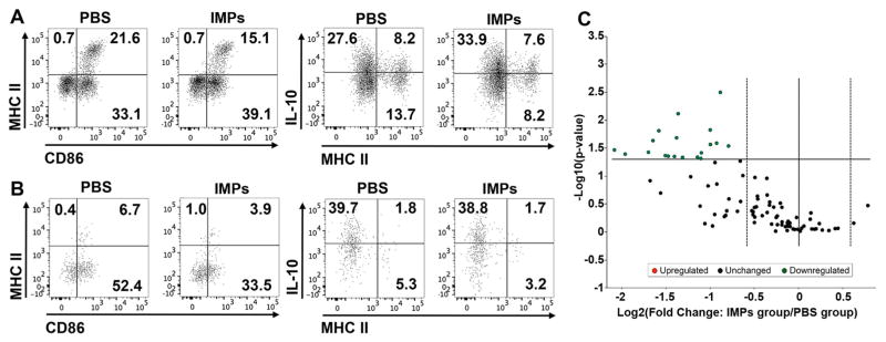
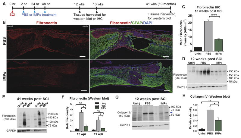
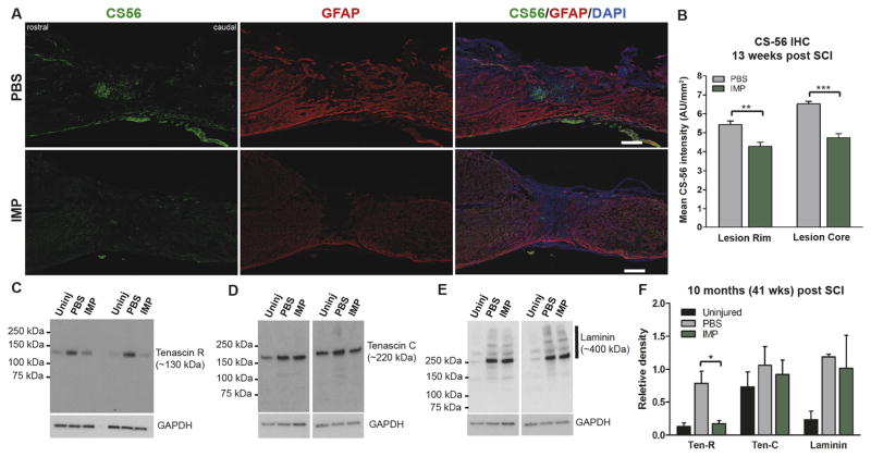
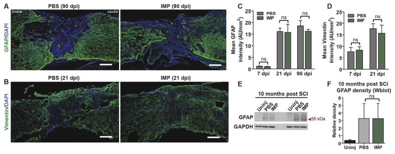
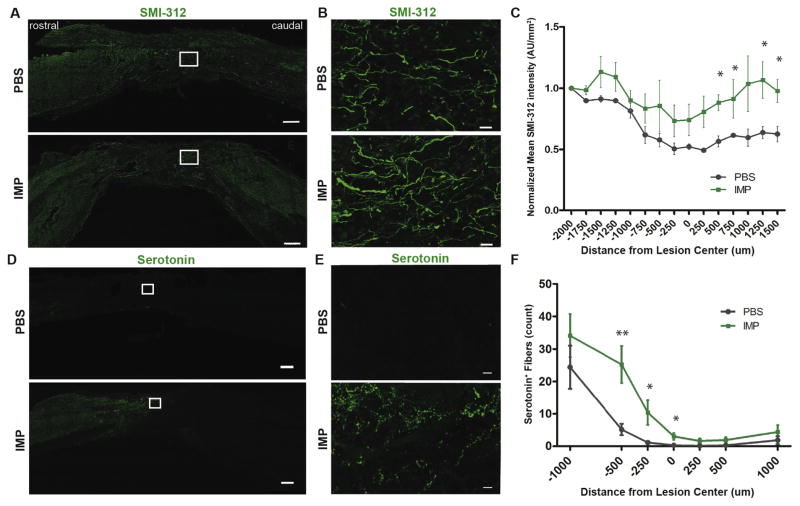
Intravenously infused synthetic 500nm nanoparticles composed of poly(lactide-co-glycolide) are taken up by blood-borne inflammatory monocytes via a macrophage scavenger receptor (macrophage receptor with collagenous structure), and the monocytes no longer traffic to sites of inflammation. Intravenous administration of the nanoparticles after experimental spinal cord injury in mice safely and selectively limited infiltration of hematogenous monocytes into the injury site. The nanoparticles did not bind to resident microglia, and did not change the number of microglia in the injured spinal cord. Nanoparticle administration reduced M1 macrophage polarization and microglia activation, reduced levels of inflammatory cytokines, and markedly reduced fibrotic scar formation without altering glial scarring. These findings thus implicate early-infiltrating hematogenous monocytes as highly selective contributors to fibrosis that do not play an indispensable role in gliosis after SCI. Further, the nanoparticle treatment reduced accumulation of chondroitin sulfate proteoglycans, increased axon density inside and caudal to the lesion site, and significantly improved functional recovery after both moderate and severe injuries to the spinal cord. These data provide further evidence that hematogenous monocytes contribute to inflammatory damage and fibrotic scar formation after spinal cord injury in mice. Further, since the nanoparticles are simple to administer intravenously, immunologically inert, stable at room temperature, composed of an FDA-approved material, and have no known toxicity, these findings suggest that the nanoparticles potentially offer a practical treatment for human spinal cord injury.

Keywords: Fibrosis; Gliosis; Macrophage; Monocyte; Nanotechnology; Spinal cord injury.


Figures

Fig. 1
Fig. 1
Treatment with IMPs reduces influx of inflammatory cells into the lesion after SCI. Mice received IMPs (black bars) or vehicle (PBS; grey bars) 2 h and 24 h after a SCI and were sacrificed at 48 h to analyze the extent of peripheral cell infiltration at the site of the injury. Uninjured control mice (white bars) also were analyzed. Cells within the lesion were counted and their profiles evaluated by FACS. IMPs treatment did not alter the number of microglia (CD45int CD11b+ LyClo) but significantly reduced the influx of dendritic cells (CD45hi CD11b+ Ly6G CD11c+), macrophages/monocytes (CD45hi CD11b+ Ly6G CD11c), inflammatory monocytes (inflammatory φ; CD45+ CD11b+ Ly6G CD11c Ly6chi), non-inflammatory monocytes (non-inflammatory φ; CD45+ CD11b+ Ly6G CD11c Ly6clo) and CD4+ T cells (CD45hi CD11b CD4+). No significant difference was observed on neutrophils (CD45+ CD11b+ Ly6G+). Values are the means of 3 independent experiments (n = 4–6 mice per experiment). All data are presented as mean ± SEM. Groups were compared by ANOVA followed by Tukey’s multiple comparison test. *p < 0.05, **p < 0.01.
Fig. 2
Fig. 2
IMPs treatment reduces M1 macrophage and microglia activation in the lesions of SCI mice. Mice received IMPs or vehicle 2 h and 24 h after a SCI and were sacrificed at 48 h to (A) analyze the macrophage profile of infiltrating cells, (B) evaluate the microglia profile and (C) quantify the chemokines and cytokines in the injured part of the CNS. (A and B) Macrophages (CD45hi CD11b+ Ly6G CD11c) show a reduced M1 profile (MHCII+ CD86+) along with increased IL-10 expression in mice treated with IMPs. In the same mice, microglia (CD45int CD11b+ LyClo) had reduced expression of the costimulatory molecule CD86. Data shown are representative of n = 2 independent experiments with 5–6 mice pooled for each experiment. (C) Volcano plot for differential gene expression showing statistical significance vs. fold change. Each point represents the results of one gene in which the x-axis is the log 2 fold change for the ratio IMPs treated mice vs PBS treated mice, whereas the y-axis is the −Log10 of p-value. Vertical dash lines represent differential expression differences of ±1.5 fold and statistically significant genes are observed above the horizontal line, which corresponds to a p-value of 0.05 (Student’s t-test, n = 5 mice per group). A complete list of genes and p values are provided in the Supporting material (Supplementary Table 2).
Fig. 3
Fig. 3
IMPs treatment attenuates chronic fibrotic scarring. (A) Experimental timeline. (B) At 13 weeks post SCI, 16 μm thick mid-sagittal sections, identified by the central canals, were obtained from PBS treated or IMPs treated mice and stained with fibronectin and GFAP. Scale bar = 250 μm. (C) Quantification of fibronectin staining inside the lesion. Lesions were identified as GFAP areas (n = 4–5 mice per group, ***p < 0.001, ANOVA with Bonferroni’s multiple comparison test). (D, E) Western blot analysis of the 1 mm lesion-containing segments of uninjured (Uninj), PBS treated, and IMPs treated mice harvested (D) 12 weeks and (E) 41 weeks post SCI (n = 4 mice per group). (F) Western blot quantifications for fibronectin at 12 weeks and 41 weeks post SCI. (G, H) Western blot analysis of the spinal lesions from uninjured (Uninj), PBS treated, and IMPs treated mice at 12 weeks post SCI show significantly reduced levels of collagen type IV (*p < 0.05, ANOVA followed by Tukey’s multiple comparison test).
Fig. 4
Fig. 4
IMPs treatment reduces chondroitin sulfate proteoglycan (CSPG) accumulation. (A) Representative images of 16 μm thick mid-sagittal sections of PBS or IMPs treated animals 13 weeks post SCI stained with CS-56 and GFAP. Scale bar = 250 μm (B) Quantifications of CS-56 intensities in the GFAP lesion core (Lesion Core) and in the 250 μm wide GFAP+ glial border area that immediately surrounds the lesion (Lesion Rim) (n = 3 mice per group, **p < 0.01, ***p < 0.001, Student’s t-test). (C) Western blot analysis of the protein samples obtained from the 1 mm lesion-containing segments of spinal cords treated with PBS or IMPs at 41 weeks post SCI (n = 3–5 per group) show reduced levels of tenascin R after IMPs treatment. (D, E) IMPs treatment did not alter levels of either tenascin C or laminin 41 weeks post SCI (n = 3–5 per group) (F) Quantification shows that IMPs treatment reduced levels of tenascin R but not tenascin C or laminin 41 weeks post SCI (n = 3 mice per group).
Fig. 5
Fig. 5
IMPs treatment does not affect astrocyte activation or glial scarring. (A, B) 16 μm thick mid-sagittal sections were taken at various time points after SCI and stained with GFAP and vimentin. Quantification of GFAP (C) and vimentin (D) staining inside the lesion core demonstrates no difference in signal intensity between PBS and IMPs treated animals at a range of time points from 7 to 90 days post-SCI. (n = 3–4 per group, Student’s t-test). (E, F) Immunoblots of protein harvested 41 weeks post SCI show unchanged GFAP levels between treatment groups. (n = 3 per group, ANOVA followed by Tukey’s multiple comparison test). All data are mean ± SEM.
Fig. 6
Fig. 6
IMPs treated animals have higher axonal density both within and caudal to the lesion site. (A) Representative images of 30 μm thick mid-sagittal sections of PBS or IMPs treated animals 26 weeks post SCI stained with SMI-312. Scale bar = 250 μm (B) Enlarged images from the outlined lesion areas. Scale bar = 20 μm. (C) SMI-312 staining intensity is increased caudal to the lesion site in IMP-treated mice. (n = 4 mice for PBS and n = 3 mice for IMPs group. *p < 0.05 by unpaired Student’s t-test) (D) Representative images of 16 μm thick mid-sagittal sections of PBS or IMPs-treated animals 13 weeks post SCI stained for serotonin (5-HT). Scale bar = 250 μm (E) Enlarged images from the outlined lesion areas. Scale bar = 10 μm. (F) 5-HT+ fibers are more numerous within and immediately rostral to the lesion core in IMP-treated animals as compared to control. (n = 8 mice per group) All data are presented as mean ± SEM. *p < 0.05, **p < 0.01 groups compared by Student’s t-test. Distances rostral to the lesion center are defined as negative numbers, while distances caudal to the lesion center are defined as positive.
Fig. 7
Fig. 7
IMPs treatment significantly improves motor recovery after SCI. Mice were treated with PBS or IMPs at 2, 24, and 48 h post SCI, and locomotor function was evaluated once every week using the Basso Mouse Scale (BMS). (A) BMS scores following a severe spinal contusion injury, 100 kdyn and 60 s dwell time. (B) BMS scores following a moderate spinal contusion injury, 60 kdyn with 0 s dwell time. All data are presented as mean ± SEM. *p < 0.05, **p < 0.01, ***p < 0.001 with groups compared by two-way repeated measures ANOVA.

References

    1. Anderson MA, Burda JE, Ren Y, Ao Y, O’Shea TM, Kawaguchi R, et al. Astrocyte scar formation aids central nervous system axon regeneration. Nature. 2016;532(7598):195–200. - PMC - PubMed
    1. Basso DM, Fisher LC, Anderson AJ, Jakeman LB, McTigue DM, Popovich PG. Basso Mouse Scale for locomotion detects differences in recovery after spinal cord injury in five common mouse strains. J Neurotrauma. 2006;23(5):635–659. - PubMed
    1. Beck KD, Nguyen HX, Galvan MD, Salazar DL, Woodruff TM, Anderson AJ. Quantitative analysis of cellular inflammation after traumatic spinal cord injury: evidence for a multiphasic inflammatory response in the acute to chronic environment. Brain. 2010;133(Pt 2):433–447. - PMC - PubMed
    1. Berton G, Lowell CA. Integrin signalling in neutrophils and macrophages. Cell Signal. 1999;11(9):621–635. - PubMed
    1. Boros P, Ochando JC, Chen SH, Bromberg JS. Myeloid-derived suppressor cells: natural regulators for transplant tolerance. Hum Immunol. 2010;71(11):1061–1066. - PMC - PubMed

MeSH terms