. 2017 Nov:164:129-138.
doi: 10.1016/j.exer.2017.08.012. Epub 2017 Aug 18.

Norrin treatment improves ganglion cell survival in an oxygen-induced retinopathy model of retinal ischemia

Affiliations

Norrin treatment improves ganglion cell survival in an oxygen-induced retinopathy model of retinal ischemia

Wendy A Dailey et al. Exp Eye Res. 2017 Nov.

Abstract

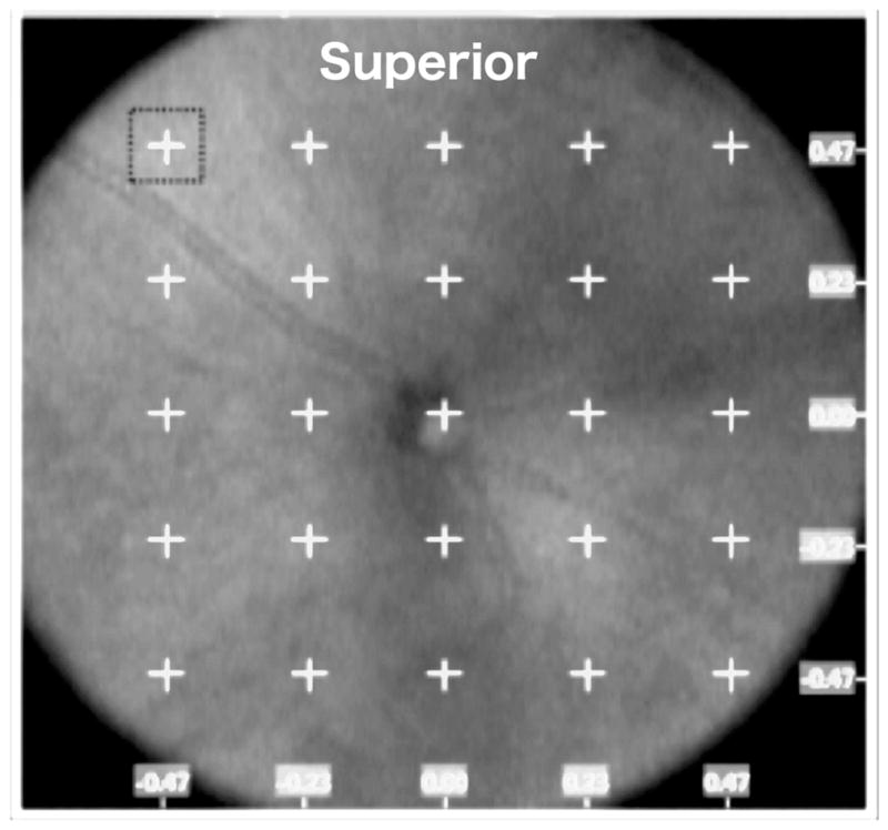
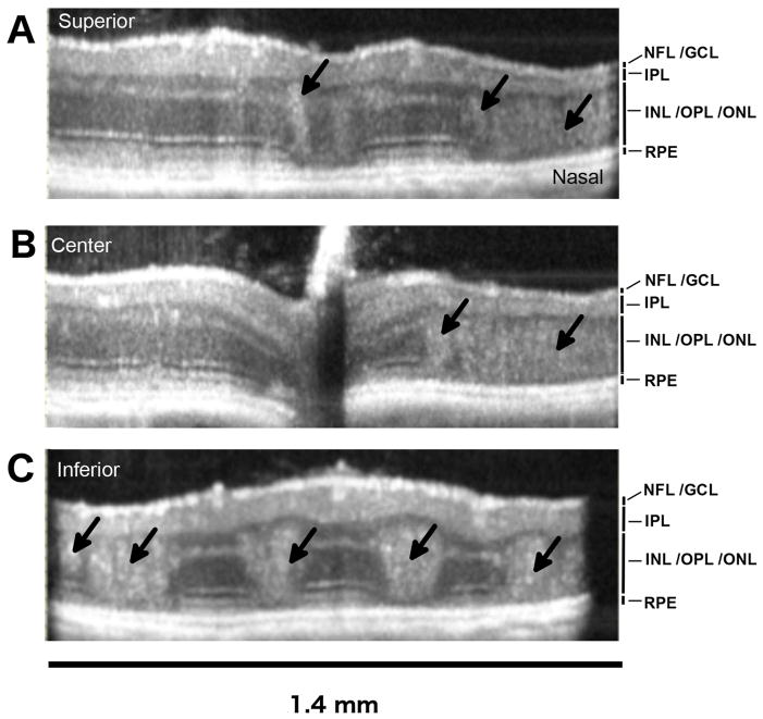
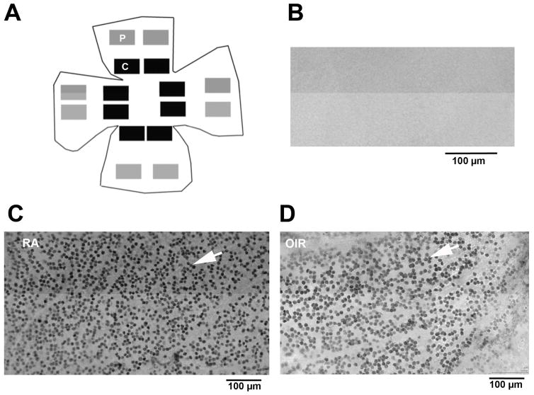
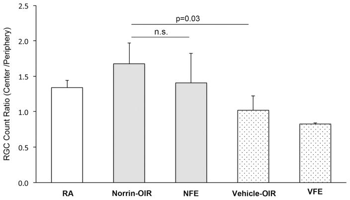
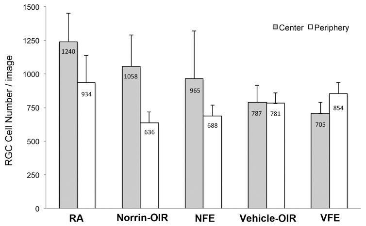
Treatment of a mouse model of oxygen-induced retinopathy (OIR) with recombinant human Norrin (Norrie Disease Protein, gene: NDP) accelerates regrowth of the microvasculature into central ischemic regions of the neural retina, which are generated after treatment with 75% oxygen. While this reduces the average duration and severity of ischemia overall, we do not know if this accelerated recovery of the microvasculature results in any significant survival of retinal ganglion cells (RGCs). The purpose of this study was to investigate ganglion cell survival with and without the intravitreal injection of Norrin in the murine model of oxygen induced retinopathy (OIR), using two strains of mice: C57BL/6J and Thy1-YFP mice. Intravitreal injections of Norrin or vehicle were done after five days of exposure to 75% oxygen from ages P7 to P12. The C57BL/J mice were followed by Spectral-Domain Optical Coherence Tomography (SD-OCT), and the average nerve fiber layer (NFL) and inner-plexiform layer (IPL) thicknesses were measured at twenty-four locations per retina at P42. Additionally, some C57BL/J retinas were flat mounted and immunostained for the RGC marker, Brn3a, to compare the population density of surviving retinal ganglion cells. Using homozygous Thy1-YFP mice, single intrinsically fluorescent RGCs were imaged in live animals with a Micron-III imaging system at ages P21, 28 and P42. The relative percentage of YFP-fluorescent RGCs with dendritic arbors were compared. At age P42, the NFL was thicker in Norrin-injected OIR eyes, 14.4 μm, compared to Vehicle-injected OIR eyes, 13.3 μm (p = 0.01). In the superior retina, the average thickness of the IPL was greater in Norrin-injected OIR eyes, 37.7 μm, compared to Vehicle-injected OIR eyes, 34.6 μm (p = 0.04). Retinas from Norrin injected OIR mice had significantly more surviving RGCs (p = 0.03) than vehicle-injected mice. Based upon NFL thickness and counts of RGCs, we conclude that Norrin treatment, early in the ischemic phase, increased the relative population density of surviving RGCs in the central retinas of OIR mice.

Keywords: Ganglion cell; Neovascular; Nerve fiber layer; Norrin; Optical Coherence Tomography; Oxygen induced retinopathy; Retinal ischemia.


Figures

Figure 1
Figure 1. SD-OCT imaging and analysis of the OIR disruption of retinal laminar layers in the OIR mouse retina
A. SD-OCT en face view (1.41 x 1.41 mm) from a P42 OIR eye, showing a 5 X 5 grid with 0.23 mm interval spacing, centered on the optic disc and used to mark 24 locations for measurements of retinal layer thickness. The horizontal axis (x) and vertical axis (y) coordinates are indicated (mm). Retinal layer measurements were not taken from the central grid location (0,0).
Figure 2
Figure 2. Laminar disruption of the bipolar and photoreceptor cell layers in the OIR mouse retina in vivo
Examples of horizontal B-scans from the P42, OIR eye shown en face in Figure-1. Laminar disruptions (black arrows) were detected in the living eye, demonstrating that disruptions and intermixing of nuclei between the inner nuclear layer (INL) and outer nuclear layer (ONL), seen in fixed retinal tissue sections, are not artifacts of histology processing. The numbers of larger disruptions were greater in the inferior retina compared to the superior retina. The optical back-scattering density of tissue disruptions was also greater in the inferior retina. Horizontal B-scan locations shown are (A) superior (y = +0.47mm), (B) central (y = 0.0 mm) and (C) inferior (y = −0.47 mm). The retinal layers visible are labeled: NFL/GCL (nerve fiber layer / ganglion cell layer), IPL (inner plexiform layer), INL/OPL/INL (inner nuclear layer / outer plexiform layer / outer nuclear layer), RPE (retinal pigment epithelium).
Figure 3
Figure 3. Diagram showing the image field locations used for measurement of RGC density in flat-mounted retinas
Fields were selected from virtual microscopy scans of complete flat-mounted retinas (age P42) after immuno-labeling for Brn3a, a transcription factor present in the nuclei of retinal ganglion cells. (A) The number of Brn3a positive RGCs were counted by computer image analysis and averaged in 8 central (C, black boxes) and 8 peripheral (P, grey boxes) images. Examples of immuno-labeling quality from central regions are shown: (B) no-primary anti-body, negative control, (C) Brn3a positive RGC nuclei (white arrow) in a normal, room-air, retina, and (D) Brn3a positive RGC nuclei (white arrow) in an OIR retina.
Figure 4
Figure 4. Norrin increases the number of Brn3a-labeled RGCs in OIR eyes
The ratio (center/periphery) of the RGC cell density was significantly higher in Norrin-injected OIR eyes (Norrin) compared to vehicle-injected OIR eyes (Vehicle) (p=0.03). Other groups: room-air (RA), fellow non-injected eye of Norrin-treated mice (NFE), fellow non-injected eye of vehicle-treated mice (VFE).
Figure 5
Figure 5. Evidence of a fellow eye effect with Norrin-treatment
Plot of the average RGC cell numbers counted per image in the Central and Peripheral image sets. Room-air (RA), Norrin-injected OIR eyes (Norrin-OIR), Fellow of Norrin-injected OIR eye (NFE), Vehicle-injected OIR eyes (The number of central cell counts were higher in the Norrin fellow eyes (NFE) compared to vehicle-injected OIR eyes (Vehicle-OIR) or vehicle-injected OIR fellow eyes (VFE).
Figure 6
Figure 6. OIR-induced reduction in the percentage of RGC's with dendritic arborization
(A) Example in vivo images of Thy1-YFP RGCs obtained using a Micron-III imaging system equipped with a YFP excitation and emission filter set. Images captured at P28 in RA (left) and OIR (right) mice. (B) The percentage of RGCs with dendritic arbors was significantly lower in OIR eyes (n=23) compared to RA eyes (n=30) at ages P21 and P28.

References

    1. Åkerblom H, Holmstrom G, Eriksson U, Larsson E. Retinal nerve fibre layer thickness in school-aged prematurely-born children compared to children born at term. Br J Ophthalmol. 2012;96:956–960. doi: 10.1136/bjophthalmol-2011-301010. - DOI - PubMed
    1. Akula JD, Hansen RM, Martinez-Perez ME, Fulton AB. Rod photoreceptor function predicts blood vessel abnormality in retinopathy of prematurity. Invest Ophthalmol Vis Sci. 2007;48:4351–4359. doi: 10.1167/iovs.07-0204. - DOI - PubMed
    1. Avery RL, Pearlman J, Pieramici DJ, Rabena MD, Castellarin AA, Nasir MA, Giust MJ, Wendel R, Patel A. Intravitreal Bevacizumab (Avastin) in the Treatment of Proliferative Diabetic Retinopathy. Ophthalmology. 2006:113. doi: 10.1016/j.ophtha.2006.05.064. - DOI - PubMed
    1. Bakbak B, Ozturk BT, Gonul S, Yilmaz M, Gedik S. Comparison of the effect of unilateral intravitreal bevacizumab and ranibizumab injection on diabetic macular edema of the fellow eye. J Ocul Pharmacol Ther. 2013;29:728–732. doi: 10.1089/jop.2013.0049. - DOI - PubMed
    1. Barnaby AM, Hansen RM, Moskowitz A, Fulton AB. Development of scotopic visual thresholds in retinopathy of prematurity. Investig Ophthalmol Vis Sci. 2007;48:4854–4860. doi: 10.1167/iovs.07-0406. - DOI - PMC - PubMed

Publication types