. 2017 Sep;33(3):380-388.
doi: 10.1007/s12288-016-0717-4. Epub 2016 Aug 18.

A Combined Ultrasonographic and Conventional Radiographic Assessment of Hemophilic Arthropathy

Affiliations

A Combined Ultrasonographic and Conventional Radiographic Assessment of Hemophilic Arthropathy

So-Won Chung et al. Indian J Hematol Blood Transfus. 2017 Sep.

Abstract

Hemophilic arthropathy (HA) can be diagnosed by a number of imaging studies. However, it is difficult with conventional radiography to find soft tissue structures around joints, and ultrasonography has limited effectiveness in evaluating internal bony structures. We attempt to determine whether a combination of ultrasonography for soft tissue around joints and conventional radiography for bony structures can be used as a cost-effective imaging tool for evaluating HA and whether it reflects the functional status of hemophilic patients. Thirty-six males (median age 16.5 years; severe 34, mild 2) with hemophilia were recruited. We evaluated the severity of HA using combined imaging score that consisted of modified Petterson X-ray score (mPXS) and the modified ultrasonographic score (mUS). Joint impairment was clinically assesses using the World Federation of Hemophilia-Physical Examination (WFH-PE) scale and the Hemophilic joint health score (HJHS). We assessed the Hemophilia activities list (HAL) for the functional level. We performed a comparative analysis between the combined imaging score and the joint impairment scores as well as the functional scores. The mean mUS was 4.97 ± 3.99 points, and the mean mPXS was 2.85 ± 2.91 points; the combined imaging score was 7.83 ± 6.31 points. The combined imaging score was significantly correlated with the HJHS (p = 0.006) and WFH-PE scores (p = 0.019) as well as the HAL score (p = 0.002). A combination of conventional radiological and ultrasongraphic study might ultimately impact the optimal evaluation of joint impairment and functional status in hemophilic patients.

Keywords: Arthropathy; Function; Hemophilia; Musculoskeletal; Radiography; Ultrasonography.


Conflict of interest statement

Jae-Hyung Kim has received research Grants from Eulji University (No. 2013-EMBRI-DJ0003).

Figures

Fig. 1
Fig. 1
Figure a, b show longitudinal ultrasonographic findings of early hemophilic arthropathy in knee and ankle joint. These demonstrate a small effusion, no fibrotic septa, minimal synovial hypertrophy (<1.5 mm) without flags on PDUS, no hemosiderin deposition and no calcification. The suprapatellar recess (white arrow, a) is minimally distended; Figure c, d show moderate stage of hemophilic arthropathy in knee and ankle joint. These demonstrate a moderate effusion, no fibrotic septa, synovial hypertrophy (1.5–2.5 mm) with 3flags, no hemosiderin deposition and irregular cartilage profile (white arrow, d). Full amount of suprapatellar recess distension (white arrow, c) is observed; Figure e, f show advanced stage of hemophilic arthropathy in knee and ankle joint. These demonstrate a large effusion, no fibrotic septa, synovial hypertrophy (>2.5 mm) with over 3 flags (f), no hemosiderin deposition and cartilage calcification (white arrow, f). Marked distension of the suprapatellar recess with synovium is demonstrated (white arrow, e)
Fig. 2
Fig. 2
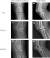
Figure a, b show early stage of hemophilic arthropathy in knee joint: AP and lateral view. Note no osteoporosis, no enlarged epiphysis, no irregular subchondral surface, no narrowing of joint space, no subchondral cyst formation, no joint margin erosion, no gross incongruence of articulating bone ends and no joint deformity; Figure c, d show moderate stage of hemophilic arthropathy in knee joint: AP and lateral view. Note the osteoporosis, no enlarged epiphysis, no irregular subchondral surface, narrowing of joint space (>1 mm), no subchondral cyst formation, no joint margin erosion, no gross incongruence of articulating bone ends and no joint deformity. This is Petterson grade 2; Figure e, f show advanced stage of hemophilic arthropathy in knee joint: AP and lateral view. Note the osteoporosis, no enlarged epiphysis, partially irregular subchondral surface, narrowing of joint space (<1 mm), 1 subchondral cyst formation (black arrow, e), no joint margin erosion, slight gross incongruence of articulating bone ends and no joint deformity. This is Petterson grade 6
Fig. 3
Fig. 3
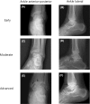
Figure a, b show early stage of hemophilic arthropathy in ankle joint: AP and lateral view. Note no osteoporosis, no enlarged epiphysis, no irregular subchondral surface, no narrowing of joint space, no subchondral cyst formation, no joint margin erosion, no gross incongruence of articulating bone ends and no joint deformity; Figure c, d show moderate stage of hemophilic arthropathy in ankle joint: AP and lateral view. Note the osteoporosis, no enlarged epiphysis, no irregular subchondral surface, narrowing of joint space (>1 mm), no subchondral cyst formation, no joint margin erosion, no gross incongruence of articulating bone ends and no joint deformity. This is Petterson grade 2; Figure e, f show advanced stage of hemophilic arthropathy in ankle joint: AP and lateral view. Note the osteoporosis, no enlarged epiphysis, partially irregular subchondral surface, narrowing of joint space (<1 mm), 2 subchondral cyst formation (black arrow, e), joint margin erosion (black arrow, f), gross incongruence of articulating bone ends and no joint deformity. This is Petterson grade 7
Fig. 4
Fig. 4
This figure shows that there were significant correlation between combined imaging score and Hemophilic Joint Health Score (HJHS), World Federation of Hemophilia-Physical Examination (WFH-PE) score as well as Hemophilic activities lists (HAL) score, however, this method was no significant correlation with Functional Independence Score in Hemophilia (FISH)

References

    1. Lobet S, Hermans C, Lambert C. Optimal management of hemophilic arthropathy and hematomas. J Blood Med. 2014;5:207–218. doi: 10.2147/JBM.S50644. - DOI - PMC - PubMed
    1. Chen YC, Chen LC, Cheng SN, Pan RY, Chang ST, Li TY. Hemophilic arthropathy of shoulder joints: clinical, radiographic, and ultrasonographic characteristics of seventy patients. J Bone Joint Surg Am. 2013;95(7):e43. doi: 10.2106/JBJS.K.01646. - DOI - PubMed
    1. Cross S, Vaidya S, Fotiadis N. Hemophilic arthropathy: a review of imaging and staging. Semin Ultrasound CT MR. 2013;34(6):516–524. doi: 10.1053/j.sult.2013.05.007. - DOI - PubMed
    1. Doria AS. State-of-the-art imaging techniques for the evaluation of hemophilic arthropathy: present and future. Haemoph Off J World Fed Hemophilia. 2010;16(Suppl 5):107–114. doi: 10.1111/j.1365-2516.2010.02307.x. - DOI - PubMed
    1. Keen HI, Wakefield RJ, Grainger AJ, Hensor EM, Emery P, Conaghan PG. Can ultrasonography improve on radiographic assessment in osteoarthritis of the hands? A comparison between radiographic and ultrasonographic detected pathology. Ann Rheum Dis. 2008;67(8):1116–1120. doi: 10.1136/ard.2007.079483. - DOI - PubMed

LinkOut - more resources