. 2017 Jan 12;27(2):1603524.
doi: 10.1002/adfm.201603524. Epub 2016 Nov 7.

Advanced Functional Nanomaterials for Theranostics

Affiliations

Advanced Functional Nanomaterials for Theranostics

Haoyuan Huang et al. Adv Funct Mater. .

Abstract

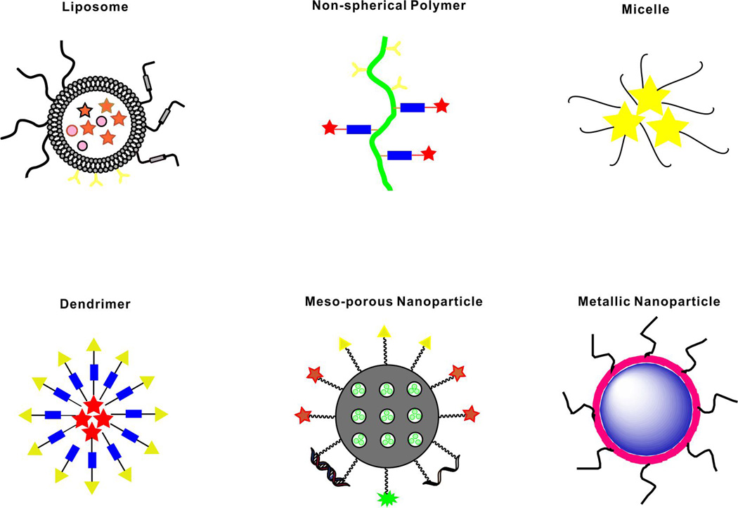
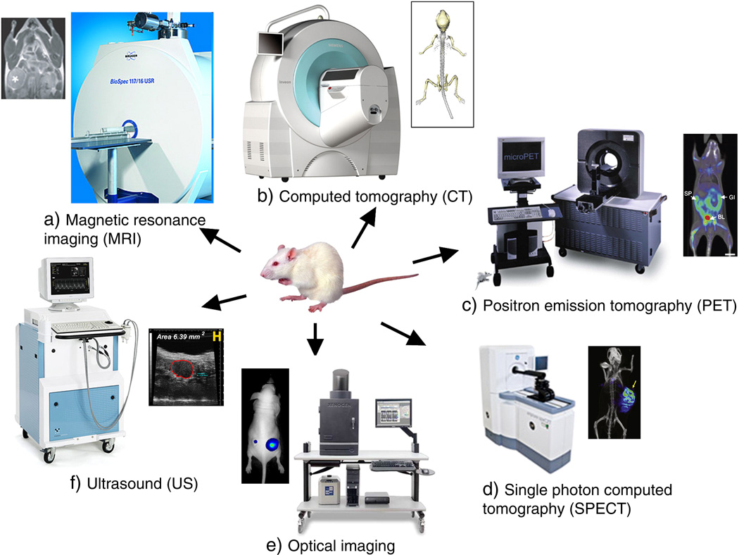
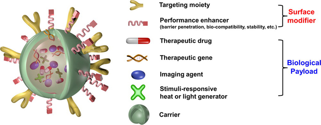
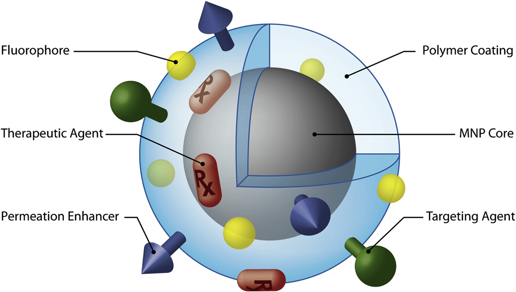
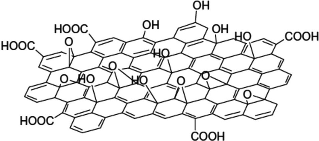
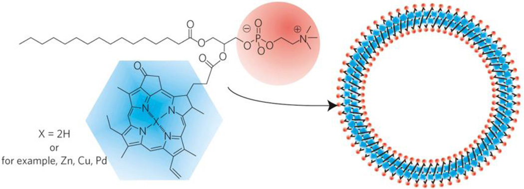
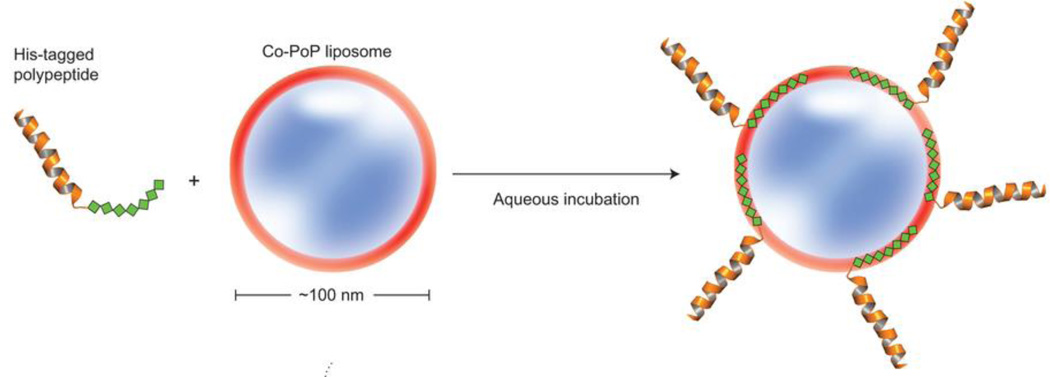
Nanoscale materials have been explored extensively as agents for therapeutic and diagnostic (i.e. theranostic) applications. Research efforts have shifted from exploring new materials in vitro to designing materials that function in more relevant animal disease models, thereby increasing potential for clinical translation. Current interests include non-invasive imaging of diseases, biomarkers and targeted delivery of therapeutic drugs. Here, we discuss some general design considerations of advanced theranostic materials and challenges of their use, from both diagnostic and therapeutic perspectives. Common classes of nanoscale biomaterials, including magnetic nanoparticles, quantum dots, upconversion nanoparticles, mesoporous silica nanoparticles, carbon-based nanoparticles and organic dye-based nanoparticles, have demonstrated potential for both diagnosis and therapy. Variations such as size control and surface modifications can modulate biocompatibility and interactions with target tissues. The needs for improved disease detection and enhanced chemotherapeutic treatments, together with realistic considerations for clinically translatable nanomaterials will be key driving factors for theranostic agent research in the near future.

Keywords: Imaging; Nanoparticles; Theranostic; Therapy.


Figures

Figure 1
Figure 1
Schematic representation of selected nanocarriers. Stars, circles and triangles represent various possible and hypothetical modifications such as payloads, cargo, imaging agents, and targeting moieties.
Figure 2
Figure 2
Molecular imaging instrumentation and images for common diagnostic modalities. Reproduced with permission.[5] Copyright 2010, Elsevier.
Figure 3
Figure 3
A prototypical multifunctional nanoparticle. Reproduced with permission.[67] Copyright 2015, American Chemical Society.
Figure 4
Figure 4
Schematic figure of multifunctional magnetic nanoparticles with surface coating. Reproduced with permission.[72] Copyright 2008, Elsevier.
Figure 5
Figure 5
69Ge-labled SPION for PET/MRI. a) Schematic figure of 69Ge labeling SPIONs; b) biodistribution monitoring post i.v. injection through PET and MRI. Contrast agents demonstrated long-term signals 36 h post injection with PET imaging and 24 h with MRI; c) Long-term lymph node mapping with PET and MRI. The green circle areas for MRI images are contrast agents injected position and the red areas are control areas. Reproduced with permission[90]. Copyright 2014, Wiley.
Figure 6
Figure 6
Structures of some common Gd chelators. Reproduced with permission.[111] Copyright 2012, Wiley.
Figure 7
Figure 7
The left figure shows a schematic figure of phospholipid-PEG encapsulated silicon quantum dots; the right figures show NIR imaging applications including lymph node mapping, multiplex imaging and tumor specific imaging after conjugation with RGD. Reproduced with permission.[177] Copyright 2011, American Chemical Society.
Figure 8
Figure 8
Ag2Se@Mn QD labeling of cell-derived microvesicles (MVs), and their real-time dual-modality whole body monitoring and quantitative biodistribution analysis. Reproduced with permission.[187] Copyright 2016, American Chemical Society.
Figure 9
Figure 9
Schematic figure of (a) conventional photoluminescence and (b) upconversion luminescence processes. Reproduced with permission.[192] Copyright 2015, American Chemical Society.
Figure 10
Figure 10
In vivo lymphatic imaging using porphyrin-phospholipid (PoP)-UCNPs. Accumulation of PoP-UCNPs in the first draining lymph node is indicated with yellow arrows. a) Fluorescence and b) upconversion images with the injection site cropped out of frame. c) Full body PET, d) merged PET/CT, and e) Cerenkov luminescence images. f) Photoacoustic images before and g) after injection show endogenous PA blood signal compared to the contrast enhancement that allowed visualization of the previously undetected lymph node. Reproduced with permission.[195] Copyright 2015, Wiley.
Figure 11
Figure 11
Schematic figure of a) MWCNT and b) SWNCT. Reproduced with permission.[271] Copyright 2009, American Chemical Society.
Figure 12
Figure 12
Dynamic NIR-II fluorescence images and contrast-enhanced images based on PCA analysis: (a–f) NIR-II fluorescence images of a 4T1 tumor bearing mouse after injection of a 200 µL solution containing 0.35 mg/mL SWNTs; (g) positive pixels from PCA, showing lungs, kidneys, and major vessels in the tumor; (h) negative pixels from PCA, showing the body of the tumor; (i) overlaid image showing the absolute value of both positive and negative pixels, from which both the vessels in the tumor and the tumor outline can be seen. Yellow arrows in the images highlight the tumor. Reproduced with permission.[292] Copyright 2012, American Chemical Society.
Figure 13
Figure 13
Schematic figure of GO. Reproduced with permission.[327] Copyright 2015, Royal Society of Chemistry.
Figure 14
Figure 14
Schematic figure of pyropheophorbide–lipid porphysome. The phospholipid headgroup (red) and porphyrin (blue) are highlighted in the subunit (left) and assembled nanovesicle (right). Reproduced with permission.[49] Copyright 2011, Nature Publishing Group.
Figure 15
Figure 15
Schematic figure of his-tagged polypeptide insertion into Co-PoP liposomes. Reproduced with permission.[387] Copyright 2015, Nature Publishing Group.

References

    1. Jokerst JV, Gambhir SS. Acc. Chem. Res. 2011;44:1050. - PMC - PubMed
    1. Lammers T, Aime S, Hennink WE, Storm G, Kiessling F. Acc. Chem. Res. 2011;44:1029. - PubMed
    1. Huynh E, Zheng G. WIRES: Nanomed. Nanobiotechnol. 2013;5:250. - PubMed
    1. Peer D, Karp JM, Hong S, Farokhzad OC, Margalit R, Langer R. Nat. Nano. 2007;2:751. - PubMed
    1. Janib SM, Moses AS, MacKay JA. Adv. Drug Deliv. Rev. 2010;62:1052. - PMC - PubMed

LinkOut - more resources