Review
. 2017 Jul 31:10:236.
doi: 10.3389/fnmol.2017.00236. eCollection 2017.

Recent Advancements in the Regeneration of Auditory Hair Cells and Hearing Restoration

Affiliations
Review

Recent Advancements in the Regeneration of Auditory Hair Cells and Hearing Restoration

Rahul Mittal et al. Front Mol Neurosci. .

Abstract

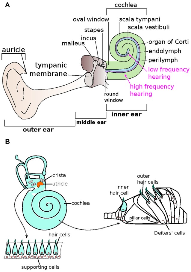
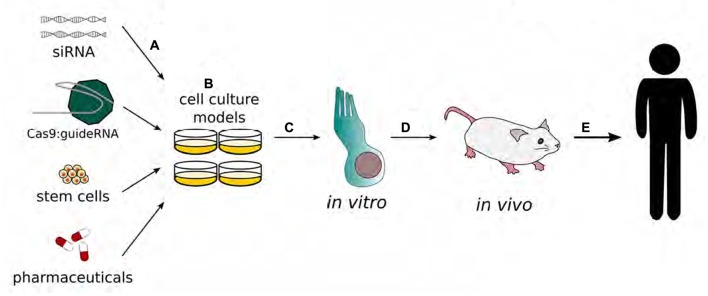
Neurosensory responses of hearing and balance are mediated by receptors in specialized neuroepithelial sensory cells. Any disruption of the biochemical and molecular pathways that facilitate these responses can result in severe deficits, including hearing loss and vestibular dysfunction. Hearing is affected by both environmental and genetic factors, with impairment of auditory function being the most common neurosensory disorder affecting 1 in 500 newborns, as well as having an impact on the majority of elderly population. Damage to auditory sensory cells is not reversible, and if sufficient damage and cell death have taken place, the resultant deficit may lead to permanent deafness. Cochlear implants are considered to be one of the most successful and consistent treatments for deaf patients, but only offer limited recovery at the expense of loss of residual hearing. Recently there has been an increased interest in the auditory research community to explore the regeneration of mammalian auditory hair cells and restoration of their function. In this review article, we examine a variety of recent therapies, including genetic, stem cell and molecular therapies as well as discussing progress being made in genome editing strategies as applied to the restoration of hearing function.

Keywords: auditory hair cells; gene therapy; hair cell regeneration; hearing loss; stem cell therapy.


Figures

Figure 1
Figure 1
Schematic representation of the auditory system. (A) The human ear is composed of three sections—the outer, middle and inner ears. The inner ear is made up of the spiral-shaped cochlea, where endolymph (blue) and perilymph (green) reside. The organ of Corti (purple), responsible for relaying sound via specialized hair cells, is arranged tonotopically, where high (base) and low (apex) frequencies are processed in separate locations. (Adapted from Géléoc and Holt, 2014). (B) The mammalian inner ear consists of two types of sensory receptor organs, which include the hair cells (light blue) and supporting cells (white). The hair cells that make up the auditory sensory epithelia in the cochlea is also known as the organ of Corti, whereas the saccule, utricle and cristae make up the vestibular epithelia. The vestibular epithelia include alternating outer hair cells and supporting cells. In the organ of Corti within the cochlea, one row of inner hair cells is associated with three rows of outer hair cells. Supporting cells such as the pillar and Deiters’ cells make up the rest of the organ of Corti. (Adapted from Bermingham-McDonogh and Reh, 2011).
Figure 2
Figure 2
Schematic representation of the development of gene therapy in the inner ear. Gene therapy begins with transfection of adenovirus vectors (A) or drug applications (B) to cell cultures. Modified functions are evaluated in in vitro models, specifically inner and/or outer hair cells. (C) The results with the best outcomes will move on to be tested in in vivo systems (D) to validate therapeutic benefits before being tested in human clinical trials (E). (Adapted from Rousset et al., 2015).
Figure 3
Figure 3
Potential routes for delivery of therapeutics to the inner ear. The round window membrane (A) is accessible from the middle ear space or delivery via cochleostomy (B) is also possible as it directly accesses to inner ear tissues. (Adapted from Rousset et al., 2015).

References

    1. Ahmed Z. M., Riazuddin S., Riazuddin S., Wilcox E. R. (2003). The molecular genetics of Usher syndrome. Clin. Genet. 63, 431–444. 10.1034/j.1399-0004.2003.00109.x - DOI - PubMed
    1. Ahmed Z. M., Yousaf R., Lee B. C., Khan S. N., Lee S., Lee K., et al. (2011). Functional null mutations of MSRB3 encoding methionine sulfoxide reductase are associated with human deafness DFNB74. Am. J. Hum. Genet. 88, 19–29. 10.1016/j.ajhg.2010.11.010 - DOI - PMC - PubMed
    1. Akil O., Seal R. P., Burke K., Wang C., Alemi A., During M., et al. (2012). Restoration of hearing in the VGLUT3 knockout mouse using virally-mediated gene therapy. Neuron 75, 283–293. 10.1016/j.neuron.2012.05.019 - DOI - PMC - PubMed
    1. Alagramam K. N., Gopal S. R., Geng R., Chen D. H., Nemet I., Lee R., et al. (2016). A small molecule mitigates hearing loss in a mouse model of Usher syndrome III. Nat. Chem. Biol. 12, 444–451. 10.1038/nchembio.2069 - DOI - PMC - PubMed
    1. An L., Hu Y., Chang S., Zhu X., Ling P., Zhang F., et al. (2016). Efficient generation of FVII gene knockout mice using CRISPR/Cas9 nuclease and truncated guided RNAs. Sci. Rep. 6:25199. 10.1038/srep25199 - DOI - PMC - PubMed