

Wernekink Commissure Syndrome Secondary to Bilateral Caudal Paramedian Midbrain Infarction Presenting with a Unique "Heart or V" Appearance Sign: Case Report and Review of the Literature
- PMID: 28824533
- PMCID: PMC5540952
- DOI: 10.3389/fneur.2017.00376
Wernekink Commissure Syndrome Secondary to Bilateral Caudal Paramedian Midbrain Infarction Presenting with a Unique "Heart or V" Appearance Sign: Case Report and Review of the Literature
Abstract
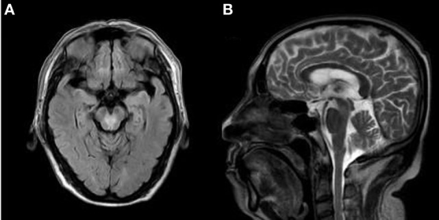
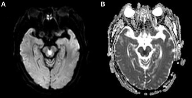
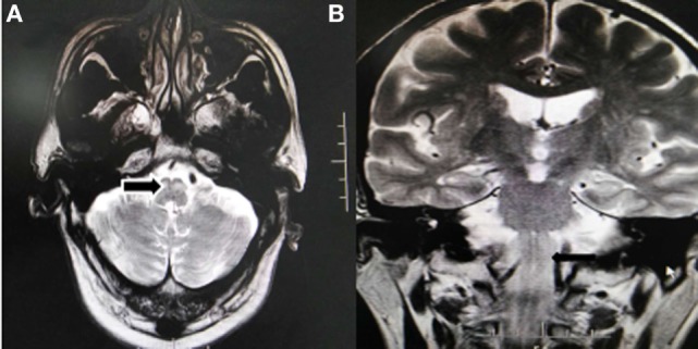
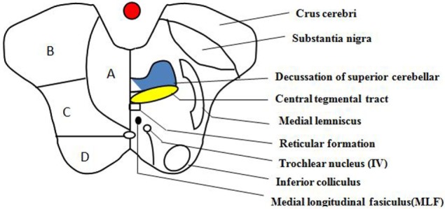
Wernekink commissure syndrome secondary to caudal paramedian midbrain infarction (CPMI) is a rare midbrain syndrome involving the decussation of the superior cerebellar peduncle in the caudal paramedian midbrain tegmentum. The central characteristics are constant bilateral cerebellar dysfunction, variable eye movement disorders, and rare delayed palatal myoclonus. Following is a description of the case of a 60-year-old man who presented with dizziness, slurred speech, and difficulty walking. Neurological examination revealed bilateral cerebellar dysfunction and bilateral internuclear ophthalmoplegia (bilateral INO). Serial magnetic resonance imaging (MRI) revealed a lesion in the caudal paramedian midbrain with a "heart-shaped" sign on fluid-attenuation inversion recovery images and a "V-shaped" appearance on diffusion-weighted imaging (DWI). An acute CPMI with a "heart or V" appearance sign was diagnosed. Upon follow-up evaluation 3 months later, a palatal tremor accompanied by involuntary head tremor was discovered. Hypertrophy and increased signal of the bilateral inferior olivary nucleus, compatible with hypertropic olivary degeneration (HOD) were revealed during a subsequent MRI study.
Keywords: Palatal myoclonus; Wernekink commissure syndrome; bilateral internuclear ophthalmolegia; caudal paramedian midbrain infarction; hypertrophic olivary degeneration; “heart-shaped” sign.
Figures
References
-
- Lhermitte F. [The cerebellar syndrome: anatomo-clinical study in the adult]. Rev Neurol (Paris) (1958) 98(6):435–77. - PubMed
Publication types
LinkOut - more resources
Full Text Sources
Other Literature Sources