Review
. 2017 Jul 26;9(7):600-608.
doi: 10.4330/wjc.v9.i7.600.

Cardiac and pericardial tumors: A potential application of positron emission tomography-magnetic resonance imaging

Affiliations
Review

Cardiac and pericardial tumors: A potential application of positron emission tomography-magnetic resonance imaging

Ahmed Fathala et al. World J Cardiol. .

Abstract

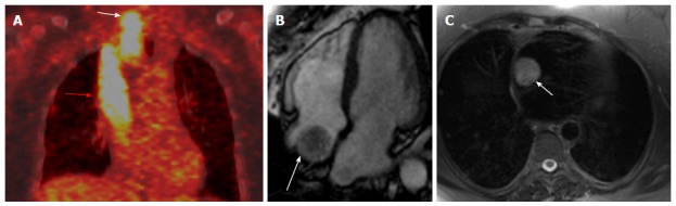
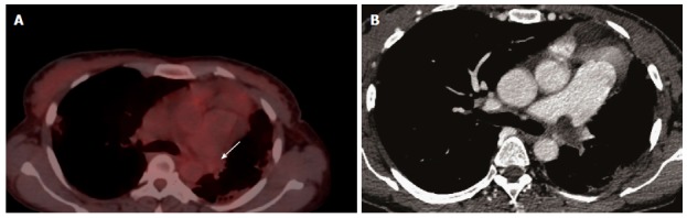
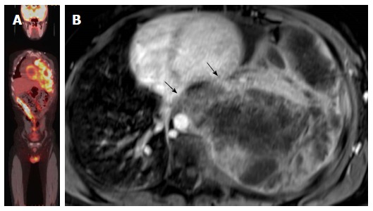
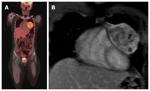
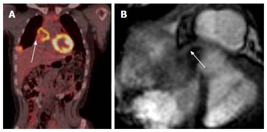
Cardiac and pericardial masses may be neoplastic, benign and malignant, non-neoplastic such as thrombus or simple pericardial cysts, or normal variants cardiac structure can also be a diagnostic challenge. Currently, there are several imaging modalities for diagnosis of cardiac masses; each technique has its inherent advantages and disadvantages. Echocardiography, is typically the initial test utilizes in such cases, Echocardiography is considered the test of choice for evaluation and detection of cardiac mass, it is widely available, portable, with no ionizing radiation and provides comprehensive evaluation of cardiac function and valves, however, echocardiography is not very helpful in many cases such as evaluation of extracardiac extension of mass, poor tissue characterization, and it is non diagnostic in some cases. Cross sectional imaging with cardiac computed tomography provides a three dimensional data set with excellent spatial resolution but utilizes ionizing radiation, intravenous iodinated contrast and relatively limited functional evaluation of the heart. Cardiac magnetic resonance imaging (CMR) has excellent contrast resolution that allows superior soft tissue characterization. CMR offers comprehensive evaluation of morphology, function, tissue characterization. The great benefits of CMR make CMR a highly useful tool in the assessment of cardiac masses. (Fluorine 18) fluorodeoxygluocse (FDG) positron emission tomography (PET) has become a corner stone in several oncological application such as tumor staging, restaging, treatment efficiency, FDG is a very useful imaging modality in evaluation of cardiac masses. A recent advance in the imaging technology has been the development of integrated PET-MRI system that utilizes the advantages of PET and MRI in a single examination. FDG PET-MRI provides complementary information on evaluation of cardiac masses. The purpose of this review is to provide several clinical scenarios on the incremental value of PET and MRI in the evaluation of cardiac masses.

Keywords: Cardiac; Echocardiography; Pericardial tumors.


Conflict of interest statement

Conflict-of-interest statement: We declare we do not have any conflict of interest.

Figures

Figure 1
Figure 1
Localization of abnormal fluorodeoxygluocse activity in the heart. A: Axial fused PET-CT image shows focal intense FDG uptake in the heart (arrow) in patient with melanoma that thought represents cardiac metastases; B: Selected delayed enhancement short axis image of the same patient with other images (not shown) shows no abnormal enhancement or tissue infiltration, the FDG uptake was corresponding to hypertrophic papillary muscle, follow PET-CT was normal. PET: Positron emission tomography; CT: Computed tomography; FDG: Fluorodeoxygluocse.
Figure 2
Figure 2
Localization and characterization of abnormal fluorodeoxygluocse activity in the heart. A: Axial PET-CT images in patient with history of invasive thymoma underwent surgical resection shows intense FDG focal uptake in the cardiac apex highly suspicious for cardiac metastases, review CT component of PET-CT shows area of and soft tissue; B: Four-chamber DE image with triple inversion recovery shows complete fat suppression of the apical activity in keeping with brown fat (arrow), with high signal fluid intensity consistent with post-operative lobulated fluid collection (arrowhead). PET: Positron emission tomography; CT: Computed tomography; FDG: Fluorodeoxygluocse.
Figure 3
Figure 3
Localization and characterization of abnormal fluorodeoxygluocse activity adjacent to the heart. A: Localization and characterization of abnormal FDG activity adjacent to the heart. Axial PET-CT image of the heart shows a non-FDG avid nodule near the base of the heart (arrow), PET-CT was performed for incidentally discovered nodule mass on Echocardiography. The study was otherwise unremarkable; B: Axial first pass perfusion image shows highly vascular intrapericardial tumors with no evidence of tissue invasion (arrow); this lesion was surgically removed and pathologically proven schwannoma. PET: Positron emission tomography; CT: Computed tomography; FDG: Fluorodeoxygluocse.
Figure 4
Figure 4
Localization and characterization of abnormal fluorodeoxygluocse activity adjacent to the heart. A: Axial Ga-68 DOTATATE PET/CT shows a focal intense radiotracer uptake adjacent to the heart with difficulty to accurately localize; patient is known to have history of ileocecal area carcinoid and underwent surgery; B: Coronal first pass perfusion images show highly vascular lesion in the tip of the left lobe of the liver corresponding to abnormal radiotracer uptake, this location is very common for PET-CT misregistration and there was no focal lesion identified in CT component of PET-CT. PET: Positron emission tomography; CT: Computed tomography; FDG: Fluorodeoxygluocse.
Figure 5
Figure 5
Differnation between tumoral thrombus from bland thrombus. A: Differnation between tumoral thrombus from bland thrombus in patient with history of thyroid cancer treated with total thyroidectomy presented with neck mass. Coronal fused FDG-PET image shows intense linear FDG uptake from in the thyroid bed (white arrow) and spreading through SCV to the right atrium (red arrow); B: Four chamber cine image shows irregularly defined mass in the right atrium (arrow), the rest of cardiac examination was unremarkable; C: Axial HASTE at the junction of superior vena cava and right atrium shows a high signal intensity mass (arrow). PET: Positron emission tomography; CT: Computed tomography; FDG: Fluorodeoxygluocse.
Figure 6
Figure 6
Differnation between tumoral thrombus from bland thrombus. A: Differnation between tumoral thrombus from bland thrombus in female patient with history ovarian cancer, patient was known to have chronic pulmonary embolism, there was a bland thrombus almost occulting the left pulmonary artery was no FDG uptake (arrow); B: Axial CT image shows a left pulmonary artery thrombus. CT: Computed tomography; FDG: Fluorodeoxygluocse.
Figure 7
Figure 7
M staging. A: M staging: Coronal PET-CT image of patient with lymphoma with unexpected cardiac involvement; B: Axial T1-weighted image at the level of interatrial septum shows well defined mass attached to the atrial septum and nearly fills the right atrium and was proved to be lymphoma. PET: Positron emission tomography; CT: Computed tomography.
Figure 8
Figure 8
Evaluation of the aggressiveness of the lesion and assessment of cardiac involvement. A: Evaluation of the aggressiveness of the lesion and assessment of cardiac involvement; whole body PET-CT image of patient with extensive Ewing sarcoma of the left hemithorax, PET-CT images are not sufficient to evaluate local extension of the tumor to the heart; B: Axial delayed enhancement image shows large necrotic mass occupying the left hemithorax with direct left ventricle (upper arrow) the arrow without circle and left atrial invasion (lower arrow). PET: Positron emission tomography; CT: Computed tomography.
Figure 9
Figure 9
Evaluation of the aggressiveness of the lesion and assessment of cardiac involvement. A: Evaluation of the aggressiveness of the lesion and assessment of cardiac involvement; Coronal PET-CT image in a patent with Ewing sarcoma of the left chest wall with direct compression of the left side of the heart; B: Coronal Post contrast T1-weighted image of the heart shows no evidence of cardiac invasion with clear separation of the mass from the heart, the mass was surgically removed and there was no evidence of cardiac invasion. PET: Positron emission tomography; CT: Computed tomography.
Figure 10
Figure 10
Evaluation of coronary artery involvement by the tumor. A: Coronal PET-CT image in a female patient with history of breast cancer with mediastinal and lung metastasis and recurrent chest pain; B: Axial cine image at the base of the heart shows metastatic lesion invading the heart causing mechanical obstruction of the right coronary artery (arrow). PET: Positron emission tomography; CT: Computed tomography.

References

    1. Amano J, Nakayama J, Yoshimura Y, Ikeda U. Clinical classification of cardiovascular tumors and tumor-like lesions, and its incidences. Gen Thorac Cardiovasc Surg. 2013;61:435–447. - PMC - PubMed
    1. Butany J, Leong SW, Carmichael K, Komeda M. A 30-year analysis of cardiac neoplasms at autopsy. Can J Cardiol. 2005;21:675–680. - PubMed
    1. Douglas PS, Garcia MJ, Haines DE, Lai WW, Manning WJ, Patel AR, Picard MH, Polk DM, Ragosta M, Parker Ward R, et al. ACCF/ASE/AHA/ASNC/HFSA/HRS/SCAI/SCCM/SCCT/SCMR 2011 Appropriate Use Criteria for Echocardiography. A Report of the American College of Cardiology Foundation Appropriate Use Criteria Task Force, American Society of Echocardiography, American Heart Association, American Society of Nuclear Cardiology, Heart Failure Society of America, Heart Rhythm Society, Society for Cardiovascular Angiography and Interventions, Society of Critical Care Medicine, Society of Cardiovascular Computed Tomography, Society for Cardiovascular Magnetic Resonance American College of Chest Physicians. J Am Soc Echocardiogr. 2011;24:229–267. - PubMed
    1. den Harder AM, Willemink MJ, de Jong PA, Schilham AM, Rajiah P, Takx RA, Leiner T. New horizons in cardiac CT. Clin Radiol. 2016;71:758–767. - PubMed
    1. O’Donnell DH, Abbara S, Chaithiraphan V, Yared K, Killeen RP, Cury RC, Dodd JD. Cardiac tumors: optimal cardiac MR sequences and spectrum of imaging appearances. AJR Am J Roentgenol. 2009;193:377–387. - PubMed

LinkOut - more resources