. 2017 Jun;8(1):131-152.
doi: 10.5598/imafungus.2017.08.01.10. Epub 2017 May 23.

The Genera of Fungi - G 4: Camarosporium and Dothiora

Affiliations

The Genera of Fungi - G 4: Camarosporium and Dothiora

Pedro W Crous et al. IMA Fungus. 2017 Jun.

Abstract

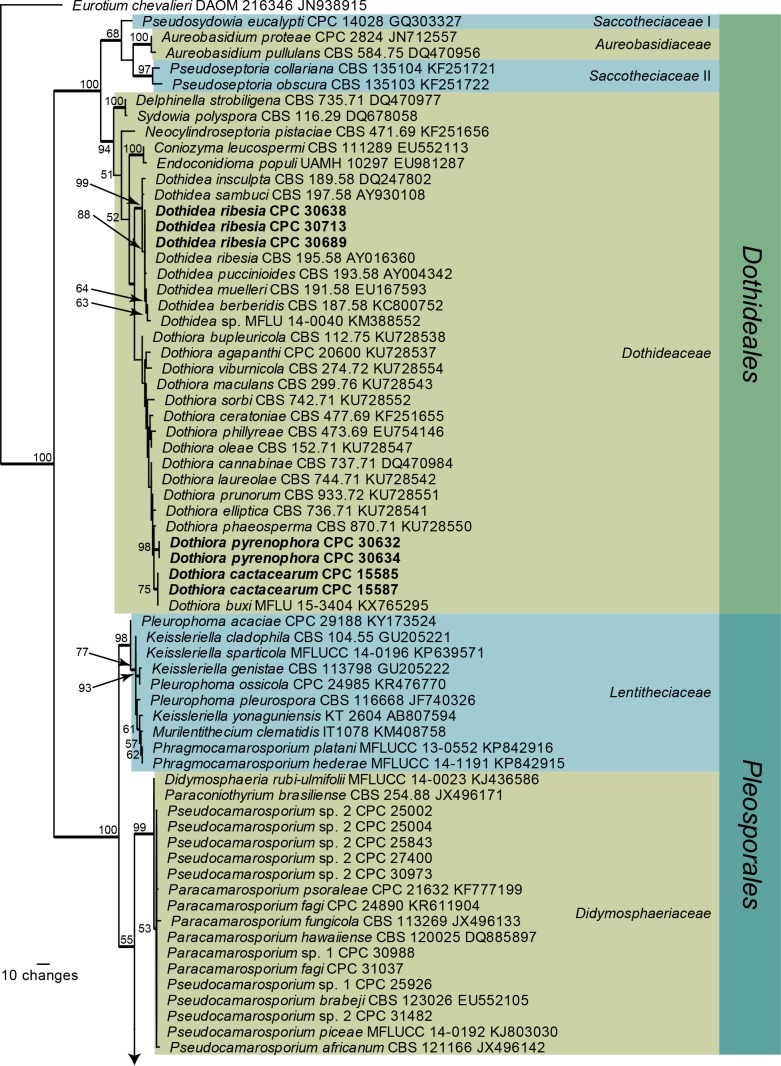
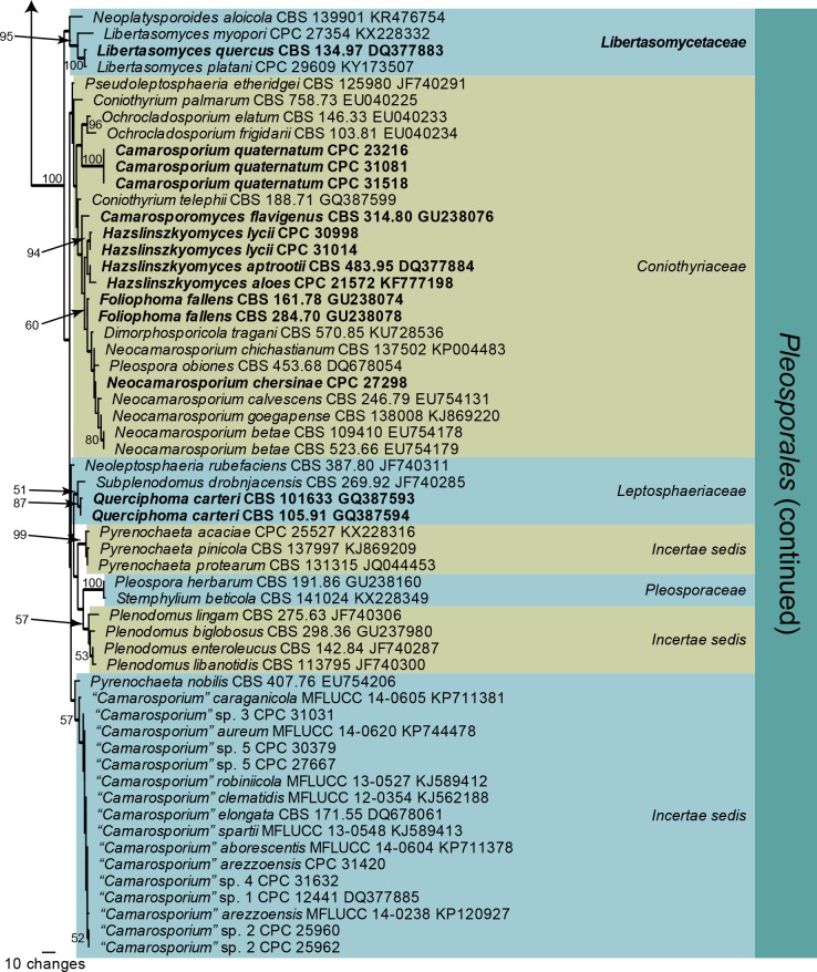
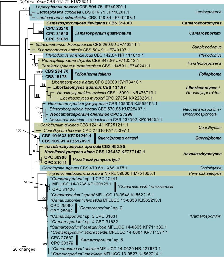
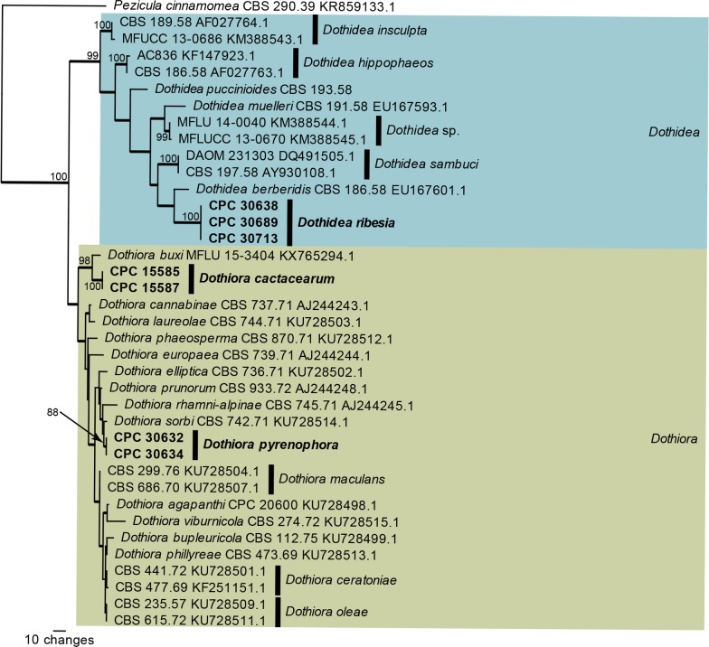
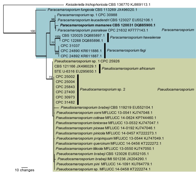
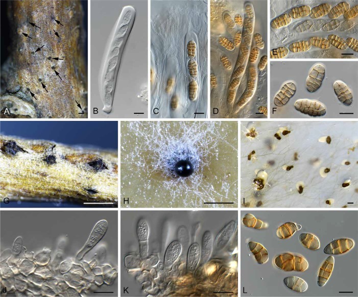
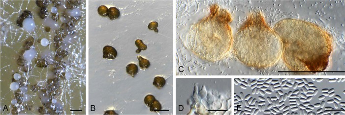
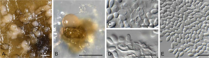
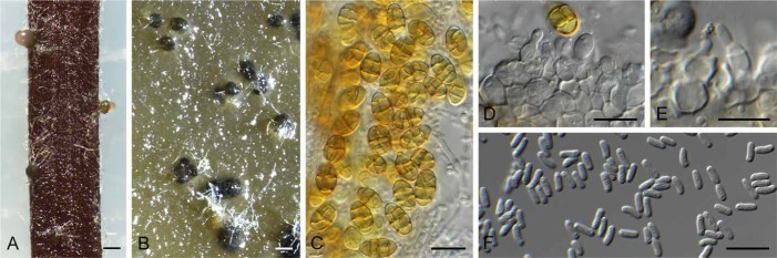
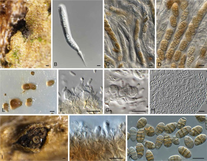
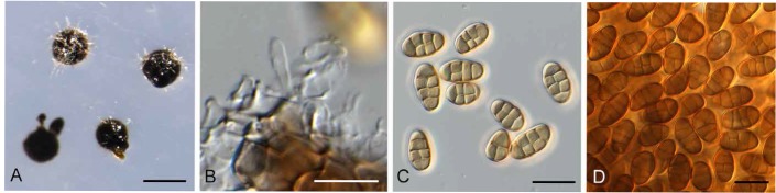
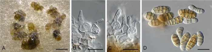
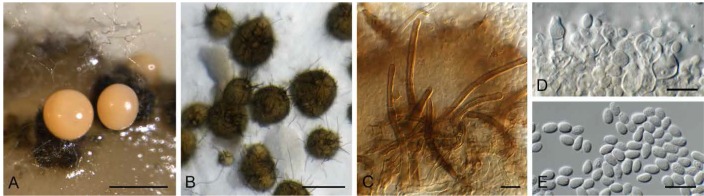
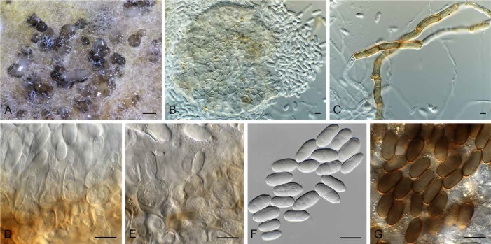
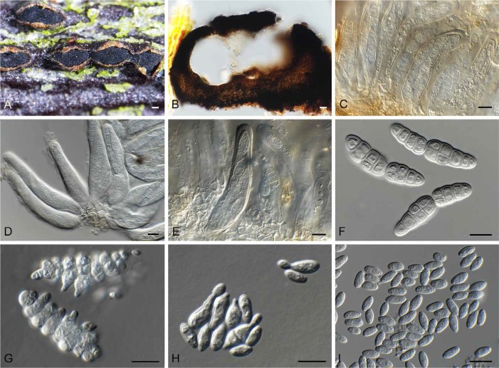
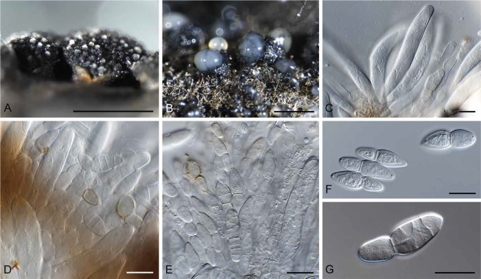
The current paper represents the fourth contribution in the Genera of Fungi series, linking type species of fungal genera to their morphology and DNA sequence data. The present paper focuses on two genera of microfungi, Camarosporium and Dothiora, which are respectively epi- and neotypified. The genus Camarosporium is typified by C. quaternatum, which has a karstenula-like sexual morph, and phoma-like synasexual morph. Furthermore, Camarosporomyces, Foliophoma and Hazslinszkyomyces are introduced as new camarosporium-like genera, while Querciphoma is introduced as a new phoma-like genus. Libertasomycetaceae is introduced as a new family to accommodate Libertasomyces and Neoplatysporoides. Dothiora, which is typified by D. pyrenophora, is shown to produce dothichiza- and hormonema-like synasexual morphs in culture, and D. cactacearum is introduced as a new species. In addition to their typification, ex-type cultures have been deposited in the Westerdijk Fungal Biodiversity Institute (CBS Culture Collection), and species-specific DNA barcodes in GenBank. Authors interested in contributing accounts of individual genera to larger multi-authored papers in this series should contact the associate editors listed on the List of Protected Generic Names for Fungi.

Keywords: DNA Barcodes; ITS; LSU; fungal systematics; typification; www.GeneraofFungi.org.


Figures

Fig. 1.
Fig. 1.
One of 1 000 equally most parsimonious trees obtained from a maximum parsimony analysis of the LSU sequence alignment. The scale bar shows 10 changes, and parsimony bootstrap support values > 49 % from 1 000 replicates are shown at the nodes. Thickened lines represent those branches also present in the strict consensus tree and families are indicated with coloured blocks and the taxonomic novelties, or species treated in the present paper, are in bold text. Orders are indicated on the right side of the tree. The tree was rooted to Eurotium chevalieri (GenBank accession number JN938915).
Fig. 1.
Fig. 1.
One of 1 000 equally most parsimonious trees obtained from a maximum parsimony analysis of the LSU sequence alignment. The scale bar shows 10 changes, and parsimony bootstrap support values > 49 % from 1 000 replicates are shown at the nodes. Thickened lines represent those branches also present in the strict consensus tree and families are indicated with coloured blocks and the taxonomic novelties, or species treated in the present paper, are in bold text. Orders are indicated on the right side of the tree. The tree was rooted to Eurotium chevalieri (GenBank accession number JN938915).
Fig. 2.
Fig. 2.
One of 284 equally most parsimonious trees obtained from a maximum parsimony analysis of the ITS sequence alignment of Camarosporium and allied genera. The scale bar shows 20 changes, and parsimony bootstrap support values > 74 % from 1 000 replicates are shown at the nodes. Thickened lines represent those branches also present in the strict consensus tree and genera are indicated with coloured blocks and the taxonomic novelties, or species treated in the present paper, are in bold text. The tree was rooted to Dothiora oleae (GenBank accession number KU728511).
Fig. 3.
Fig. 3.
One of 140 equally most parsimonious trees obtained from a maximum parsimony analysis of the ITS sequence alignment of Dothidea and Dothiora. The scale bar shows 10 changes, and parsimony bootstrap support values > 74 % from 1 000 replicates are shown at the nodes. Thickened lines represent those branches also present in the strict consensus tree and genera are indicated with coloured blocks and the taxonomic novelties, or species treated in the present paper, are in bold text. The tree was rooted to Pezicula cinnamomea (GenBank accession number KR859133).
Fig. 4.
Fig. 4.
One of 16 equally most parsimonious trees obtained from a maximum parsimony analysis of the ITS sequence alignment of Paracamarosporium and Pseudocamarosporium. The scale bar shows 10 changes, and parsimony bootstrap support values > 69 % from 1 000 replicates are shown at the nodes. Thickened lines represent those branches also present in the strict consensus tree and genera are indicated with coloured blocks and the taxonomic novelties, or species treated in the present paper, are in bold text. The tree was rooted to Keissleriella trichophoricola (GenBank accession number KJ869113).
Fig. 5.
Fig. 5.
Camarosporium quaternatum (CPC 31081). A. Immersed ascomata on twig (arrows). B–E. Asci and pseudoparaphyses. F. Ascospores. G. Conidiomata on twig. H. Camarosporium conidioma on OA. I. Phoma-like conidiomata on SNA. J–K. Conidiogenous cells giving rise to Camarosporium conidia. L. Conidia. Bars: A, I = 200 μm, G = 800 μm, H = 300 μm, all others = 10 μm.
Fig. 6.
Fig. 6.
Camarosporomyces flavigenus (CBS 314.80). A. Conidiomata on PDA. B. Conidiomata on SNA. C. Conidiomata showing darker ostiolar area. D. Conidiogenous cells. E. Phoma-like conidia. Bars: A–C = 90 μm, D–E = 10 μm.
Fig. 7.
Fig. 7.
Foliophoma fallens (CBS 284.70). A. Conidiomata on PDA. B. Coniiomata on SNA. C–D. Conidiogenous cells. E. Conidia. Bars: A–B = 250 μm, all others = 10 μm.
Fig. 8.
Fig. 8.
Hazslinszkyomyces aptrootii (CBS 483.95). A. Conidiomata on PNA. B. Conidiomata on PDA. C. Hazslinszkyomyces and phoma-like conidia. D–E. Conidiogenous cells. F. Phoma-like conidia. Bars: A–B = 170 μm, all others = 10 μm.
Fig. 9.
Fig. 9.
Hazslinszkyomyces lycii (CPC 30998). A. Ascoma immersed in twig. B–D. Asci and pseudoparaphyses. E. Phoma-like conidiomata on SNA. F–G. Conidiogenous cells. H. Conidia. I. Hazslinszkyomyces conidioma on twig. J. Conidiogenous cells. K. Conidia. Bars: A = 130 μm, I = 250 μm, all others = 10 μm.
Fig. 10.
Fig. 10.
Neocamarosporium chersinae (CPC 27298). A. Conidiomata on SNA. B. Conidiogenous cells. C–D. Conidia. Bars: A = 300 μm, all others = 10 μm.
Fig. 11.
Fig. 11.
Libertasomyces quercus (CBS 134.97). A. Conidiomata on OA. B, C. Conidiogenous cells. D. Conidia. Bars: A = 200 μm, all others = 10 μm.
Fig. 12.
Fig. 12.
Querciphoma carteri (CBS 105.91). A. Conidiomata on PDA. B. Conidiomata on SNA. C. Setae. D. Conidiogenous cells. E. Conidia. Bars: A, B = 250 μm, all others = 10 μm.
Fig. 13.
Fig. 13.
Dothiora cactacearum (CPC 15585). A. Conidiomata on OA. B. Conidioma. C. Brown hyphae forming on SNA. D, E. Conidiogenous cells. F. Conidia. G. Conidia turning brown and verruculose with age. Bars: A = 150 μm, all others = 10 μm.
Fig. 14.
Fig. 14.
Dothiora pyrenophora (CPC 30632). A. Ascostroma on twig. B. Section through ascoma. C–E. Asci. F. Ascospores. G. Ascospores undergoing microcyclic conidiation. H. Conidia undergoing budding. I. Conidia. Bars: A, B = 200 μm, all others = 10 μm.
Fig. 15.
Fig. 15.
Dothidea ribesia (CPC 30638). A. Ascostromata on twig. B. Ostioles with exuding ascospores. C–E. Asci with ascospores that become muriformly septate. F. One-septate ascospores. G. Muriformly septate ascospore. Bars: A = 3 mm, B = 100 μm, all others = 10 μm.

References

    1. Boerema GH, De Gruyter J, Noordeloos ME, Hamers MEC. (2004) Phoma Identification Manual: Differentiation of specific and infra-specific taxa in culture. Wallingford: CABI Publishing.
    1. Carbone I, Kohn LM. (1999) A method for designing primer sets for speciation studies in filamentous ascomycetes. Mycologia 91: 553–556.
    1. Cheewangkoon R, Crous PW, Hyde KD, Groenewald JZ, To-anan C. (2008) Species of Mycosphaerella and related anamorphs on Eucalyptus leaves from Thailand. Persoonia 21: 77–91. - PMC - PubMed
    1. Crous PW, Gams W, Stalpers JA, Robert V, Stegehuis G. (2004) MycoBank: an online initiative to launch mycology into the 21st century. Studies in Mycology 50: 19–22.
    1. Crous PW, Giraldo A, Hawksworth DL, Robert V, Kirk PM, et al. (2014a) The Genera of Fungi: fixing the application of type species of generic names. IMA Fungus 5: 141–160. - PMC - PubMed

LinkOut - more resources