Review
. 2017 Jul 27;3(3):149-159.
doi: 10.3233/BLC-170119.

Image-Guided Transurethral Resection of Bladder Tumors - Current Practice and Future Outlooks

Affiliations
Review

Image-Guided Transurethral Resection of Bladder Tumors - Current Practice and Future Outlooks

Timothy C Chang et al. Bladder Cancer. .

Abstract

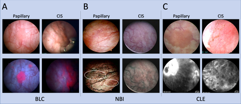
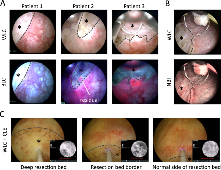
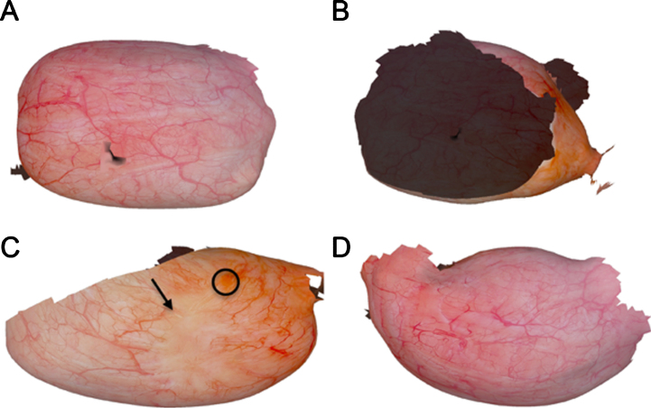
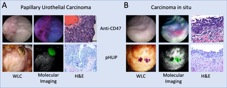
Transurethral resection of bladder tumor (TURBT) under white light cystoscopy (WLC) is the cornerstone for the diagnosis, removal and local staging of non-muscle invasive bladder cancer (NMIBC). Despite technological improvements over the decades, significant shortcomings remain with WLC for tumor detection, thereby impacting the surgical quality and contributing to tumor recurrence and progression. Enhanced cystoscopy modalities such as blue light cystoscopy (BLC) and narrow band imaging (NBI) aid resections by highlighting tumors that might be missed on WLC. Optical biopsy technologies such as confocal laser endomicroscopy (CLE) and optical coherence tomography (OCT) characterize tissue in real-time to ensure a more thorough resection. New resection techniques, particularly en bloc resection, are actively under investigation to improve the overall quality of resections and aid pathologic interpretation. Moreover, new image processing computer algorithms may improve perioperative planning and longitudinal follow-up. Clinical translation of molecular imaging agents is also on the horizon to improve optical diagnosis of bladder cancer. This review focuses on emerging technologies that can impact the quality of TURBT to improve the overall management of NMIBC.

Keywords: Urinary bladder neoplasms; antigens CD47; computer-assisted; confocal microscopy; cystoscopy; hexaminolevulinic acid; laser surgery; molecular imaging; narrow band imaging; optical coherence tomography.


Figures

Fig.1
Fig.1
Imaging modalities for improved bladder tumor detection. Papillary and CIS bladder lesions visualized with BLC, NBI, and CLE with corresponding white light images. (A) Positive, red fluorescence of small, satellite papillary tumors seen on BLC that may be missed on WLC. For CIS, red fluorescence also noted on BLC of what appears to be normal urothelium on WLC. (B) NBI improves visualization of aberrant tumor vasculature. Two papillary tumors are more easily visualized on NBI (encircled). For CIS detection, a patch of erythema is more pronounced under NBI compared to a relatively normal appearing urothelium on WLC. CIS images for NBI obtained with permission from [80]. (C) CLE of papillary tumors and CIS provide microscopic detail that can augment macroscopic imaging. A fibrovascular stalk may be visualized as noted in the top left of the papillary CLE example. CIS is notable for a disorganized architecture with pleomorphic cells and indistinct cellular borders.
Fig.2
Fig.2
Imaging of resection bed. (A) BLC can be used to detect residual tumor during TURBT as positive fluorescence is noted at the outside edges of the resection beds in these examples. There are residual tumors noted for patients 2 and 3 even within the resection bed on BLC that is not noted on WLC. (B) Residual tumor at the edge of the resection bed is noted on NBI. NBI images obtained with permission from [80]. (C) CLE can be used to interrogate the resection bed to determine adequate depth of resection. Features such as elastin strands, muscle fibers and perivesical fat can be visualized in the deep resection bed to verify adequate resection to the muscle layer in real time. All three WLC + CLE images with picture-in-picture are from the same resection bed. In the first image, elastin strands are noted on CLE in the deep resection bed. At the resection bed border, cautery artifact is noted with mostly absence of features seen in the muscle layer. On the normal side of the resection bed, CLE visualizes a capillary network typically seen in normal lamina propria. * – Indicates area within the resection bed. Dashed lines delineate the resection bed border.
Fig.3
Fig.3
Reconstruction from a clinical dataset of human bladder. Cystoscopy image reconstruction views from the (A) anterior, (B) posterior, (C) left lateral, and (D) right lateral walls. Black circle and arrow in (C) show regions of a papillary tumor and scarring, respectively. Regions that appear dark represent the interior of the bladder. From [59] with permission.
Fig.4
Fig.4
Molecular imaging of human bladder tumors. Ex vivo molecular imaging of human bladder using anti-CD47-Qdot625 (Anti-CD47) imaged with BLC and indocyanine green with pH low insertion peptide (pHLIP) agent imaged with da Vinci Si NIRF imaging system. The respective imaging systems for the two molecular imaging strategies are capable of detecting both (A) papillary tumors and (B) CIS with high sensitivity and specificity. Anti-CD47 images from [75], reprinted with permission from AAAS. pHLIP images obtained from [79] with permission from PNAS.

References

    1. Beer E. Landmark article May 28, Removal of neoplasms of the urinary bladder. By Edwin Beer. Jama 1983;250(10):1324–5. - PubMed
    1. Babjuk M, Bohle A, Burger M, Capoun O, Cohen D, Comperat EM, et al. EAU Guidelines on non-muscle-invasive urothelial carcinoma of the bladder: Update 2016. European urology 2017;71(3):447–61. - PubMed
    1. Zainfeld D, Daneshmand S. Transurethral resection of bladder tumors: Improving quality through new techniques and technologies. Current Urology Reports 2017;18(5):34. - PubMed
    1. Witjes JA. Bladder carcinoma in situ in State of the art. European Urology 2004;45(2):142–6. - PubMed
    1. Schwaibold HE, Sivalingam S, May F, Hartung R. The value of a second transurethral resection for T1 bladder cancer. BJU International 2006;97(6):1199–201. - PubMed