. 2017 Oct;37(7):707-714.
doi: 10.1007/s10875-017-0431-5. Epub 2017 Aug 21.

Detection of Sp110 by Flow Cytometry and Application to Screening Patients for Veno-occlusive Disease with Immunodeficiency

Affiliations

Detection of Sp110 by Flow Cytometry and Application to Screening Patients for Veno-occlusive Disease with Immunodeficiency

Florian A Marquardsen et al. J Clin Immunol. 2017 Oct.

Abstract

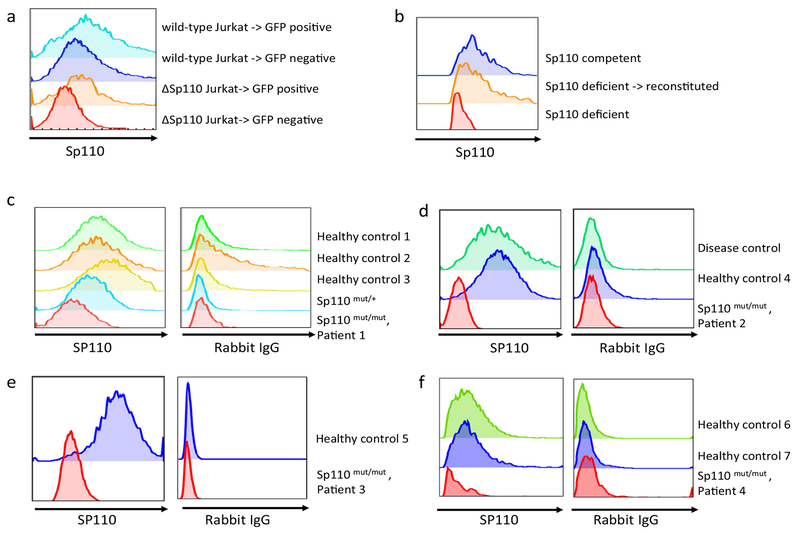
Mutations in Sp110 are the underlying cause of veno-occlusive disease with immunodeficiency (VODI), a combined immunodeficiency that is difficult to treat and often fatal. Because early treatment is critically important for patients with VODI, broadly usable diagnostic tools are needed to detect Sp110 protein deficiency. Several factors make establishing the diagnosis of VODI challenging: (1) Current screening strategies to identify severe combined immunodeficiency are based on measuring T cell receptor excision circles (TREC). This approach will fail to identify VODI patients because the disease is not associated with severe T cell lymphopenia at birth; (2) the SP110 gene contains 17 exons, making it a challenge for Sanger sequencing. The recently developed next-generation sequencing (NGS) platforms that can rapidly determine the sequence of all 17 exons are available in only a few laboratories; (3) there is no standard functional assay to test for the effects of novel mutations in Sp110; and (4) it has been difficult to use flow cytometry to identify patients who lack Sp110 because of the low level of Sp110 protein in peripheral blood lymphocytes. We report here a novel flow cytometric assay that is easily performed in diagnostic laboratories and might thus become a standard assay for the evaluation of patients who may have VODI. In addition, the assay will facilitate investigations directed at understanding the function of Sp110.

Keywords: Combined immunodeficiency; Flow cytometry; Newborn screening; Pneumocystis; Primary immunodeficiency; Sp110; VODI; Veno-occlusive disease with immunodeficiency.


Conflict of interest statement

Compliance with Ethical Standards

Conflict of Interest The authors declare that they have no conflict of interest.

Figures

Fig. 1
Fig. 1
a Rabbit anti-Sp110 antiserum reacts with Sp110, but not with Sp140, in immunoblot assays. An adenovirus encoding Sp110 was used to express the protein in HEp-2 cells. Sp110 was detected in cells expressing either Sp110 alone or Sp110 together with Sp140. A very low level of Sp110 (or a cross-reacting, unrelated co-migrating protein) was detected in HEp-2 cells that were infected with a control adenovirus (lane labeled GFP). Rabbit anti-Sp110 antiserum did not cross-react with Sp140. Rabbit anti-Sp110 and anti-Sp140 antisera detected larger isoforms of the corresponding proteins, suggesting the presence of posttranslational modifications of both proteins. Dark arrows indicate the presence of the major isoform of both proteins, with smaller arrows indicating slower migrating, presumably posttranslationally modified, Sp110 and Sp140. b Rabbit anti-Sp110 reacts with Sp110, but not with Sp140, as detected by indirect immunofluorescence. In HEp-2 cells infected with an adenovirus encoding Sp140, the protein was detected in nuclear bodies (green, a) and co-localized in nuclear bodies with Sp100 (red, b). Ad.Sp110 infection of HEp-2 cells in the absence of Sp140, and subsequent staining with rabbit anti-Sp110 antibody, revealed the presence of Sp110 in fine speckles throughout the nucleus (green, d). Sp110 did not co-localize with Sp100 (red, e). Co-expression of Sp140 and Sp110 in HEp-2 cells resulted in localization of Sp110 to nuclear bodies (green, g) and co-localization with Sp100 (red, h). To further test whether rabbit anti-Sp110 antiserum was specific for Sp110 and did not cross-react with Sp140, HEp-2 cells were infected with Ad.Sp140 at a multiplicity of infection sufficient to infect more than 75% of the cells. Rabbit anti-Sp140 antiserum detected Sp140 in nuclear bodies (green, j) and Sp140 co-localized with Sp100 (red, k). Rabbit anti-Sp110 antiserum did not stain the nuclei of Ad.Sp140-infected HEp-2 cells (green, m). Merge of panels a and b, d and e, g and h, j and k, and m and n is shown, together with DAPI staining to indicate the location of nuclei (blue), in panels c, f, i, l, and o, respectively. White arrows in g–i indicate representative nuclear bodies that contain both Sp110 and Sp100. White bar indicates 10 μm. c T cell blasts from two patients with VODI, from a healthy control, and from the mother of a VODI patient were stained with rabbit anti-Sp110 antibody and analyzed for Sp110 expression by flow cytometry. CD3 was stained in parallel as indicated. Individual histograms are shown. d Sp110 was stained by flow cytometry in Jurkat T cells following siRNA knockdown of Sp110 (blue) or treatment with nonspecific siRNA (red) as a control for 24 h. CD3 was stained in parallel as indicated. e Sp110 was stained by flow cytometry in Jurkat T cells that lack Sp110 expression due to CRISPR/Cas9-mediated knockout (blue) or nonmodified control Jurkat T cells (red)
Fig. 2
Fig. 2
a, b ΔSp110-Jurkat T cells (a) or Sp110-deficient T cell blasts from a patient with VODI (b) were transfected with an expression plasmid encoding Sp110 (and co-expressing green fluorescent protein, GFP, as a marker of successful transfection) for 24 h. GFP-positive (or GFP-negative, nontransfected cells used as a control) were flow-sorted and assessed for Sp110 expression by flow cytometry. Sp110-competent (wild-type) Jurkat T cells or Sp110-competent T cell blasts from healthy controls are shown as a control. c–f EDTA-treated peripheral blood (c–e) or frozen PBMCs (f) from VODI patients (patients 1–4, see Table 1 for patient characteristics) and controls were shipped by courier in a process that required 48 h. Following Ficoll-based isolation of PBMCs (c–e) or resting of thawed PBMCs in medium overnight (f), PBMCs were stained for Sp110 or nonspecific rabbit IgG as a control and analyzed by flow cytometry after gating on CD3+ T cells. Disease control refers to a patient with recurrent infections but normal SP110 gene sequence
Fig. 3
Fig. 3
ΔSp110-Jurkat T cells were transfected with an empty plasmid or Sp110 expression plasmids expressing either wild-type SP110 or a missense-mutated SP110 (c.78_79delinsAT, Sp110Ile27Leu). Each of the plasmids co-expressed green fluorescent protein, GFP, which was used as a marker of successful transfection. GFP-positive cells within the “live” cell gate based on forward/sideways scatter characteristics were flow-sorted and assessed for Sp110 mRNA by real-time PCR and for Sp110 protein by flow cytometry. The Sp110Ile27Leu mutation was associated with very low Sp110 protein expression as assessed by flow cytometry. SP110 mRNA expression was similar to that produced by wild-type Sp110

References

    1. Mellis C, Bale PM. Familial hepatic venoocclusive disease with probable immune deficiency. J Pediatr. 1976;88:236–42. - PubMed
    1. Roscioli T, et al. Mutations in the gene encoding the PML nuclear body protein Sp110 are associated with immunodeficiency and hepatic veno-occlusive disease. Nat Genet. 2006;38:620–2. - PubMed
    1. Roscioli T, Ziegler JB, Buckley M & Wong M Hepatic venoocclusive disease with immunodeficiency. In: Pagon RA, et al., editors. GeneReviews(R) Seattle; 1993). - PubMed
    1. Cliffe ST, et al. Clinical, molecular, and cellular immunologic findings in patients with SP110-associated veno-occlusive disease with immunodeficiency syndrome. J Allergy Clin Immunol. 2012;130: 735–742. e6. - PubMed
    1. Ganaiem H, et al. The role of hematopoietic stem cell transplantation in SP110 associated veno-occlusive disease with immunodeficiency syndrome. Pediatr Allergy Immunol. 2013;24:250–6. - PubMed

MeSH terms

Substances

LinkOut - more resources