. 2017 Jan-Dec:13:1744806917726255.
doi: 10.1177/1744806917726255.

Post-discharge hyperpolarization is an endogenous modulatory factor limiting input from fast-conducting nociceptors (AHTMRs)

Affiliations

Post-discharge hyperpolarization is an endogenous modulatory factor limiting input from fast-conducting nociceptors (AHTMRs)

M Danilo Boada et al. Mol Pain. 2017 Jan-Dec.

Abstract

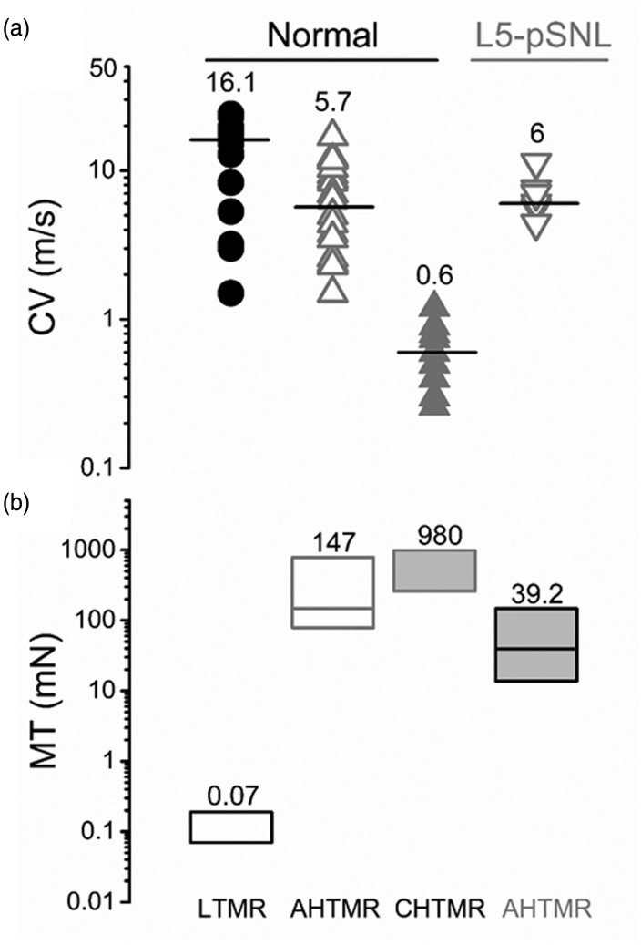
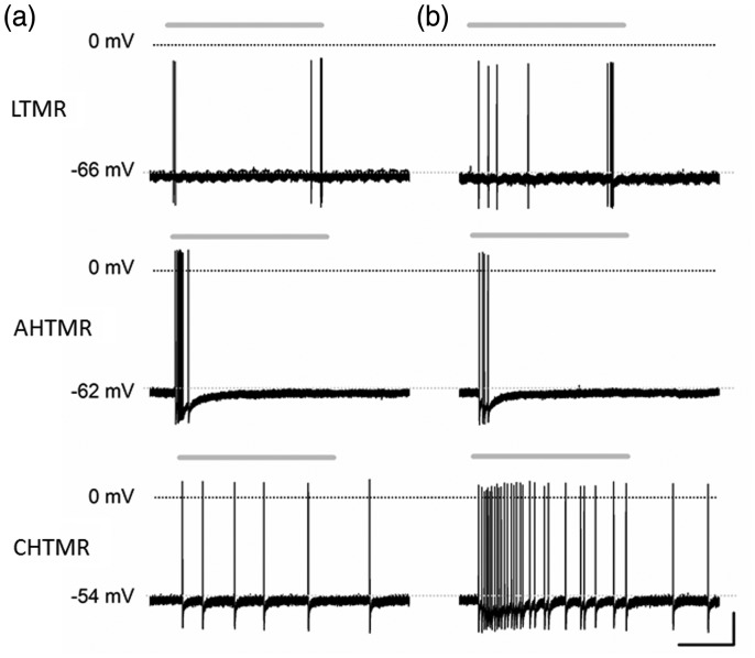
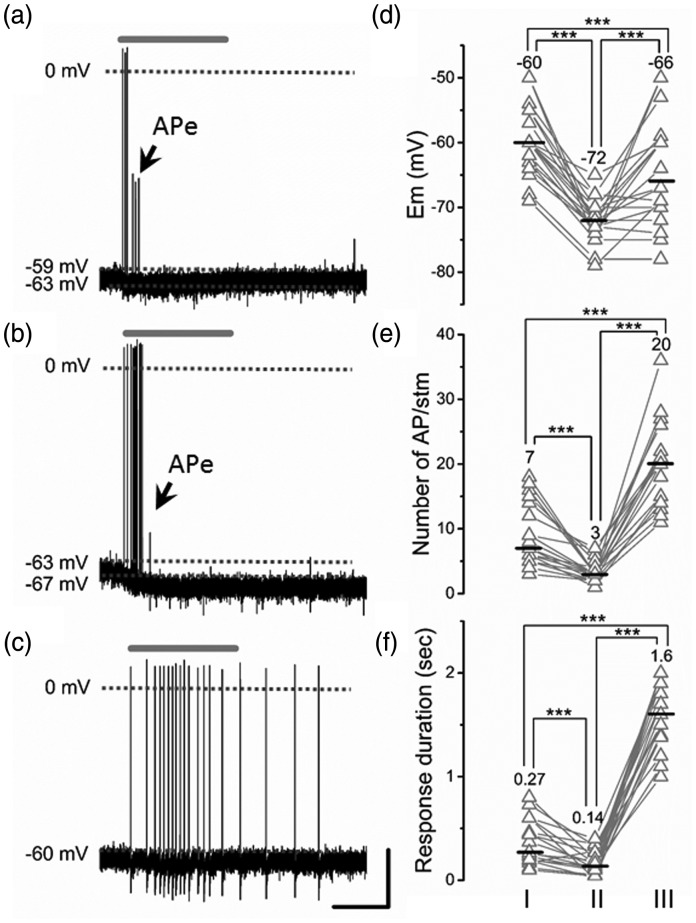
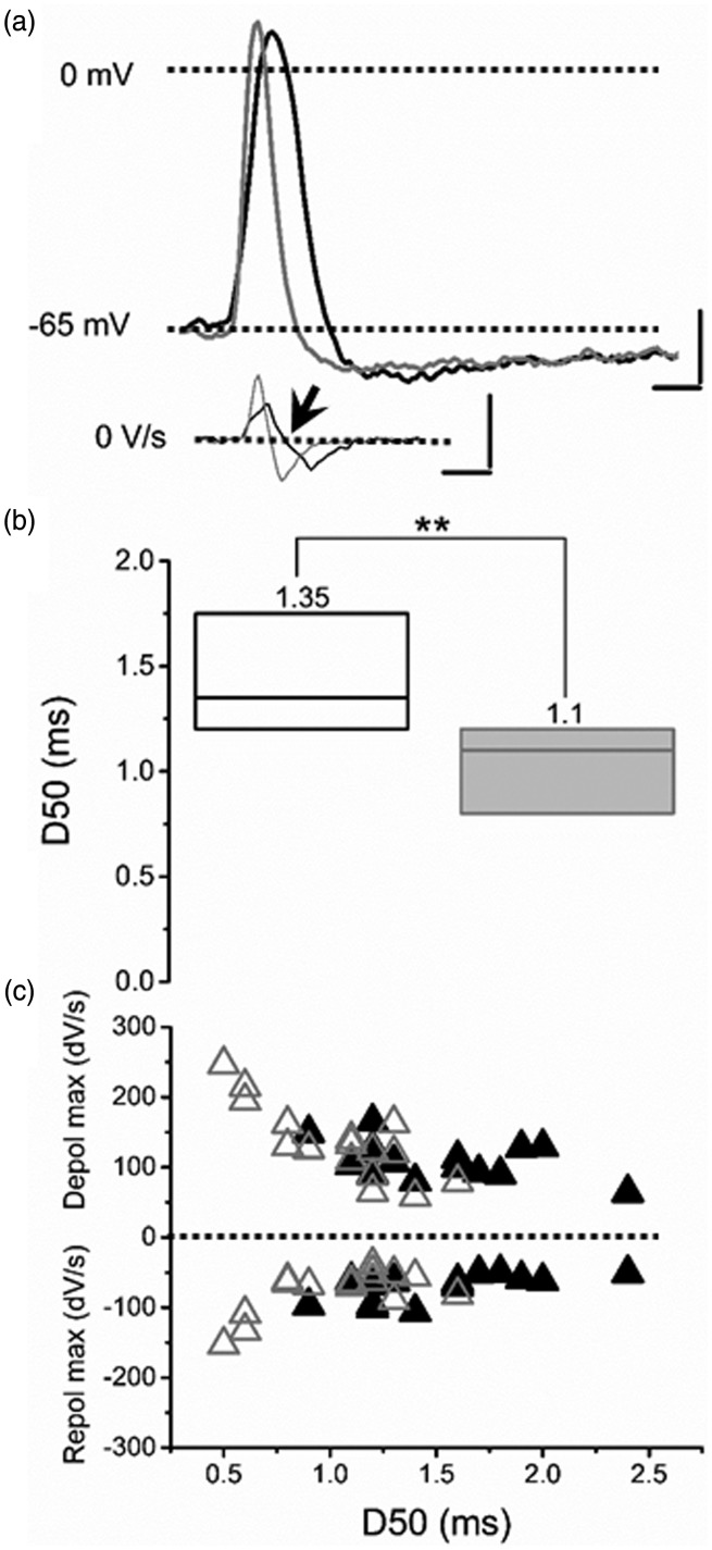
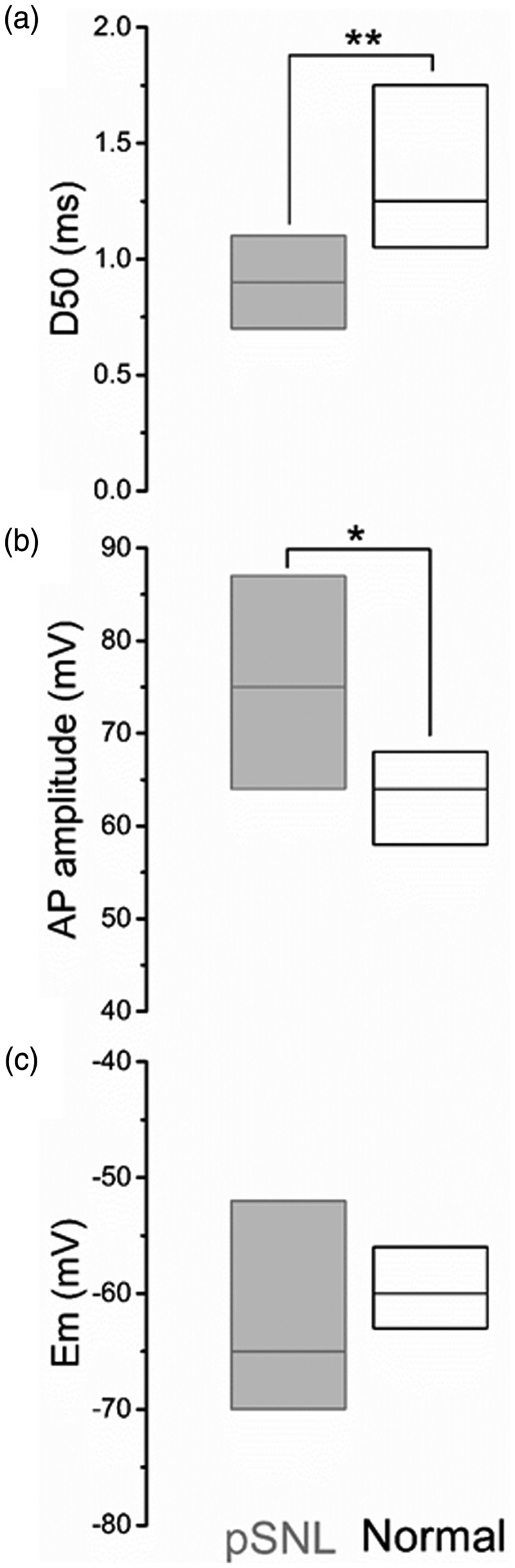
Peripheral somatosensory neurons are frequently exposed to mechanical forces. Strong stimuli result in neuronal activation of high-threshold mechanosensory afferent neurons, even in the absence of tissue damage. Among these neurons, fast-conducting nociceptors (A-fiber high-threshold mechanoreceptors (AHTMRs)) are normally resistant to sustained activation, transiently encoding the mechanical stimulus intensity but not its full duration. This rapidly adapting response seems to depend on changes in the electrical excitability of the membrane of these afferent neurons during sustained stimulation, a restraint mechanism that disappears following sensitization. Here, we examine the mechanism by which strong peripheral activation of mechanoreceptors elicits this control process in the absence of tissue injury and temporally silences afferent neurons despite ongoing stimulation. To study this, mechanoreceptors in Sprague-Dawley rats were accessed at the soma in the dorsal root ganglia from T11 and L4/L5. Neuronal classification was performed using receptive field characteristics and passive and active electrical properties. Sustained mechanical nociceptive stimulation in the absence of tissue damage of AHTMRs induces a rapid membrane hyperpolarization and a period of reduced responsiveness to the stimuli. Moreover, this phenomenon appears to be unique to this subset of afferent neurons and is absent in slow-conducting C-mechanonociceptors (C-fiber high-threshold mechanoreceptors) and rapidly adapting fast-conducting low-threshold mechanoreceptors. Furthermore, this mechanism for rapid adaptation and reducing ongoing input is ablated by repeated strong stimuli and in sensitized AHTMRs after chronic neuropathic injury. Further studies to understand the underling molecular mechanisms behind this phenomenon and their modulation during the development of pathological conditions may provide new targets to control nociceptive hyperexcitability and chronic pain.

Keywords: Primary sensory neurons; in vivo electrophysiology; membrane hyperpolarization.


Figures

Figure 1.
Figure 1.
Flow diagram of the distribution of neurons recorded and analyzed by group in the study. LTMR: low-threshold mechanoreceptor; AHTMR: A-fiber high-threshold mechanoreceptor; CHTMR: C-fiber high-threshold mechanoreceptor; L4-AHTMR: A-fiber high-threshold mechanoreceptor recorded from the L4 ganglia.
Figure 2.
Figure 2.
(a) Conduction velocity by classification. Values and bars on the scatter points are medians. LTMR, AHTMR, CHTMR, L4 AHTMR after L5-pSNL. (b) Mechanical threshold of neurons by classification. Numbers indicate medians (on top), with boxes representing the 25 and 75 percentiles. LTMR: low-threshold mechanoreceptor; AHTMR: A-fiber high-threshold mechanoreceptor; CHTMR: C-fiber high-threshold mechanoreceptor; L4-AHTMR: A-fiber high-threshold mechanoreceptor recorded from the L4 ganglia.
Figure 3.
Figure 3.
Typical examples of responses of different types of DRG neurons to (a) threshold (von Frey filaments) and (b) supra-threshold (pinch) stimuli. Both stimuli were applied consecutively (<1 min apart) for approximately 2 s (gray above each trace) in normal animals. LTMR: low-threshold mechanoreceptors; ATHMR: A-fiber high-threshold mechanoreceptor; CHTMR: C-fiber high-threshold mechanoreceptors. Scale bars: 0.67 s, 20 mV.
Figure 4.
Figure 4.
Representative AHTMR response after three consecutive pinching stimuli. (a) Initial pinch (I), (b) second pinch hyperpolarization (II), and (c) third pinch (III). Note the cumulative nature of the hyperpolarization and initial incomplete development of AP (APe: electrotonic propagated APs) concluding with membrane potential recovery and the shift of the response for phasic (RA) to tonic (SA). Effect of the latest described patterns of activation on the AHTMR response and properties: membrane potential (Em) (d), number of AP/stimuli (stm) (e), and duration of response (f). ATHMR. Values and middle bars on the cell’s scatter points are means for the Em and medians for the others. Scale bars: 1 s, 20 mV. One-way repeated measures ANOVA with Bonferroni correction for multiple comparisons was used for the Em while the Friedman test was used for AP/stm and duration of the response with Bonferroni corrections for multiple comparisons, ***p < 0.001.
Figure 5.
Figure 5.
Example of the effects of consecutive pinching (three stimuli) on the active electrical properties of AHTMR afferent neurons. (a) Typical AP shape before (black) and after stimulation (gray) presented with their dV/dt. Arrow shows the inflection point in the first derivative of the voltage Scale bars. 20 mV, 100 V/s, 1 ms. (b) Cumulative effect of repeated pinching stimulation on the duration of AP at 50% of their amplitude (D50). No change in amplitude was noted despite the shorter duration of the D50. Data are medians (on top), with boxes representing the 25 and 75 percentiles, Wilcoxon signed rank was used to test significance, **p < 0.001. (c) Changes in the maximal depolarizing and hyperpolarizing rates before (black triangles) and after pinching stimulation (open triangles) and it relations with the AP duration at 50% of their amplitude (D50).
Figure 6.
Figure 6.
Comparison of somatic electrical properties of nociceptive AHTMR afferent neurons, obtained after L5 pSNL (gray) and normal naïve (black). Data show as medians (bars) and 25th and 75th percentiles (boxes). *p < 0.05; **p < 0.01.

References

    1. Andrew D, Greenspan JD. Peripheral coding of tonic mechanical cutaneous pain: comparison of nociceptor activity in rat and human psychophysics. J Neurophysiol 1999; 82: 2641–2648. - PubMed
    1. Dubin AE, Patapoutian A. Nociceptors: the sensors of the pain pathway. J Clin Invest 2010; 120: 3760–3772. DOI: 10.1172/JCI42843. - PMC - PubMed
    1. Boada MD, Gutierrez S, Aschenbrenner CA, et al. Nerve injury induces a new profile of tactile and mechanical nociceptor input from undamaged peripheral afferents. J Neurophysiol 2015; 113: 100–109. DOI: 10.1152/jn.00506.2014. - PMC - PubMed
    1. Boada MD, Martin TJ, Peters CM, et al. Fast-conducting mechanoreceptors contribute to withdrawal behavior in normal and nerve injured rats. Pain 2014; 155: 2646–2655. DOI: 10.1016/j.pain.2014.09.030. - PMC - PubMed
    1. Boada MD. Relationship between electrophysiological signature and defined sensory modality of trigeminal ganglion neurons in vivo. J Neurophysiol 2013; 109: 749–757. DOI: 10.1152/jn.00693.2012. - PubMed

Publication types

LinkOut - more resources