. 2017 Nov;187(11):2399-2412.
doi: 10.1016/j.ajpath.2017.07.018. Epub 2017 Aug 19.

Reduced Glutathione Level Promotes Epithelial-Mesenchymal Transition in Lens Epithelial Cells via a Wnt/β-Catenin-Mediated Pathway: Relevance for Cataract Therapy

Affiliations

Reduced Glutathione Level Promotes Epithelial-Mesenchymal Transition in Lens Epithelial Cells via a Wnt/β-Catenin-Mediated Pathway: Relevance for Cataract Therapy

Zongbo Wei et al. Am J Pathol. 2017 Nov.

Abstract

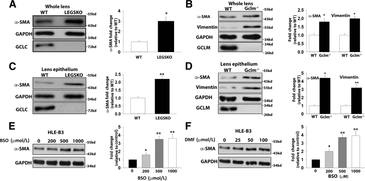
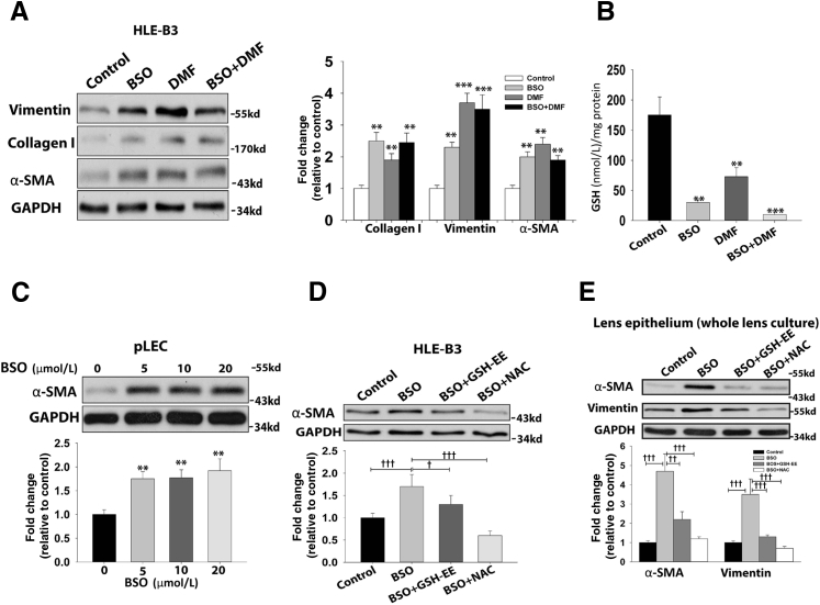
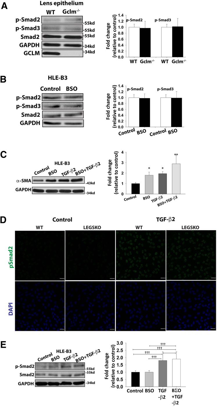
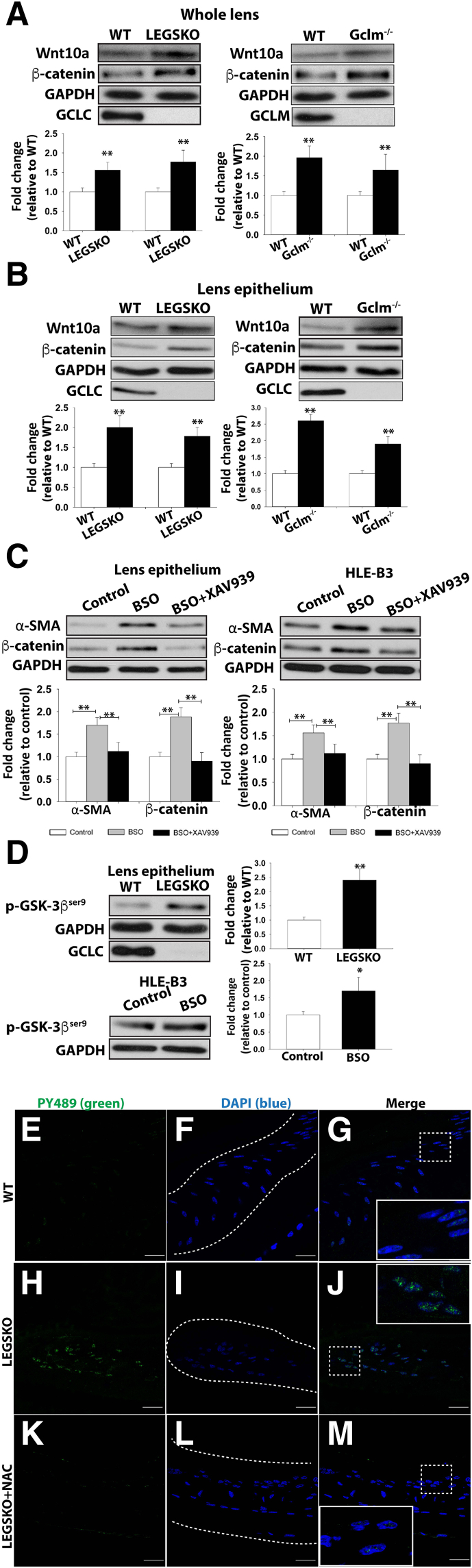
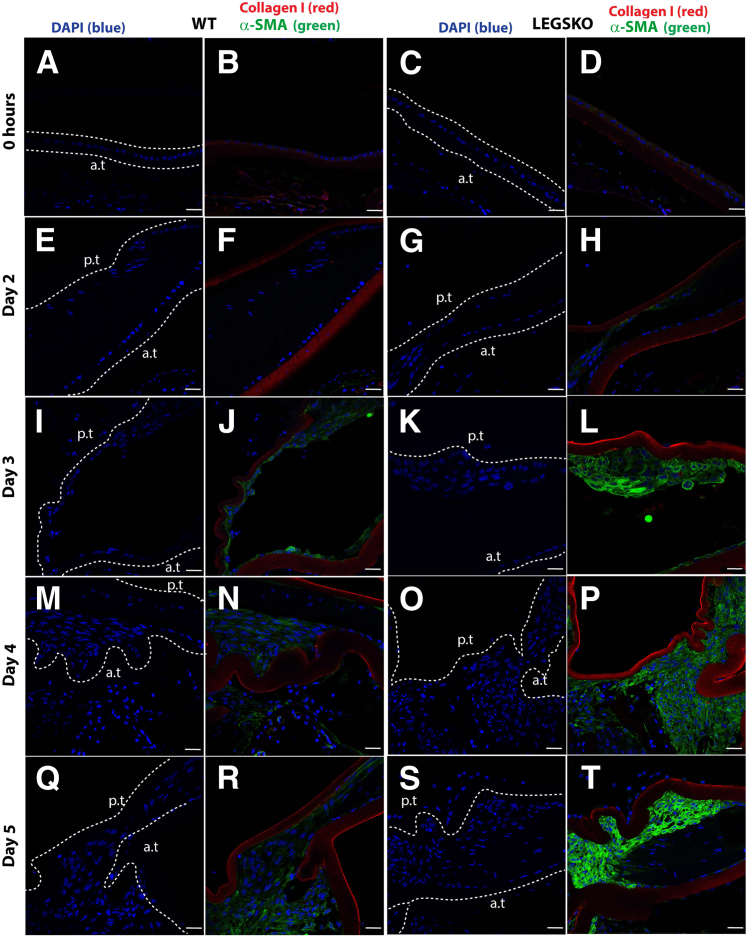
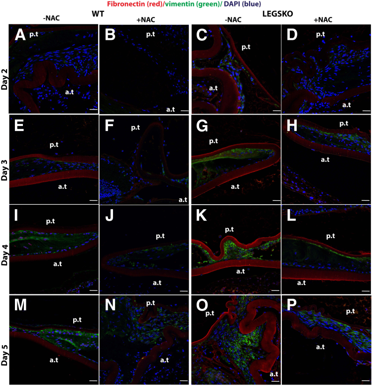
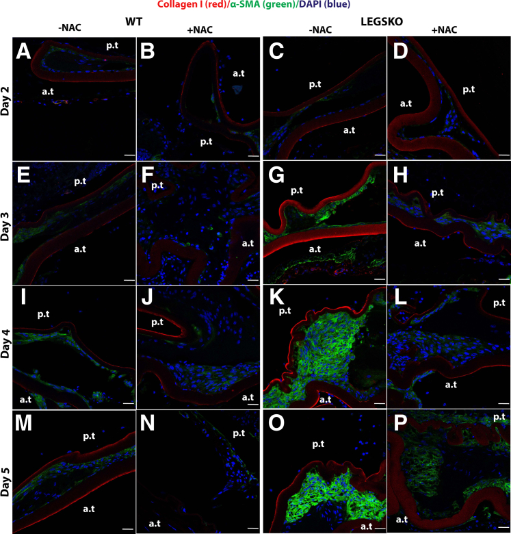
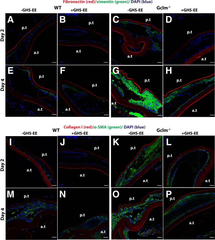
The epithelial-mesenchymal transition (EMT) process plays a pivotal role in the pathogenesis of posterior capsular opacification because of remnant lens epithelial cell proliferation, migration, and transformation after cataract surgery. The latter, we hypothesize, may result in posterior capsule wrinkling and opacification because of a profound change in the lens growth environment via a 1000-fold reduction of extracellular glutathione (GSH) levels. To test this hypothesis, we investigated the EMT process in cell culture and GSH biosynthesis deficiency mouse models. Our data indicate a dramatic increase of pro-EMT markers, such as type I collagen, α-smooth muscle actin, vimentin, and fibronectin, under conditions of lens GSH depletion. Further study suggests that decreased GSH triggers the Wnt/β-catenin signal transduction pathway, independent of transforming growth factor-β. Equally important, the antioxidants N-acetyl cysteine and GSH ethyl ester could significantly attenuate the EMT signaling stimulated by decreased GSH levels. These findings were further confirmed by mock cataract surgery in both gamma glutamyl-cysteine ligase, catalytic subunit, and gamma glutamyl-cysteine ligase, modifier subunit, knockout mouse models. Remarkably, increased EMT marker expression, β-catenin activation, and translocation into the nucleus were found in both knockout mice compared with the wild type, and such increased expression could be significantly attenuated by N-acetyl cysteine or GSH ethyl ester treatment. This study, for the first time we believe, links oxidative stress to lens fibrosis and posterior capsular opacification formation via EMT-mediated mechanisms.


Figures

Figure 1
Figure 1
Epithelial-mesenchymal transition (EMT) markers are enhanced in LEGSKO and GCLM knockout (KO) mice at 4 months. Lens or cell homogenate was analyzed by immunoblot probing α-smooth muscle actin (α-SMA), vimentin, GCLC, GCLM, and glyceraldehyde-3-phosphate dehydrogenase (GAPDH). A: α-SMA is increased in the whole lens homogenate of LEGSKO mice compared with WT. B: α-SMA and vimentin are increased in the whole lens homogenate of Gclm KO mice compared with WT. C: α-SMA is increased in LEGSKO lens epithelium compared with wild-type (WT). D: α-SMA and vimentin levels are increased in Gclm KO mice lens epithelium compared with WT. E: α-SMA expression is increased in human lens epithelial (HLE-B3) cells after treatment with an increased concentration of buthionine sulfoximine (BSO) for 24 hours F: α-SMA expression is increased in HLE-B3 cells after treatment with an increasing concentration of dimethyl fumarate (DMF) for 24 hours. GAPDH was used as a loading control. GCLC and GCLM blotting served as additional genotype verification besides PCR genotype conclusion. A t-test was used to compare WT and KO groups, and P < 0.05 is considered significant. Data are expressed as means ± SEM (AF). n = 6 (whole lens); n = 3 (epithelium). P < 0.05, ∗∗P < 0.01 versus WT or control.
Figure 2
Figure 2
EMT markers are elevated under GSH depletion condition. A: Significantly increased expression of collagen I, vimentin, and α-smooth muscle actin (α-SMA) is seen in 500 μmol/L buthionine sulfoximine (BSO), 25 μmol/L dimethyl fumarate (DMF), and 500 μmol/L BSO plus 25 μmol/L DMF stimulated cells for 24 hours. B: The intracellular GSH concentration after 24 hours of treatment with 500 μmol/L BSO, 25 μmol/L DMF, or 500 μmol/L BSO combined with 25 μmol/L DMF. C: α-SMA expression increases in porcine lens epithelial cells (pLECs) after treatment with an increased concentration of BSO for 24 hours D: α-SMA production is alleviated in human lens epithelial (HLE-B3) cells when cotreated with either 10 mmol/L N-acetyl cysteine (NAC) or 0.5 mol/L GSH ethyl ester (GSH-EE) with 500 μmol/L BSO for 24 hours. E: α-SMA and vimentin production are alleviated in the lens epithelium from cultured whole lens when cotreated with 10 mmol/L NAC or 0.5 mmol/L GSH-EE with 500 μmol/L BSO for 48 hours. Glyceraldehyde-3-phosphate dehydrogenase (GAPDH) was used as a loading control. One-way analysis of variance and t-test were used to compare WT and knockout groups, and P < 0.05 is considered significant. n = 5 for each immunoblot. P < 0.05, ∗∗P < 0.01, and ∗∗∗P < 0.001 versus control; P <0.05, ††P< 0.01, and †††P < 0.001.
Figure 3
Figure 3
EMT signaling triggered by decreased GSH levels is independent of canonical transforming growth factor (TGF)-β signaling. A: No remarkable changes of phosphorylated Smad2 and Smad3 (p-Smad2 and p-Smad3, respectively) are seen in lens epithelium from Gclm knockout (KO) mice compared with WT. B: No remarkable changes of p-Smad2 and p-Smad3 are seen in human lens epithelial (HLE-B3) cells challenged by 500 μmol/L buthionine sulfoximine (BSO) for 24 hours. C: Synergistic effect is seen in HLE-B3 cells challenged by 500 μmol/L BSO, 1 ng/mL TGF-β2, and 500 μmol/L BSO plus 1 ng/mL TGF-β2 for 24 hours. D: TGF-β2 stimulation increased Smad2 phosphorylation, but no remarkable change was seen between LEGSKO and WT mice. E: TGF-β2 stimulation increased Smad2 phosphorylation, but no remarkable change was seen between HLE-B3 cells treated with or without BSO. Glyceraldehyde-3-phosphate dehydrogenase (GAPDH) was used as a loading control. One-way analysis of variance and t-test were used to compare WT and KO groups, and P < 0.05 is considered significant. n = 5 for each immunoblot. P < 0.05, ∗∗P < 0.01, and ∗∗∗P < 0.001 versus control; †††P < 0.001. Scale bars = 25 μm. α-SMA, α-smooth muscle actin.
Figure 4
Figure 4
Oxidative stress–modulated EMT signaling is mediated via the Wnt/β-catenin signaling pathway. A: Wnt10a and β-catenin are elevated in the whole lens protein extract in both LEGSKO and Gclm knockout (KO) mice compared with WT. B: Similarly, Wnt10a and β-catenin are significantly (P < 0.01) elevated in the lens epithelium protein extract in both LEGSKO and Gclm KO mice compared with WT. C: The α-smooth muscle actin (α-SMA) and β-catenin production is blocked when XAV939, a Wnt inhibitor, is coincubated with 500 μmol/L buthionine sulfoximine (BSO) in lens ex vivo culture (lens epithelium was extracted for immunoblot analysis) and human lens epithelial (HLE-B3) cells. D: The Ser9 phosphorylation of glycogen synthase kinase (GSK)-3β (p-GSK-3βSer9) is significantly elevated in LEGSKO lens epithelium compared with WT and also in HLE-B3 cells after stimulation with 500 μmol/L BSO for 24 hours. GSH ethyl ester and N-acetyl cysteine (NAC) attenuate β-catenin expression and GSK-3βSer9 phosphorylation, respectively. EM: Immunofluorescence image of activated β-catenin colocalization within the nucleus. The active β-catenin, Tyr489 phosphorylated β-catenin, was labeled in green, and the lens epithelial cell nucleus was labeled in blue by DAPI. Boxed areas are shown at higher magnification in the insets. Dotted lines illustrate the boundary of the lens capsule. EG: WT lens capsule 72 hours after surgery demonstrates mild β-catenin nuclear translocation. HJ: LEGSKO mice lens capsule demonstrates robust nucleus translocation of β-catenin. KM: The active β-catenin nuclear translocation was significantly attenuated when 10 mmol/L NAC was applied immediately after surgery. For immunoblot, all data have been adjusted for glyceraldehyde-3-phosphate dehydrogenase (GAPDH) level. One-way analysis of variance and t-test were used to compare WT and KO groups, and P < 0.05 is considered significant. n = 5 in each assay. P < 0.05, ∗∗P < 0.01 versus WT or control. Scale bars = 25 μm (EM).
Figure 5
Figure 5
LEGSKO mouse promotes type I collagen and α-smooth muscle actin (α-SMA) production in the mock cataract surgery in vivo model. Type I collagen (red fluorescence) and α-SMA (green) are costained in mouse lens capsule 2 to 5 days after mock cataract surgery by removing lens fiber mass. The lens nucleus was stained with DAPI (blue). Dotted lines illustrate the boundary of the lens capsule. A, E, I, M, Q and C, G, K, O, S: Nucleus DAPI stain. B, F, J, N, R and D, H, L, P, T: Merged images colocalized for collagen I (red), α-SMA (green), and nucleus (blue). A and B: WT lens capsule at time 0. C and D: LEGSKO lens capsule at time 0. E and F: WT lens capsule at post-surgery day 2. G and H: LEGSKO lens capsule at post-surgery day 2. I and J: WT lens capsule at post-surgery day 3. K and L: LEGSKO lens capsule at post-surgery day 3. M and N: WT lens capsule at post-surgery day 4. O and P: LEGSKO lens capsule at post-surgery day 4. Q and R: WT lens capsule at post-surgery day 5. S and T: LEGSKO lens capsule at post-surgery day 5. Six mice at each time point were used. The same confocal parameters in each channel were used to capture the image from all of the samples. Scale bars = 25 μm (AT). a.t., anterior capsule; p.t., posterior capsule.
Figure 6
Figure 6
The antioxidant N-acetyl cysteine (NAC) significantly attenuates the EMT signaling in vivo. The fibronectin (red fluorescence) and vimentin (green) were costained in mouse lens capsule 2 to 5 days after mock cataract surgery by removing lens fiber mass. The lens nucleus was stained with DAPI (blue). A, E, I, M and C, G, K, O: Merged images colocalized for fibronectin (red), vimentin (green), and nucleus (blue) of either WT or LEGSKO mice without 10 mmol/L NAC treatment. B, F, J, N and D, H, L, P: Merged images colocalized for fibronectin (red), vimentin (green), and nucleus (blue) of either WT or LEGSKO mice with 10 mmol/L NAC treatment. A and B: WT lens capsule at post-surgery day 2 with or without NAC treatment. C and D: LEGSKO lens capsule at post-surgery day 2 with or without NAC treatment. E and F: WT lens capsule at post-surgery day 3 with or without NAC treatment. G and H: LEGSKO lens capsule at post-surgery day 3 with or without NAC treatment. I and J: WT lens capsule at post-surgery day 4 with or without NAC treatment. K and L: LEGSKO lens capsule at post-surgery day 4 with or without NAC treatment. M and N: WT lens capsule at post-surgery day 5 with or without NAC treatment. O and P: LEGSKO lens capsule at post-surgery day 5 with or without NAC treatment. Six mice at each time point were used. The same confocal parameters in each channel were used to capture the image from all of the samples. Scale bars = 25 μm (AP). a.t., anterior capsule; p.t., posterior capsule.
Figure 7
Figure 7
The antioxidant N-acetyl cysteine (NAC) could significantly attenuate the EMT signaling in vivo. The type I collagen (red fluorescence) and α-smooth muscle actin (α-SMA; green) were costained in mouse lens capsule 2 to 5 days after mock cataract surgery by removing lens fiber mass. The lens nucleus was stained with DAPI (blue). A, E, I, M and C, G, K, O: Merged images colocalized for collagen I (red), α-SMA (green), and nucleus (blue) of either WT or LEGSKO mice without 10 mmol/L NAC treatment. B, F, J, N and D, H, L, P: Merged images colocalized for collagen I (red), α-SMA (green), and nucleus (blue) of either WT or LEGSKO mice with 10 mmol/L NAC treatment. A and B: WT lens capsule at post-surgery day 2 with or without NAC treatment. C and D: LEGSKO lens capsule at post-surgery day 2 with or without NAC treatment. E and F: WT lens capsule at post-surgery day 3 with or without NAC treatment. G and H: LEGSKO lens capsule at post-surgery day 3 with or without NAC treatment. I and J: WT lens capsule at post-surgery day 4 with or without NAC treatment. K and L: LEGSKO lens capsule at post-surgery day 4 with or without NAC treatment. M and N: WT lens capsule at post-surgery day 5 with or without NAC treatment. O and P: LEGSKO lens capsule at post-surgery day 5 with or without NAC treatment. Six mice at each time point were used. The same confocal parameters in each channel were used to capture the image from all of the samples. Scale bars = 25 μm (AP). a.t., anterior capsule; p.t., posterior capsule.
Figure 8
Figure 8
Gclm knockout (KO) mouse modulates EMT signaling in the mock cataract surgery in vivo model. AH: All images are merged for colocalization of fibronectin (red), vimentin (green), and nucleus (blue). A and B: WT lens capsule at post-surgery day 2 with or without 2 mmol/L GSH ethyl ester (GSH-EE) treatment. C and D: Gclm KO lens capsule at post-surgery day 2 with or without 2 mmol/L GSH-EE treatment. E and F: WT lens capsule at post-surgery day 4 with or without 2 mmol/L GSH-EE treatment. G and H: Gclm KO lens capsule at post-surgery day 4 with or without 2 mmol/L GSH-EE treatment. IP: All images are merged for colocalization of collagen I (red), α-smooth muscle actin (α-SMA; green), and nucleus (blue). I and J: WT lens capsule at post-surgery day 2 with or without 2 mmol/L GSH-EE treatment. K and L: Gclm KO lens capsule at post-surgery day 2 with or without 2 mmol/L GSH-EE treatment. M and N: WT lens capsule at post-surgery day 4 with or without 2 mmol/L GSH-EE treatment. O and P: Gclm KO lens capsule at post-surgery day 4 with or without 2 mmol/L GSH-EE treatment. Six mice at each time point were used. The same confocal parameters in each channel were used to capture the images from all of the samples. Scale bars = 25 μm (AP). a.t., anterior capsule; p.t., posterior capsule.

References

    1. Liu Y.C., Wilkins M., Kim T., Malyugin B., Mehta J.S. Cataracts. Lancet. 2017;390:600–612. - PubMed
    1. Eldred J.A., Dawes L.J., Wormstone I.M. The lens as a model for fibrotic disease. Philos Trans R Soc Lond B Biol Sci. 2011;366:1301–1319. - PMC - PubMed
    1. Dawes L.J., Sleeman M.A., Anderson I.K., Reddan J.R., Wormstone I.M. TGFbeta/Smad4-dependent and -independent regulation of human lens epithelial cells. Invest Ophthalmol Vis Sci. 2009;50:5318–5327. - PubMed
    1. Walker J.L., Wolff I.M., Zhang L., Menko A.S. Activation of SRC kinases signals induction of posterior capsule opacification. Invest Ophthalmol Vis Sci. 2007;48:2214–2223. - PubMed
    1. Mamuya F.A., Wang Y., Roop V.H., Scheiblin D.A., Zajac J.C., Duncan M.K. The roles of alphaV integrins in lens EMT and posterior capsular opacification. J Cell Mol Med. 2014;18:656–670. - PMC - PubMed