Review
. 2017 Nov 10;429(22):3363-3375.
doi: 10.1016/j.jmb.2017.08.008. Epub 2017 Aug 19.

RING-Between-RING E3 Ligases: Emerging Themes amid the Variations

Affiliations
Review

RING-Between-RING E3 Ligases: Emerging Themes amid the Variations

Katja K Dove et al. J Mol Biol. .

Abstract

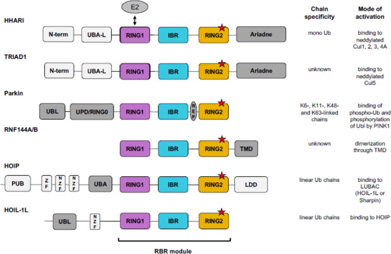
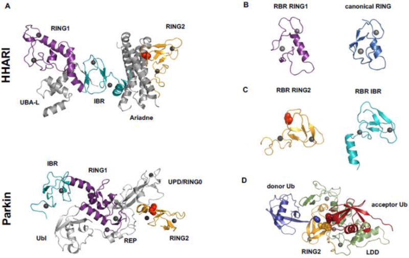
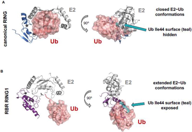
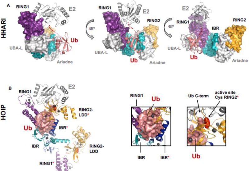
Covalent, reversible, post-translational modification of cellular proteins with the small modifier, ubiquitin (Ub), regulates virtually every known cellular process in eukaryotes. The process is carried out by a trio of enzymes: a Ub-activating (E1) enzyme, a Ub-conjugating (E2) enzyme, and a Ub ligase (E3) enzyme. RING-in-Between-RING (RBR) E3s constitute one of three classes of E3 ligases and are defined by a RING-HECT-hybrid mechanism that utilizes a E2-binding RING domain and a second domain (called RING2) that contains an active site Cys required for the formation of an obligatory E3~Ub intermediate. Albeit a small class, RBR E3s in humans regulate diverse cellular process. This review focuses on non-Parkin members such as HOIP/HOIL-1L (the only E3s known to generate linear Ub chains), HHARI and TRIAD1, both of which have been recently demonstrated to work together with Cullin RING E3 ligases. We provide a brief historical background and highlight, summarize, and discuss recent developments in the young field of RBR E3s. Insights reviewed here include new understandings of the RBR Ub-transfer mechanism, specifically the role of RING1 and various Ub-binding sites, brief structural comparisons among members, and different modes of auto-inhibition and activation.

Keywords: RING-between-RING ubiquitin E3 ligases; protein ubiquitylation; ubiquitin.


Figures

Figure 1
Figure 1
RBR E3s have diverse domain architectures. All RBR E3s contain an RBR module comprised of RING1 (purple), IBR (blue), and RING2 (orange). An RBR module can appear at any position relative to other domains, but its three domains are always found in the order: RING1-IBR-RING2. RING1 binds the E2 (grey oval) and RING2 contains the conserved active-site Cys (red stars). Domains not shared among RBR family members are colored grey, with dark grey indicating domains implicated in autoinhibition/activation. (UBA-L = Ubiquitin Associated-Like domain, UPD = Unique Parkin Domain; UBL = Ubiquitin Like domain; REP = Repressor Element; TMD = Transmembrane Domain; PUB = PNGase/Ubiquitin-associated domain; ZF = Zinc Finger domain; NZF = NPL4 Zinc Finger domain; LDD = Linear chain Determining Domain). Current information available regarding chain specificity (based on rigorous biochemical evidence) and mode of activation for each RBR is summarized.
Figure 2
Figure 2
Features of RBR E3 structures. A) Structures of auto-inhibited HHARI (top, PDB 4KBL) and Parkin (bottom, PDB 4K95) shown in cartoon representation. RING1 is in purple, IBR in cyan, and RING2 in orange. Red spheres represent the RING2 active-site Cys that is partially buried by other domains (Ariadne domain in the case of HHARI and UPD/RING0 in the case of Parkin). The relative arrangement of the sub-domains of the RBR module differs between the two RBRs. In the auto-inhibited state of HHARI the E2-binding RING1 domain is accessible to bind E2, but the active-site Cys-containing RING2 domain is far away. Compared to HHARI, Parkin’s RING1 and RING2 domains are positioned closer together, but the REP element of Parkin blocks the E2 binding site on RING1. Hence, both structures suggest that major domain rearrangements must occur prior to Ub transfer onto the active site. B) RBR RING1 domains (left, HHARI, PDB 5UDH) are structurally similar to canonical RINGs (right, BRCA1, PDB 1JM7) as demonstrated by the typical cross-brace fold. C) Structures of RBR RING2 domains (left, HHARI, PDB 5UDH) do not resemble RING domains, but are structurally similar to IBR domains (right, HHARI, PDB 5UDH). D) Crystal structure of HOIP RING2-LDD (RING2 in orange, LDD in green, PDB 4LJO) in complex with a donor Ub (blue) covalently bound to the active site Cys (orange spheres) through its C-term (blue spheres). The acceptor Ub (red) is non-covalently bound to the LDD. The N-term of the acceptor Ub (red spheres) is in proximity to the thioester of RING2~Ub donor poised for Ub transfer. The LDD (green) is tightly integrated structurally with RING2 (orange) generating a compact RING2-LDD unit.
Figure 3
Figure 3
Comparison of E2~Ub conformations when bound to canonical RING and RBR RING1. A) Cartoon and surface representations of a canonical RING/E2~Ub complex (E3 = BIRC7; E2 = UbcH5; PDB 4AUQ). RING domains (blue cartoon) induce closed E2~Ub conformations. In RING-induced closed E2~Ub states the hydrophobic surface of Ub (teal) is buried in an interface with the E2. B) Cartoon and surface representations of a RBR RING1/E2~Ub (E3 = RING1; E2 = UbcH7; PDB 5UDH). RING1 domains (purple cartoon) promote open/extended E2~Ub conformations that expose the Ub hydrophobic surface (teal). E2s are shown in grey cartoon representations. Ub is shown in salmon-colored surface representation.
Figure 4
Figure 4
Comparison of an auto-inhibited RBR/E2~Ub complex and an “activated” RBR/E2~Ub complex. A) HHARI bound to E2~Ub (E2 = UbcH7; PDB 5UDH) is still auto-inhibited. Ub (salmon cartoon) makes no contacts with any domains of its cognate HHARI molecule. In the auto-inhibited state the active-site Cys (red spheres, but not visible in the surface representation of the auto-inhibited structure) is occluded by the Ariadne domain and therefore not visible in the surface representation shown here. RING1 (purple surface) and RING2 (orange surface) domains are far apart (as seen in apo-HHARI, Fig. 2A). IBR is shown in cyan surface representation. Density for the C-terminus of Ub is missing in the crystal structure and is instead indicated by red dots. B) HOIP RBR module bound to E2~Ub (E2 = UbcH5; PDB 5EDV). The Ub moiety (salmon surface) of E2~Ub contacts IBR and RING2 domains of two different HOIP molecules (left box, all domains belonging to the second (non-cognate) polypeptide are marked with a [*]). Domain colors are the same as A). The domain swap in the crystal affords a view of what an activated version of HOIP RBR could look like, with the C-term of ~Ub conjugated to the E2 (right box, black arrow) in proximity to the active-site Cys of HOIP (right box; black arrow and red spheres).

References

    1. Marin I, Lucas JI, Gradilla AC, Ferrus A. Parkin and relatives: the RBR family of ubiquitin ligases. Physiol Genomics. 2004;17:253–63. - PubMed
    1. Pickrell AM, Youle RJ. The roles of PINK1, parkin, and mitochondrial fidelity in Parkinson’s disease. Neuron. 2015;85:257–73. - PMC - PubMed
    1. Eiyama A, Okamoto K. PINK1/Parkin-mediated mitophagy in mammalian cells. Curr Opin Cell Biol. 2015;33:95–101. - PubMed
    1. Kitada T, Asakawa S, Hattori N, Matsumine H, Yamamura Y, Minoshima S, et al. Mutations in the parkin gene cause autosomal recessive juvenile parkinsonism. Nature. 1998;392:605–8. - PubMed
    1. Matsumine H, Saito M, Shimoda-Matsubayashi S, Tanaka H, Ishikawa A, Nakagawa-Hattori Y, et al. Localization of a gene for an autosomal recessive form of juvenile Parkinsonism to chromosome 6q25.2–27. Am J Hum Genet. 1997;60:588–96. - PMC - PubMed

Publication types

Substances

LinkOut - more resources