. 2017 Oct 1;144(19):3612-3624.
doi: 10.1242/dev.152736. Epub 2017 Aug 21.

A fixation method to preserve cultured cell cytonemes facilitates mechanistic interrogation of morphogen transport

Affiliations

A fixation method to preserve cultured cell cytonemes facilitates mechanistic interrogation of morphogen transport

William J Bodeen et al. Development. .

Abstract

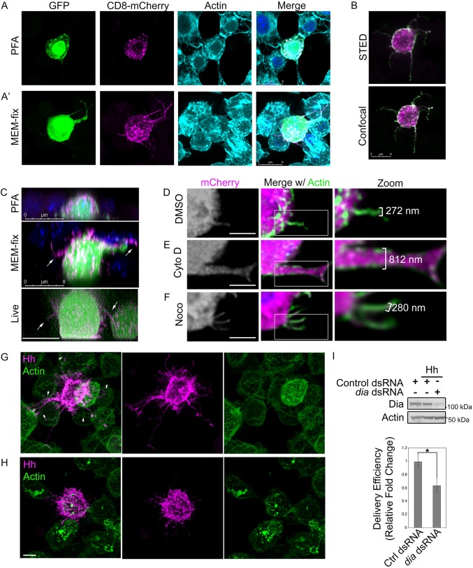
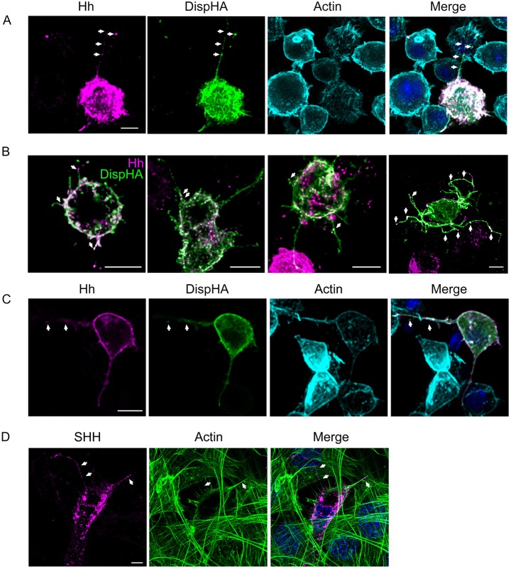
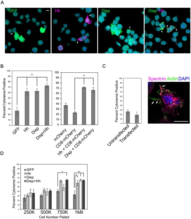
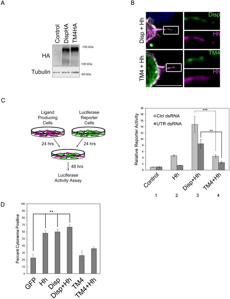
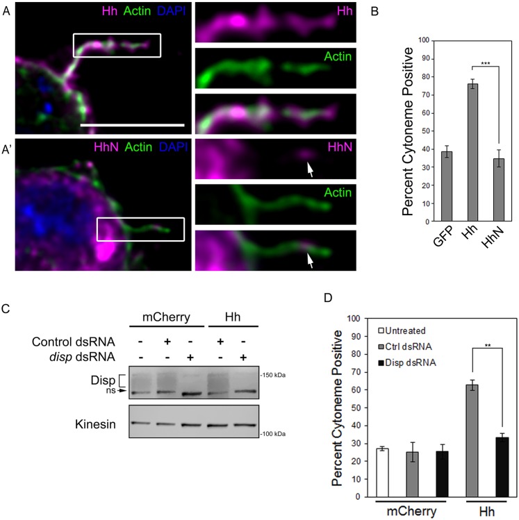
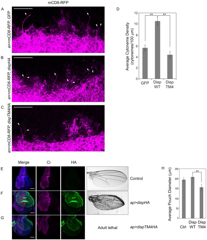
During development, extracellular cues guiding cell fate determination are provided by morphogens. One mechanism by which morphogens are proposed to traverse extracellular space is by traveling along specialized filopodia called cytonemes. These cellular highways extend between signal-producing and -receiving cells to enable direct morphogen delivery. Although genetic studies support cytoneme involvement in morphogen transport, mechanistic insight into how they are regulated is limited owing to technical challenges associated with performing cell biological analysis of the delicate filopodial structures. Here, we introduce a fixation method whereby cultured cell cytonemes can be preserved for imaging studies, allowing investigation of cytoneme regulation using standard cell biological techniques. Using this method, we examined Hedgehog-containing cytonemes and identified a role for the Hedgehog deployment protein Dispatched in cytoneme stabilization. We demonstrate that Hedgehog and Dispatched colocalize in cytonemes, and that cholesterol-modified Hedgehog acts through Dispatched to increase cytoneme occurrence. Live imaging suggests that this occurs through Dispatched-mediated slowing of cytoneme retraction rates. Dispatched-induced cytoneme modulation was recapitulated in wing imaginal discs of transgenic Drosophila, providing evidence that cultured cell cytoneme analysis is predictive of in vivo functionality.

Keywords: Cytoneme; Dispatched; Hedgehog; Morphogen; Signal transduction.


Conflict of interest statement

Competing interestsThe authors declare no competing or financial interests.

Figures

Fig. 1.
Fig. 1.
MEM-fix preserves cultured cell cytonemes. (A,A′) S2 cells expressing GFP and CD8-mCherry were fixed in 4% PFA (A) or MEM-fix (A′) and stained with phalloidin to label actin. (B) S2 cells expressing Hh (green) and CD8-mCherry (magenta) were MEM-fixed, immunostained for Hh and imaged by STED and confocal microscopy. (C) z-stack images of S2 cells expressing GFP and CD8-mCherry were collected from PFA-fixed, MEM-fixed and live cells. DAPI is shown in blue in fixed samples. (D-F) S2 cells expressing cytoplasmic mCherry were plated and treated with DMSO vehicle (D), 10 µm cytochalasin D (E) or 10 µm nocodazole (F) for 1.5 h prior to processing for imaging. Phalloidin (green) marks actin. (G,H) Hh-expressing cells were cultured with non-expressing cells. Hh puncta (magenta) are evident in non-expressing neighboring cells contacted by Hh-positive filopodia (arrowheads). Phalloidin (green) marks actin. (I) More than 60 cells per condition were analyzed over three independent experiments to determine average Hh delivery efficiency to non-expressing cells in contact with a cytoneme. Significance was determined using a two-tailed Student's t-test (*P≤0.05). Error bars indicate s.e.m. Lysates from dsRNA-treated cell populations are shown to confirm Dia knockdown. Actin is the loading control. Scale bars: 8 µm (A-C); 5 µm (D-H).
Fig. 2.
Fig. 2.
Disp and Hh localize to cytonemes of cultured cells. (A-C) DispHA (green) was co-expressed with Hh (magenta) in S2 (A,B) and Cl-8 (C) cells. Cells were MEM-fixed, immunostained and imaged by fluorescence confocal microscopy. Colocalization is indicated by arrows. Phalloidin (cyan) marks actin in A and C. (D) SHH (magenta) was expressed in NIH3T3 cells. Cells were fixed with MEM-fix, stained for actin (green) and imaged by confocal microscopy. DAPI (blue) marks nuclei. Arrows indicate SHH-positive puncta. Scale bars: 5 µm.
Fig. 3.
Fig. 3.
Disp and Hh increase cytoneme occurrence. (A,B) S2 cells were transfected with vectors encoding the indicated proteins, and cytoneme occurrence (arrows) in transfected cells was quantified. Statistical significance was determined using a one-way ANOVA (**P≤0.01). Representative fields of cells are shown in A. Approximately 150 transfected cells were counted per condition across three independent experiments. (C) Cytoneme occurrence was quantified in untransfected S2 cells and S2 cells transfected with empty pAc5.1 vector. Cytonemes were identified by actin (green) and the membrane protein α-Spectrin (magenta, arrows). Percentage of cytoneme occurrence was calculated for approximately ∼150 cells per condition across three separate experiments, and all data were pooled. (D) Cells were plated at increasing densities and cytoneme occurrence was quantified. Approximately 75 cells per condition were counted. The experiment was repeated three times and all data pooled. Significance was determined using a two-way ANOVA (*P≤0.05, **P≤0.01). Error bars indicate s.e.m. Scale bars: 5 µm.
Fig. 4.
Fig. 4.
Cytoneme modulation correlates with Hh deployment. (A) Lysates were prepared from Cl8 cells expressing wild-type or TM4 DispHA proteins, and analyzed by western blot. Tubulin is the loading control. (B) S2 cells expressing wild-type or TM4 DispHA proteins (green) along with Hh (magenta) were fixed in MEM-fix, immunostained and imaged by confocal fluorescence microscopy. Scale bar: 5 µm. (C) Cl8 reporter cells transfected with ptcΔ136-luciferase reporter and actin-Renilla normalization control were co-cultured with Hh-producing cells in which disp expression was modulated. Ligand-producing cells were treated with control or disp3′UTR dsRNA, and transfected with empty vector control or plasmids encoding wild-type or the TM4 Disp proteins. The experiment was repeated two times in duplicate and all data pooled. Error bars indicate s.e.m. Statistical significance was determined using a one-way ANOVA (**P≤0.01, ***P≤0.001). (D) To determine cytoneme occurrence rates, approximately ∼150 cells per condition were examined over three separate experiments and all data were averaged as in Fig. 3. Error bars indicate s.e.m, (**P≤0.01).
Fig. 5.
Fig. 5.
Hh requires Disp to modulate cytonemes. (A,A′) S2 cells expressing cholesterol-modified Hh (A, magenta) or cholesterol-free HhN (A′, magenta) were fixed using MEM-fix, immunostained and imaged by confocal fluorescent microscopy. Phalloidin (green) marks actin. DAPI (blue) marks nucleus. Arrows indicate HhN present in the cytoneme. Scale bar: 5 µm. (B) Cytonemes were quantified in S2 cells expressing the indicated proteins. Cytoneme occurrence was quantified in ∼75 cells per condition over three independent experiments and all data pooled. Error bars indicate s.e.m. Significance was determined using a one-way ANOVA test (***P≤0.001). (C) Endogenous disp was knocked down in control, cytoplasmic mCherry- or Hh-expressing S2 cells. Kinesin serves as loading control. Disp is detected as a broad band (bracket). A non-specific band (ns) recognized by the antibody is indicated. (D) S2 cells were transfected with Hh or cytoplasmic mCherry expression vectors in the presence of control or disp3′UTR dsRNA. Cytoneme occurrence was determined as in B. Error bars indicate s.e.m. Significance was determined using a one-way ANOVA (**P≤0.01).
Fig. 6.
Fig. 6.
Disp influences cytonemes in vivo. (A-C) GFP or the indicated DispHA proteins were expressed with CD8-RFP (magenta) in the posterior compartment of wing imaginal discs using en-GAL4. The basolateral region of the anterior/posterior border directly adjacent to the dorsal/ventral border was analyzed. Cytonemes reaching into the anterior compartment are indicated (arrows). Six to ten discs were examined per condition. Representative discs are shown. Scale bars: 10 µm. Discs are oriented such that anterior is up. (D) Cytonemes crossing the compartment boundary in a straight posterior to anterior trajectory were counted across multiple discs (GFP n=6, DispWT n=10, DispTM4 n=10). Density was calculated as the average number of cytonemes reaching at least one cell diameter into the anterior compartment, and is presented as average cytoneme number per 100 µm. Error bars indicate s.e.m. Significance was determined using a one-way ANOVA (**P≤0.01). (E-G) DispHA proteins (green) were expressed in the dorsal compartment of wing imaginal discs using ap-GAL4. Discs are oriented such that the dorsal compartment is up and anterior is left. Ci is shown in magenta, DAPI in blue. Wings shown on the right are representative of adult phenotypes observed. Scale bars: 50 µm. Average wing pouch size is indicated in H. Error bars indicate s.e.m. Student's t-test was used to determine the statistical significance (**P≤0.01).

References

    1. Bacallao R., Sohrab S. and Phillips C. (2006). Guiding principles of specimen preservation for confocal fluorescence microscopy. In Handbook of Biological Confocal Microscopy, 3rd edn (ed. Pawley J. B.), pp. 368-380. New York: Springer Science+Business Media, LLC.
    1. Bischof J., Maeda R. K., Hediger M., Karch F. and Basler K. (2007). An optimized transgenesis system for Drosophila using germ-line-specific phiC31 integrases. Proc. Natl. Acad. Sci. USA 104, 3312-3317. 10.1073/pnas.0611511104 - DOI - PMC - PubMed
    1. Bischoff M., Gradilla A.-C., Seijo I., Andrés G., Rodríguez-Navas C., González-Mendez L. and Guerrero I. (2013). Cytonemes are required for the establishment of a normal Hedgehog morphogen gradient in Drosophila epithelia. Nat. Cell Biol. 15, 1269-1281. 10.1038/ncb2856 - DOI - PMC - PubMed
    1. Blair S. S., Brower D. L., Thomas J. B. and Zavortink M. (1994). The role of apterous in the control of dorsoventral compartmentalization and PS integrin gene expression in the developing wing of Drosophila. Development 120, 1805-1815. - PubMed
    1. Bornschlogl T., Romero S., Vestergaard C. L., Joanny J.-F., Van Nhieu G. T. and Bassereau P. (2013). Filopodial retraction force is generated by cortical actin dynamics and controlled by reversible tethering at the tip. Proc. Natl. Acad. Sci. USA 110, 18928-18933. 10.1073/pnas.1316572110 - DOI - PMC - PubMed

Publication types

LinkOut - more resources