. 2017 Aug 21;7(1):9000.
doi: 10.1038/s41598-017-09460-2.

Characterization and structural determination of a new anti-MET function-blocking antibody with binding epitope distinct from the ligand binding domain

Affiliations

Characterization and structural determination of a new anti-MET function-blocking antibody with binding epitope distinct from the ligand binding domain

Danielle M DiCara et al. Sci Rep. .

Abstract

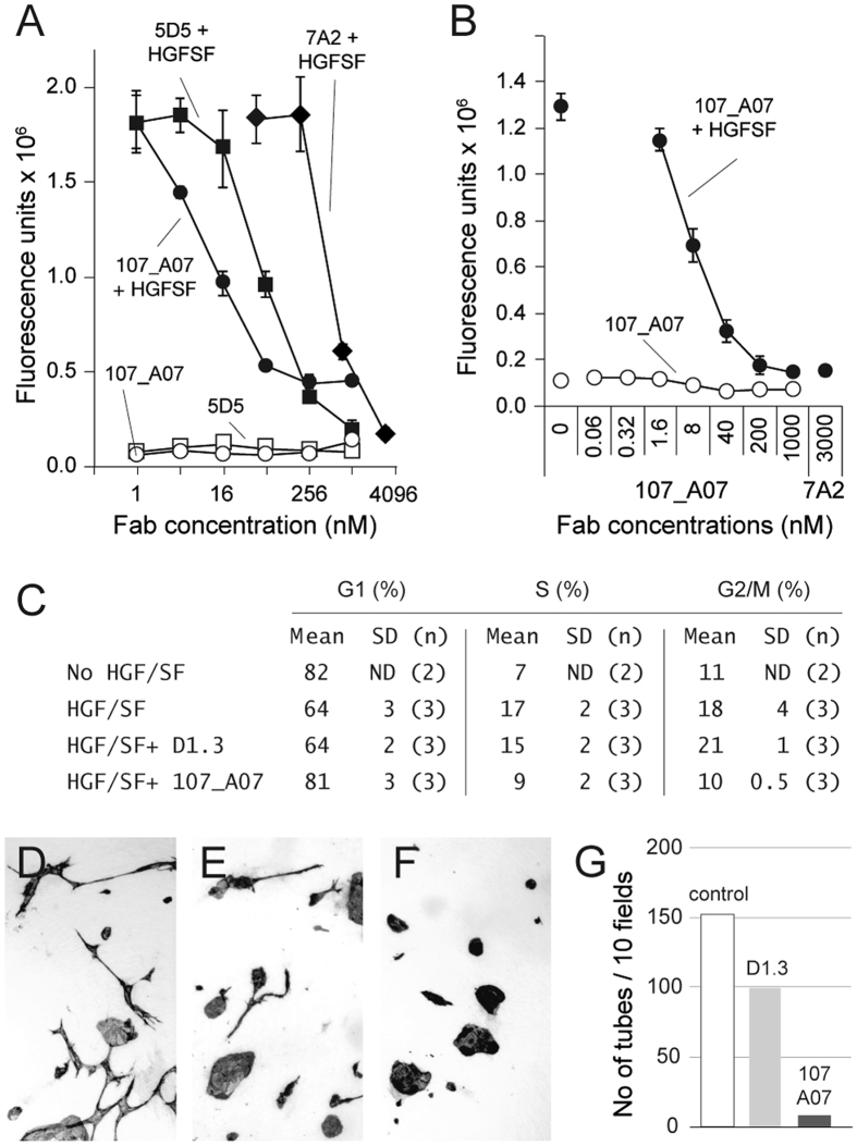
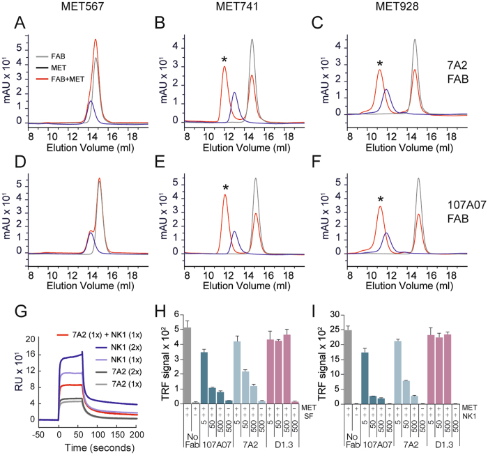
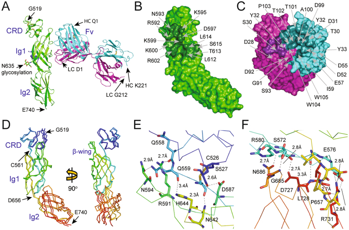
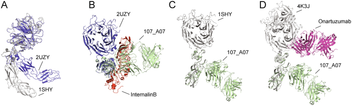
The growth and motility factor Hepatocyte Growth Factor/Scatter Factor (HGF/SF) and its receptor, the product of the MET proto-oncogene, promote invasion and metastasis of tumor cells and have been considered potential targets for cancer therapy. We generated a new Met-blocking antibody which binds outside the ligand-binding site, and determined the crystal structure of the Fab in complex with its target, which identifies the binding site as the Met Ig1 domain. The antibody, 107_A07, inhibited HGF/SF-induced cell migration and proliferation in vitro and inhibited growth of tumor xenografts in vivo. In biochemical assays, 107_A07 competes with both HGF/SF and its truncated splice variant NK1 for MET binding, despite the location of the antibody epitope on a domain (Ig1) not reported to bind NK1 or HGF/SF. Overlay of the Fab-MET crystal structure with the InternalinB-MET crystal structure shows that the 107_A07 Fab comes into close proximity with the HGF/SF-binding SEMA domain when MET is in the "compact", InternalinB-bound conformation, but not when MET is in the "open" conformation. These findings provide further support for the importance of the "compact" conformation of the MET extracellular domain, and the relevance of this conformation to HGF/SF binding and signaling.


Conflict of interest statement

The authors declare that they have no competing interests.

Figures

Figure 1
Figure 1
107_A07 FAb inhibits HGF/SF-induced cancer cell migration, DNA synthesis and angiogenesis in vitro. Effect of parental Fab-formatted 7A2 and matured 107_A07 Fab on SKOV3 (A) and U87MG (B) cell migration in the presence and absense of HGF/SF in a modified Boyden chamber assay. A Fab-formatted version of the anti-MET antibody 5D5 was used as a control. Closed and open symbols show respectively the effect of antibody in the presence and absence of HGF/SF. Data represent mean ± standard deviation of triplicate wells. (C) Cell cycle analysis by propidium iodide staining of fixed, serum-starved U87MG cells 24 hours after exposure to 300 pM HGF/SF and/or Fab as indicated. Mean and standard deviation of (n) experiments are shown. (D,E,F and G) PEGylated 107A07 blocks angiogenesis in an in vitro angiogenesis assay in which co-culture with fibroblasts promotes endothelial cell sprouting and tubulogenesis. (D) control culture, no Fab, (E) PEG-D1.3 Fab, (F) PEG-107_A07 Fab, (G) quantification of endothelial tubules three days after the addition of Fab.
Figure 2
Figure 2
Effect of 107_A07 IgG on the growth of subcutaneous U87MG xenografts. Following xenograft implantation on day 0, mice (n = 8) were given vehicle alone (PBS), control D1.3 IgG (10 mg/kg) or 107_A07 IgG (2 or 10 mg/kg) intraperitoneally at day 7 and then every 3–5 days for 26 days. Tumor volumes were measured by caliper twice weekly.
Figure 3
Figure 3
7A2 and 107_A07 bind within the Ig1-Ig2 domains of MET and compete with NK1 for MET binding. Panels A–F show size exclusion chromatography analysis of 6 µM Fab 7A2 (AC) or 107_A07 (DF) and 4 µM MET567 (A,D), MET741 (B,E) or MET928 (C,F), alone or following co-incubation. Y-axis indicates absorbance at 280 nm and x-axis elution volume. Dark blue line, MET alone; gray line, Fab alone; red line, MET/Fab mixture. Peaks present only in MET-Fab mixtures represent complex formation and are marked with an asterix. (G) Binding to a MET928-coated CM5 Biacore chip of 7A2 scFv (light gray line, 134 nM), NK1 (light blue line, 238 nM), or 7A2 scFv mixed with NK1 (red line). Binding of higher concentrations of 7A2 scFv (dark gray line, 268 nM) and NK1 (dark blue line, 268 nM) are also shown for comparison. Sensorgrams are shown aligned to the start of each 60 s injection. (H and I) Following co-incubation, untagged Fab and His-tagged MET928 were exposed to plates coated with HGF/SF (H) or NK1 (I) and bound MET928 detected with anti-5xHis peroxidase (Qiagen). Data represent mean and standard deviation of a minimum of three replicates per sample. Fab were tested at 5, 50 and 500 nM.
Figure 4
Figure 4
Crystal structure of the 107_A07-MET complex. A: Content of the asymmetric unit. The two views are related by a 90° rotation along the y axis. Complex molecule 1: MET receptor fragment (519–740) – green; MET receptor amino terminal peptide (22–32) – violet; 107_A07 Fab heavy chain – cyan; 107_A07 Fab light chain – magenta. Complex molecule 2: MET receptor fragment (519–740) – yellow; MET receptor amino terminal peptide (22–32) – orange; 107_A07 Fab heavy chain – salmon pink; 107_A07 Fab light chain – grey. The disulfide bridges are shown as sticks. The figure was generated with PYMOL. B: The view showing the position of MET receptor amino-terminal peptide in orange (22–32) relative to MET receptor fragment in yellow (519–740). The disulfide bridge between residues Cys26 of the peptide and Cys584 of MET receptor fragment is shown in sticks. The zoomed in portion of the figure is showing the final 2Fo-Fc electron density map contoured at 1.1 sigma level.
Figure 5
Figure 5
Structure of the complex between 107_A07 and MET519-740. (A) Ribbon diagram of complex 1 of the 107_A07 - MET519-740 structure showing the MET519-740 molecule (green) and the 107_A07 Fab on the right (heavy chain cyan, light chain purple). (B,C) Residues involved in contacts between MET519-740 (B) and 107_A07 Fab (VH domain in cyan, VL domain in purple). The picture highlights the polar and charged nature of the extensive VH-MET contacts (see Table S1 for further details). (D) Superposition of the structures of the MET fragment containing residues 519–740 from the structure of the 107_A07-MET complex (chain A) and the InlB-MET structure (PDB accession 2UZY, chain B). (E and F) Contacts between the CR domain and the IG1 domain (E) of MET519-740 and between the bottom of IG1 domain and the top of the IG2 domain (F). These two sets of contacts account for the rigid structure of the MET519-740 fragment of MET.
Figure 6
Figure 6
Comparison of the 107_A07 Fab with compact (InlB-bound) and extended (HGF/SFβ-bound) structures of the MET ectodomain. (A) The compact (blue) and extended (grey) conformations of extracellular MET (amino acids 25–741). The structure of compact MET is from PDB accession 2UZY (chain B), (complex of MET with InlB); the structure of MET in extended conformation is from PDB accession code 1SHY (HGF/SF beta chain in complex with SEMA domain of MET). (B) Superposition of the MET-107_A07 Fab complex (shown in green) with MET in compact conformation (2UZY), obtained by superposing the CR domains. The MET CRD-Ig1-Ig2 domains are oriented perpendicular to the image. The InternalinB structure is shown in red. (C) Superposition with MET in extended conformation (1SHY). The position of Ig1-Ig2 was obtained as described in (B). (D) Superposition with MET in extended conformation (4K3J, complex of MET and the Fab fragment of onartuzumab). The onartuzumab structure is shown in magenta.

References

    1. Valastyan S, Weinberg RA. Tumor metastasis: molecular insights and evolving paradigms. Cell. 2011;147:275–292. doi: 10.1016/j.cell.2011.09.024. - DOI - PMC - PubMed
    1. Schmidt C, et al. Scatter factor/hepatocyte growth factor is essential for liver development. Nature. 1995;373:699–702. doi: 10.1038/373699a0. - DOI - PubMed
    1. Uehara Y, et al. Placental defect and embryonic lethality in mice lacking hepatocyte growth factor/scatter factor. Nature. 1995;373:702–705. doi: 10.1038/373702a0. - DOI - PubMed
    1. Bladt F, Riethmacher D, Isenmann S, Aguzzi A, Birchmeier C. Essential role for the c-met receptor in the migration of myogenic precursor cells into the limb bud. Nature. 1995;376:768–771. doi: 10.1038/376768a0. - DOI - PubMed
    1. Chmielowiec J, et al. c-Met is essential for wound healing in the skin. J Cell Biol. 2007;177:151–162. doi: 10.1083/jcb.200701086. - DOI - PMC - PubMed

Publication types

MeSH terms