. 2017 Aug 21;7(1):8390.
doi: 10.1038/s41598-017-08273-7.

Selection of nanobodies with broad neutralizing potential against primary HIV-1 strains using soluble subtype C gp140 envelope trimers

Affiliations

Selection of nanobodies with broad neutralizing potential against primary HIV-1 strains using soluble subtype C gp140 envelope trimers

Kathrin Koch et al. Sci Rep. .

Abstract

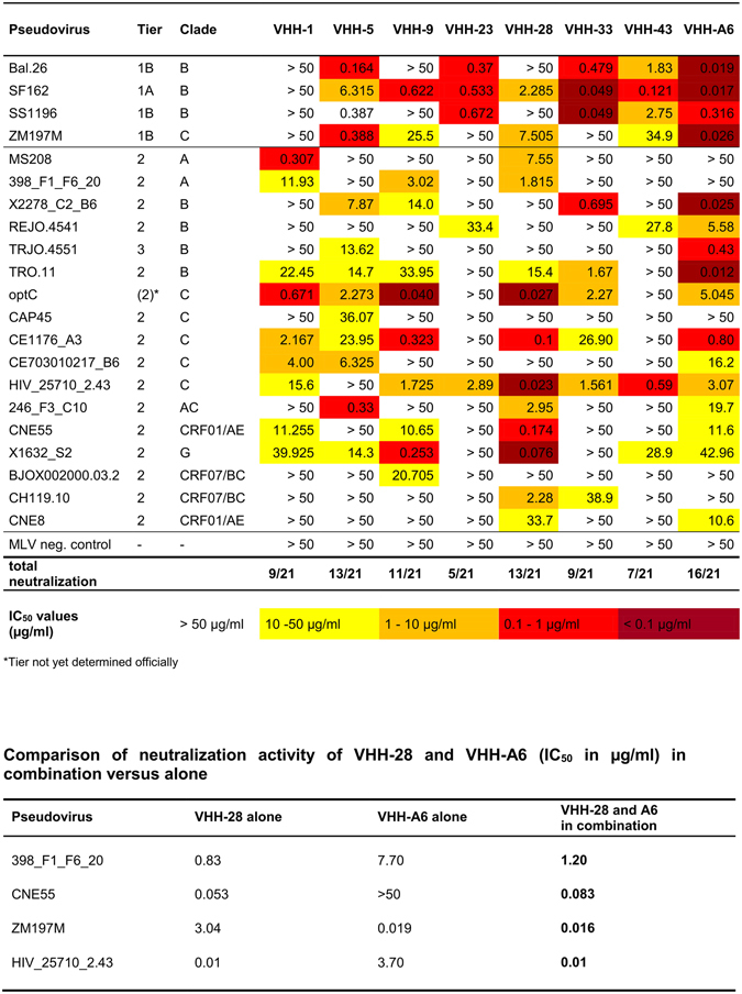
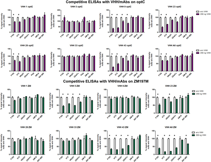
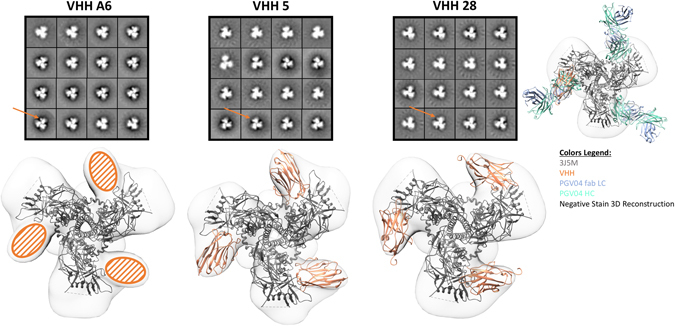
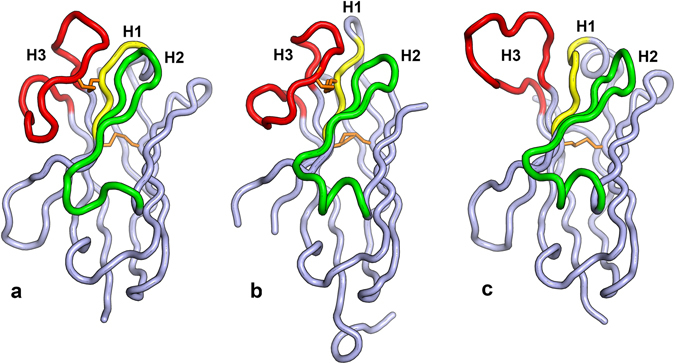
Broadly neutralizing antibodies (bnAbs) against HIV-1 protect from infection and reduce viral load upon therapeutic applications. However no vaccine was able so far to induce bnAbs demanding their expensive biotechnological production. For clinical applications, nanobodies (VHH) derived from heavy chain only antibodies from Camelidae, may be better suited due to their small size, high solubility/stability and extensive homology to human VH3 genes. Here we selected broadly neutralizing nanobodies by phage display after immunization of dromedaries with different soluble trimeric envelope proteins derived from HIV-1 subtype C. We identified 25 distinct VHH families binding trimeric Env, of which 6 neutralized heterologous primary isolates of various HIV-1 subtypes in a standardized in vitro neutralization assay. The complementary neutralization pattern of two selected VHHs in combination covers 19 out of 21 HIV-1 strains from a standardized panel of epidemiologically relevant HIV-1 subtypes. The CD4 binding site was preferentially targeted by the broadly neutralizing VHHs as determined by competition ELISAs and 3D models of VHH-Env complexes derived from negative stain electron microscopy. The nanobodies identified here are excellent candidates for further preclinical/clinical development for prophylactic and therapeutic applications due to their potency and their complementary neutralization patterns covering the majority of epidemiologically relevant HIV-1 subtypes.


Conflict of interest statement

The authors declare that they have no competing interests.

Figures

Figure 1
Figure 1
Silver staining of purified ZM197M, CAP45 and optC gp140 SOSIP proteins. Indicated Env gp140 SOSIP proteins are shown after lectin purification (1) followed by additional size exclusion and anion exchange chromatography purification steps (2). The gel was cropped below the 100 kDa marker and the marker lane was substituted by indicating the molecular weights of the marker proteins. The original gel is shown in Suppl. Figure 1a (1) together with the individual fractions collected after SEC and AEC for the three SOSIPs (2) to (4).
Figure 2
Figure 2
Immunization schedule of dromedaries with subtype C SOSIPs. Animals 54A and 6A5 received optC SOSIP, whereas DBO and O5E received a mixture of ZM197M and CAP45 SOSIPs. The first immunization cycle consisted of 7 weekly protein boosts followed by an additional boost seven months later. Red arrows indicate the timepoints of serum samples used for neutralization assays. Blood samples from timepoints t1 and t2 were also used for the generation of phage displayed nanobody libraries for all 4 animals.
Figure 3
Figure 3
Env-specific antibody response in dromedary sera during the seven immunization boosts of the first immunization cycle. ELISA experiments showing (A) optC antibody response in sera from 54A (diluted 1:1,000, open circles) and 6A5 (1:200, black circles), (B) ZM197M antibody response in sera from DBO (1:120, open squares) and O5E (1:375, black squares) and (C) CAP45 antibody response in sera from DBO (1:120, open squares) and O5E (1:375, black squares). 0: preimmune sera, 1–7: serum samples after the respective immunization boosts. One representative experiment of two is shown. Error bars indicate SEM from triplicates.
Figure 4
Figure 4
Neutralization activity of purified VHHs against a standard panel of pseudoviruses comprising different clades and neutralization sensitivities. VHHs were tested against a panel of pseudoviruses in the TZM-bl assay. Neutralization sensitivities are colour-coded according to the IC50 values: dark red corresponds to IC50 < 0.1 µg/ml, light red is from 0.1–1 µg/ml, orange is from 1–10 µg/ml and yellow is from 10–50 µg/ml. IC50 values were determined from the respective neutralization curves using GraphPad Prism and correspond to the mean IC50 values of 2–3 independent experiments. MLV pseudovirus was included as negative control. *Tier not yet determined officially. VHH28 and A6 were additionally tested in combination due to their complementary neutralization pattern.
Figure 5
Figure 5
Competitive ELISA with VHH and known mAbs on optC and ZM197M SOSIPs. On optC (upper part), CD4bs ligands compete with VHH-9, VHH-23, VHH-43, A6, less with VHH-28 and VHH-1; on ZM197M (lower part), CD4bs ligands compete with A6, VHH-43, VHH-5, VHH-23, less/not with VHH-1, VHH-28, VHH-33. Detection of competition also depends on strength of VHH binding to the respective SOSIP (see Supplementary Figure S5: VHH-9 does not bind ZM197M −> no competition detectable). Stars indicate competition, see also legend to Supplementary Table S2.
Figure 6
Figure 6
Negative stain electron microscopy reveals VHH’s target the CD4 binding site. Class averages from negative stain electron microscopy of ZM197M SOSIP trimers (version 5.2 donated by Rogier Sanders) complexed with the indicated VHH’s (upper row). In each complex three VHH’s (orange arrow) bind per trimer. A high resolution model of BG505 SOSIP.664-PGV04 Fab complex (PDB 3J5M) is docked into the 3D reconstructions of ZM197M SOSIP-VHH complexes (bottom row). Crystal structures of the VHH’s are in orange (A6 not yet available). The CD4 binding site Fab PGV04 is shown for comparison with the heavy chain in green and the light chain in blue. Alhough the VHH’s bind at a slightly different angle than PGV04, these data are consistent with the VHH’s targeting the CD4bs of the trimer.
Figure 7
Figure 7
Crystal structures for VHH-5 and VHH-28. (a) The Cα trace for VHH-5 is shown with CDR’s labeled and colored yellow (H1), green (H2) and red (H3). This coloring scheme is used throughout the figure. The disulfide bonds linking CDR3 to CDR1 and residues H22 and H92 is colored in orange. (b) The Cα trace for VHH-28. Extra residues at the C-terminus belong to the 6-Histidine tag. (c) The Cα trace of the VH domain of human broadly neutralizing anti-HIV antibody PGV04, that also binds to the CD4 binding site.

References

    1. Geiss Y, Dietrich U. Catch Me If You Can–The Race Between HIV and Neutralizing Antibodies. AIDS Rev. 2015;17:107–113. - PubMed
    1. Sanders RW, et al. Stabilization of the soluble, cleaved, trimeric form of the envelope glycoprotein complex of human immunodeficiency virus type 1. J Virol. 2002;76:8875–8889. doi: 10.1128/JVI.76.17.8875-8889.2002. - DOI - PMC - PubMed
    1. Julien JP, et al. Crystal structure of a soluble cleaved HIV-1 envelope trimer. Science. 2013;342:1477–1483. doi: 10.1126/science.1245625. - DOI - PMC - PubMed
    1. Pugach P, et al. A native-like SOSIP.664 trimer based on an HIV-1 subtype B env gene. J Virol. 2015;89:3380–3395. doi: 10.1128/JVI.03473-14. - DOI - PMC - PubMed
    1. Sanders RW, et al. A next-generation cleaved, soluble HIV-1 Env trimer, BG505 SOSIP.664 gp140, expresses multiple epitopes for broadly neutralizing but not non-neutralizing antibodies. PLoS Pathog. 2013;9:e1003618. doi: 10.1371/journal.ppat.1003618. - DOI - PMC - PubMed

Publication types

MeSH terms

Substances