. 2017 Aug 21;7(1):9002.
doi: 10.1038/s41598-017-09079-3.

Local and systemic immunomodulatory mechanisms triggered by Human Papillomavirus transformed cells: a potential role for G-CSF and neutrophils

Affiliations

Local and systemic immunomodulatory mechanisms triggered by Human Papillomavirus transformed cells: a potential role for G-CSF and neutrophils

Karla Lucia Fernandez Alvarez et al. Sci Rep. .

Abstract

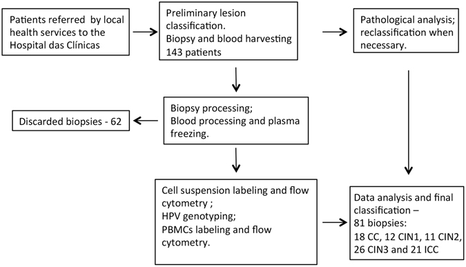
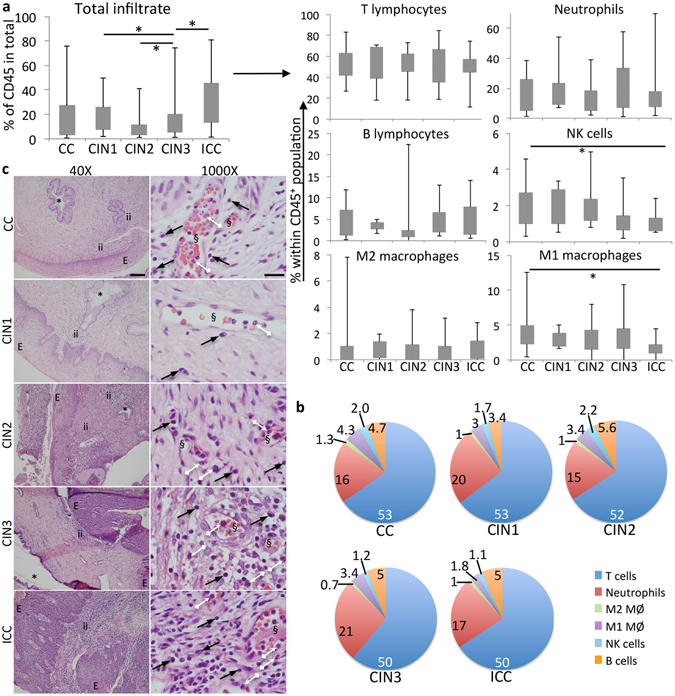
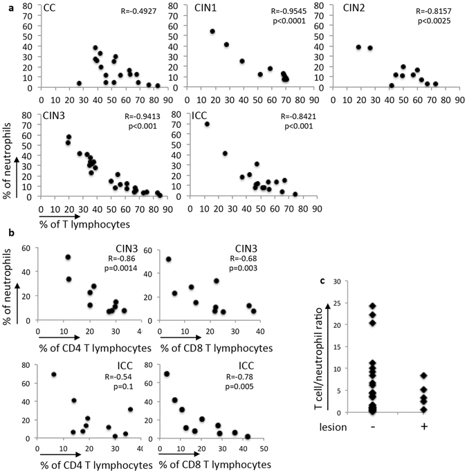
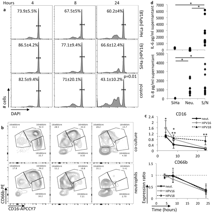
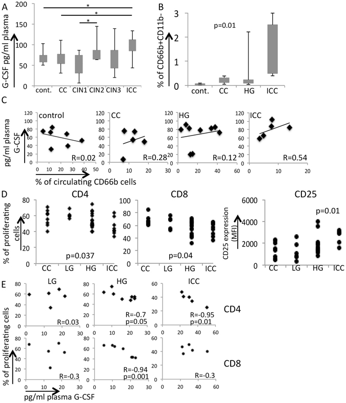
Cervical cancer is the last stage of a series of molecular and cellular alterations initiated with Human Papillomavirus (HPV) infection. The process involves immune responses and evasion mechanisms, which culminates with tolerance toward tumor antigens. Our objective was to understand local and systemic changes in the interactions between HPV associated cervical lesions and the immune system as lesions progress to cancer. Locally, we observed higher cervical leukocyte infiltrate, reflected by the increase in the frequency of T lymphocytes, neutrophils and M2 macrophages, in cancer patients. We observed a strong negative correlation between the frequency of neutrophils and T cells in precursor and cancer samples, but not cervicitis. In 3D tumor cell cultures, neutrophils inhibited T cell activity, displayed longer viability and longer CD16 expression half-life than neat neutrophil cultures. Systemically, we observed higher plasma G-CSF concentration, higher frequency of immature low density neutrophils, and tolerogenic monocyte derived dendritic cells, MoDCs, also in cancer patients. Interestingly, there was a negative correlation between T cell activation by MoDCs and G-CSF concentration in the plasma. Our results indicate that neutrophils and G-CSF may be part of the immune escape mechanisms triggered by cervical cancer cells, locally and systemically, respectively.


Conflict of interest statement

The authors declare that they have no competing interests.

Figures

Figure 1
Figure 1
Organogram of cohort enrolment. Patients with cytology indicative of HPV associated lesion were referred to Hospital das Clínicas. A total of 143 patients were enrolled with colposcopy suspicious of cervical lesion. Biopsies and peripheral blood were processed and if we obtained enough cells from biopsies, these cells were labeled with antibodies to continue the study, if not, only the blood sample was saved. From the 62 discarded biopsies, 54 did not have enough cells; the others were discarded because there was not enough patient’s clinical data. Several of the harvested and analyzed samples were reclassified according to the histopathological classification determined by the Pathology service at Hospital das Clínicas. This meant that flow cytometry analyses were performed as blind assays regarding lesion grade, which avoided any kind of bias by the student conducting the experiments. In the end of the enrolment period, we had 81 analyzed biopsies distributed in different lesion grades as indicated in the figure.
Figure 2
Figure 2
Leukocyte populations infiltrating cervical lesions. (a) Flow cytometry analyses of cervical single cell suspensions labeled with antibodies against cell surface markers. Total infiltrate corresponds to all CD45+ cells (box plot left side), all other box-plots are cells gated in the CD45+ population. Phenotype of each population is specified in Supplemental Material 1. Number of patients in each group is specified in Fig. 1. (b) Frequency of different cell populations per patient group. The values correspond the average frequency of each population; standard deviation values were omitted in the sake of space. Populations’ frequencies were compared by one-way ANOVA, *indicates significant differences in cells frequency among groups. (c) Histology of cervical tissues stained with HE. Image magnification is indicated, as well as lesion grade. E – epithelial compartment, ii – inflammatory infiltrate, * - glandular tissue, § - blood vessels, black arrows point to mononuclear cells, white arrows point to granulocytes. CC – cervicitis, CIN 1, 2, 3 – Cervical Intraepithelial Neoplasia grades 1, 2, 3, ICC – invasive cervical cancer. Scale bars indicate 250 μm and 50 μm in 40X and 1000X magnification micrographs (upper panels), respectively.
Figure 3
Figure 3
Negative correlation between neutrophils and T cells in CIN and ICC. (a) Frequencies of T cells and neutrophils in each cohort group were compared by Pearson correlation. Number of samples per group is described in Fig. 1. (b) Comparison between CD4 and CD8 and neutrophil frequencies in CIN3 and ICC samples by Pearson correlation. There were 9 CIN3 biopsies and 11 ICC biopsies in these analyses. The graphs indicate the R values and p values (significance) for each correlation. (c) T cell/neutrophil frequency ratio in patients with CIN1, 2 and 3 free of disease (−) or with persistent or recurrent disease (+) after excision procedure. Follow up period varies between 1 and 3 years.
Figure 4
Figure 4
Characteristics of cervical cancer infiltrating neutrophils. (a) CD16 expression (median fluorescence intensity, MFI) was evaluated in biopsy neutrophils, after collagenase digestion, labeling with anti-CD33, CD66b, CD11c, CD15, CD11b, CD45, CD62L, and analyzed by flow cytometry. After exclusion of debris and doublets, by SSC-A x FSC-A and FCS-A x FSC-H, cells gated on high SSC and CD45+ were evaluated for expression of the indicated makers. The only marker with significant changes among patient groups was CD16. As we had few CIN2 patients to evaluate for this parameter, we joined these patients with CIN3 patients to form a high grade lesion group (HG). CC – cervicitis, LG – low grade lesion group (same as CIN1), ICC – invasive cervical cancer. We evaluated 11 CC patients, 10 LG patients (same as CIN1), 12 HG patients and 15 ICC patients. The indicated p value, was obtained with one-way ANOVA testing. (b) Comparison of CD16, CD66b and CD62L expression in biopsy and circulating neutrophils (PB) from ICC patients. Results are displayed as median fluorescence intensity (MFI) and in this case, compared by t-test. The resulting p values are indicated. (c) ROS production by biopsy and circulating neutrophils from ICC patients, measured by DHR123 fluorescence. Neutrophils isolated from biopsies or peripheral blood were stimulated with 100 ng/ml TPA for 15 min and then incubated with DHR-123 for 15 min. Cells were then labeled with anti-CD45, CD11b and CD66b. DHR signal, displayed as median fluorescence intensity (MFI), was measured in triple positive cells, after gating out debris and doublets. In this case, our experimental group had 5 patients. Results were tested by t-test between control and treated cells, and the p values are indicated. There was no difference between ROS production in control or treated neutrophils from biopsies compared to circulating cells.
Figure 5
Figure 5
Cross-talk between cervical cancer derived cells and neutrophils. Neutrophils in co-culture with SiHa spheroids (co-culture) or in pure cultures (neutrophils) were evaluated at the indicated periods of time for: (a) viability, (b) CD66b and CD16 expression, and (c) cytokine production (only at 24 hours of culture). At each time point, cells and supernatants were harvested, cells were labeled with anti-CD45, CD66b, CD16, fixed and incubated with 10 μg/ml DAPI for 30 min before flow cytometry analysis. A. DAPI fluorescence was measured in the CD45+ population. The gated cells in the histograms indicate DNA content corresponded to whole cells, events to the left, correspond to sub-G1 dead cells. Significant differences were found only at 24 hours of culture (p value indicated). Still within the CD45+ population, we evaluated frequency (b) and expression ratio (c), depicted as expression of CD66b and CD16 in co-cultures/expression in pure neutrophil cultures. The frequency plots are representative of one experiment, and the kinetic in the right side graphs are the average ratios obtained from all experiments. A total of 5 different blood samples were used in these experiments. The U-test Mann Whitney tested differences in CD16 and CD66b expression ratios through time; *indicates significant differences in CD16 expression between experimental conditions; dotted lines indicate ratio 1 for reference. (d) Cytokine production. Supernatants from 24 hours cultures were tested for cytokine production using the Human Inflammatory CBA kit, according to the manufacture’s instructions. Although this kit has antibodies for several cytokines, only IL-6 and IL-8 were secreted in concentrations above cut off values. SiHa – tumor cells spheroid supernatant, Neu. – neutrophils supernatant, S/N – SiHa spheroids and neutrophil co-cultures. We tested the results using one-way ANOVA, *indicates significant differences between experimental groups.
Figure 6
Figure 6
Effects of neutrophils over T cell activity in a tumor-like environment. Cell proliferation dye labeled T cells were stimulated with 100 ng/ml TPA and 1 μg/ml ionomycin (TPA/Iono) for 5 hours, before they were washed and added to neutrophils cultures (in 1 T cell: 1 neutrophil or 10 T cells: 1 neutrophil ratios), or 4 days SiHa spheroid cultures (SiHa) or neutrophils previously incubated for 5 hours with 4 days SiHa spheroid cultures. After 5 days incubation, cells were harvested and labeled with anti-CD45, CD4, CD8, CD25, CD69, fixed, and labeled with 10 μg/ml DAPI and then analyzed by flow cytometry. (a) For proliferation analysis, we gated out debris and doublets, than gated on CD45+ cells and then CD4+ or CD8+ cells, where we evaluated the percentage of cells that had low Cell Proliferation dye fluorescence, compared to a non-stimulated T cell control. Experiment representative of 4 independent ones (there was variation among cells from different donors). Numbers indicated in the graph, show the average of the proliferation ratios between T cells in 1:1 and 1:10 cultures with neutrophils incubated with SiHa spheroids in 4 independent experiments. (b) Relative CD25 and CD69 expression in the same cultures described in (a), as well as concentration of IFNγ in the cultures supernatant measured with the CBA kit (BD Biosciences). Results are the average of 4 independent experiments, and were tested by one-way ANOVA, either comparing 1:1 culture conditions and 1:10 culture conditions; *indicates significant differences between experimental groups, dotted lines indicate ratio 1. (c) Depletion of SiHa cells from cultures with 10:1 T cells/neutrophils. Dot-plots on top represent the SiHa cells gate obtained after cells harvesting and flow cytometry analyzes. Total absolute cell numbers were determined by counting cells suspensions using a hemocytometer. These numbers were multiplied by the frequency of SiHa cells to obtain the absolute SiHa numbers per experimental condition. The graph bellow shows the ratio of SiHa cell numbers in SiHa/T cells cultures (TS) divided by the SiHa cell numbers in SiHa/neutrophils/ T cells cultures (TSN) in the different T cell/neutrophil experimental ratios. Results are the average of 3 independent experiments, tested by t-test, p value indicated in the graph, dotted lines indicate ratio 1.
Figure 7
Figure 7
Cervical lesion systemic effects. (A) G-CSF plasma concentrations, measured by ELISA. We used plasma from 8 control donors, from 10 CC patients, 8 CIN1, 11 CIN2, 18 CIN3 and 17 ICC patients. Results were compared by one-way ANOVA, significant differences are indicated by *. (B) Frequency of immature circulating neutrophils. The mononuclear cells from the Ficoll-Paque gradient were harvested and labeled with anti-CD66b and CD11b. The graph shows the frequency of CD66b+CD11b cells in each experimental group, which contained 5 controls, 4 CC patients, 13 high grade lesion patients (HG) and 11 ICC patients. We had only 2 patients with CIN1 lesions, therefore, had to exclude this group from this analysis. Again, results were tested with one-way ANOVA, and the p value is indicated. (C) Correlation between G-CSF plasma concentration and frequency of circulating immature neutrophils CD66b+. We used Pearson correlation to compare these parameters, R values indicated in each graph. (D) Allogeneic T cell stimulation. Cell proliferation dye labeled T cells from control donors were incubated for 4 days with MoDCs from patients of the indicated groups. After incubation, cells were harvested, labeled with CD4, CD8 and CD25 and analyzed by flow cytometry. Cell proliferation data shows the frequency of cells within the CD4+ or CD8+ T populations that diluted the proliferation dye, therefore, that proliferated during the experiments. CD25 expression, represented as median fluorescence intensity (MFI) was gated on CD4 T cells. We used one-way ANOVA to compare experimental groups. In all cases, samples from ICC patients were significantly different from the other patients. CC – cervicitis, LG – cells from patients with low grade lesions (or CIN1), HG – cells from patients with high grade lesions (CIN2/3), ICC – cells from patients with invasive cervical carcinoma. (E) Pearson correlations between T cell proliferation determined in D and plasma G-CSF concentrations, determined in B; R and p values are indicated in the plots.

References

    1. Walboomers JM, et al. Human papillomavirus is a necessary cause of invasive cervical cancer worldwide. J. Pathol. 1999;189:12–9. doi: 10.1002/(SICI)1096-9896(199909)189:1<12::AID-PATH431>3.0.CO;2-F. - DOI - PubMed
    1. Wira CR, Rodriguez-Garcia M, Patel MV. The role of sex hormones in immune protection of the female reproductive tract. Nat. Rev. Immunol. 2015;15:217–30. doi: 10.1038/nri3819. - DOI - PMC - PubMed
    1. de Jong A, et al. Human papillomavirus type 16-positive cervical cancer is associated with impaired CD4+ T-cell immunity against early antigens E2 and E6. Cancer Res. 2004;64:5449–55. doi: 10.1158/0008-5472.CAN-04-0831. - DOI - PubMed
    1. Trimble CL, et al. Human papillomavirus 16-associated cervical intraepithelial neoplasia in humans excludes CD8 T cells from dysplastic epithelium. J. Immunol. 2010;185:7107–14. doi: 10.4049/jimmunol.1002756. - DOI - PMC - PubMed
    1. Coleman N, et al. Immunological events in regressing genital warts. Am. J. Clin. Pathol. 1994;102:768–74. doi: 10.1093/ajcp/102.6.768. - DOI - PubMed

Publication types

Substances