. 2017 Aug 21;7(1):8906.
doi: 10.1038/s41598-017-08360-9.

The human IL-17A/F heterodimer: a two-faced cytokine with unique receptor recognition properties

Affiliations

The human IL-17A/F heterodimer: a two-faced cytokine with unique receptor recognition properties

Arnaud Goepfert et al. Sci Rep. .

Abstract

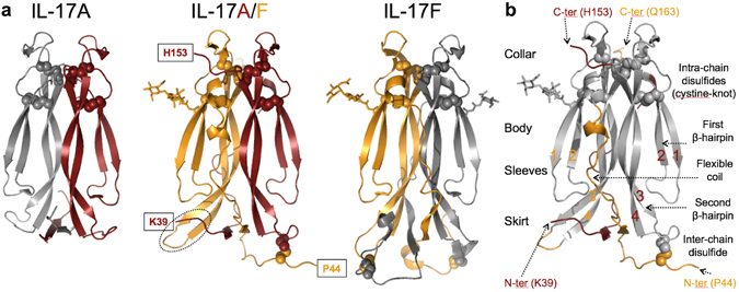
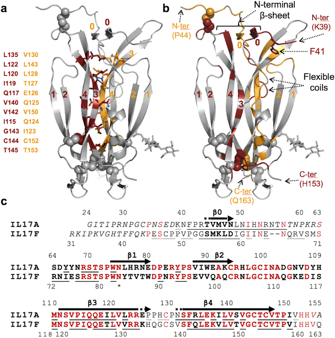
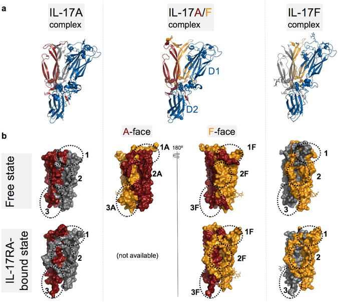
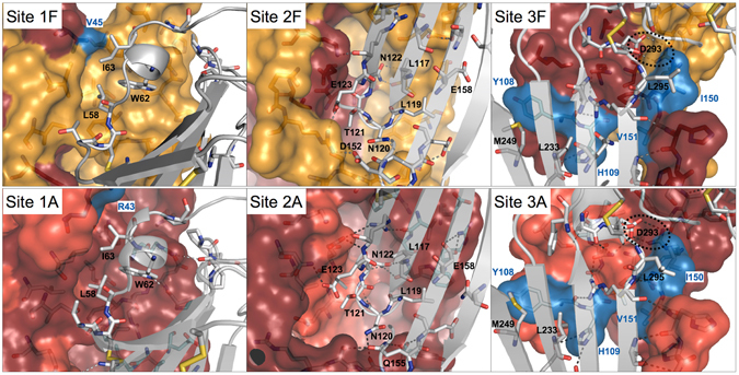
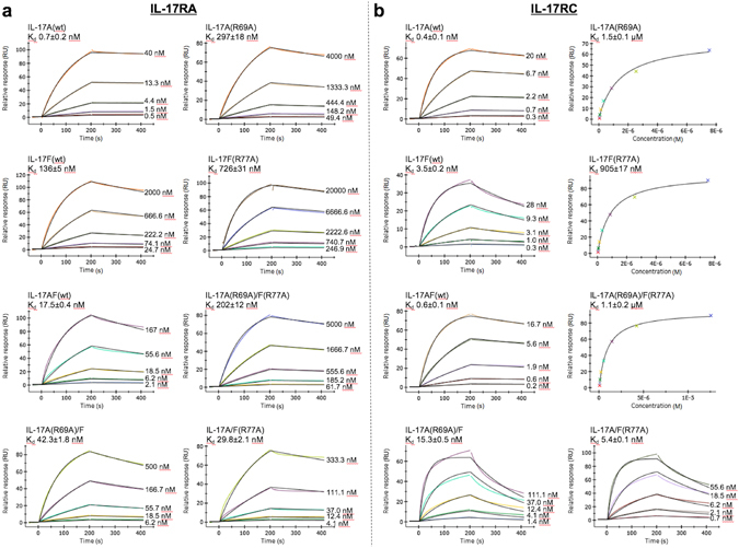
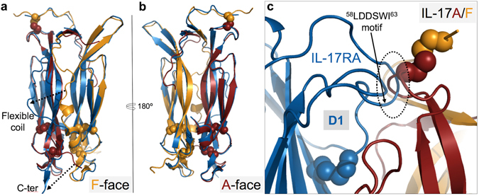
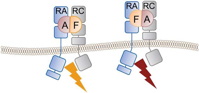
IL-17A and IL-17F are prominent members of the IL-17 family of cytokines that regulates both innate and adaptive immunity. IL-17A has been implicated in chronic inflammatory and autoimmune diseases, and anti-IL-17A antibodies have shown remarkable clinical efficacy in psoriasis and psoriatic arthritis patients. IL-17A and IL-17F are homodimeric cytokines that can also form the IL-17A/F heterodimer whose precise role in health and disease remains elusive. All three cytokines signal through the assembly of a ternary complex with the IL-17RA and IL-17RC receptors. Here we report the X-ray analysis of the human IL-17A/F heterodimer that reveals a two-faced cytokine closely mimicking IL-17A as well as IL-17F. We also present the crystal structure of its complex with the IL-17RA receptor. Unexpectedly in view of the much higher affinity of this receptor toward IL-17A, we find that IL-17RA is bound to the "F-face" of the heterodimer in the crystal. Using site-directed mutagenesis, we then demonstrate that IL-17RA can also bind to the "A-face" of IL-17A/F with similar affinity. Further, we show that IL-17RC does not discriminate between the two faces of the cytokine heterodimer either, thus enabling the formation of two topologically-distinct heterotrimeric complexes with potentially different signaling properties.


Conflict of interest statement

All authors are employees of Novartis Pharma AG, Switzerland.

Figures

Figure 1
Figure 1
Structure of human IL-17A/F. (a) Ribbon diagram of IL-17A/F (center view; A chain, carmine; F chain, orange) and comparison with IL-17A (left, 4hr9.pdb) and IL-17F (right, 1jpy.pdb). Disulfides are represented with spheres. Sugar residues are shown in stick representation. Observed N- and C-termini of IL-17A/F are boxed. The tip of the second β-hairpin of the F-chain is highlighted with a dotted ellipse. (b) Upside down orientation emphasizing the analogy between the IL-17 fold and a garment. Structurally-conserved regions in IL-17A/F with respect to the corresponding homodimers are in grey, while non-conserved structural elements are in carmine (A chain) or orange (F chain). The β-strands forming the two β-hairpins of each subunit are numbered sequentially 1 to 4.
Figure 2
Figure 2
IL-17A/F dimerization interface. (a) Core interface of the IL-17A/F heterodimer. The 22 residues forming the core interface, listed on the left, are shown in stick representation, with their positions in the structure shown in carmine and orange for the A- and F-subunit, respectively, while the rest of the heterodimer is in light grey. β-strands are labelled sequentially (0 to 4). (b) Non-core interface of the IL-17A/F heterodimer, highlighted using the same color-coding scheme as before. N-termini are labelled. Phe41 (see text) is shown in stick representation. (c) Structure-based sequence alignment of the A and F subunits of human IL-17A/F. Disordered residues in the crystal structure are italicized. Structurally equivalent residues are in bold, and conserved amino-acids are highlighted in red. Residues experiencing a reduction (>10 Å2) of their solvent accessible surface upon heterodimer formation are underlined. The N-terminal β-strand β0 as well as the first (β1–β2) and second (β3–β4) β-hairpins are indicated with arrows. The observed N-glycosylation site on the F-chain is marked with an asterisk.
Figure 3
Figure 3
Structural comparison of IL-17RA-cytokine complexes. (a) Overall structure of the IL-17A/F complex with IL-17RA (center) compared to the IL-17A (left; 4hsa.pdb) and IL-17F (right; 3jvf.pdb) complexes. The IL-17RA is shown as a blue ribbon, while IL-17A and IL-17F subunits are shown as a carmine or orange ribbon, respectively. The second IL-17 subunit of the homodimeric cytokines is in grey. Disulfide bonds are highlighted as spheres and N-linked glycans are depicted in stick representation. (b) Surface representations of IL-17A (left; carmine/grey; 4hr9.pdb), IL-17F (right; orange/grey; 1jpy.pdb) and IL-17A/F (center; carmine/orange) in the free (top row) and IL-17RA-bound states (lower row). For clarity, IL-17RA is omitted from the receptor-bound states. Sites 1–3 are indicated by dotted lines. The two faces of the IL-17A/F heterodimer in the free state are shown (top row, center). The structure of the IL-17A/F complex with IL-17RA bound to the A-face is not known. Note the occlusion of Sites 1–3 on the “F-face” of free IL-17A/F.
Figure 4
Figure 4
Conserved binding interactions and binding sites in IL-17A, IL17F and IL-17A/F. Top row: IL-17A/F complex with IL-17RA. Sites 1F-3F of IL-17A/F are depicted as surface representations, with the A- and F-subunits in carmine and orange, respectively. Non-conserved side-chains with respect to the IL-17F homodimer are in blue. The IL-17RA receptor is shown as a grey ribbon with key side-chains depicted in stick representation. Bottom row: IL-17A complex with IL-17RA (4hsa.pdb). Sites 1–3 of IL-17A are shown in surface representation with the 2 subunits in carmine and light red, respectively. Non-conserved side-chains in Sites 1A-3A of IL-17A/F are highlighted in blue. As before, the IL-17RA receptor is shown as a grey ribbon with key side-chains depicted in stick representation. Note the conservation of Site 1A-2A of IL-17A/F with respect to IL-17A.
Figure 5
Figure 5
SPR affinity measurements of wild-type and variant IL-17A, IL-17F and IL-17A/F to the immobilized extracellular domain of the IL-17 receptors IL-17RA (a) or IL-17RC (b). Representative sensorgrams are plotted as response in resonance units (RUs) versus time and shown with colored lines. The concentrations of the injected analytes are indicated on the right of the sensorgrams. The kinetic parameters are calculated using a Langmuir 1:1 binding model with the fitted curves depicted as black lines. For cytokine variants with weaker affinity, the equilibrium dissociation constants were obtained from steady-state analysis by fitting the plot of the response at equilibrium (RUs) as a function of analyte concentration (M). The curve fit is shown as a black line. The indicated Kd represents the mean from at least five independent experiments ± the standard error of the mean (sem) and are also summarized in Supplementary Table 2.
Figure 6
Figure 6
Least square superposition of the free- (carmine/orange ribbon) and IL-17RA-bound states (blue ribbon) of IL-17A/F, using a maximum matching distance of 1.2Å. (a) Receptor view of the “F-face” of the cytokine. Note the large conformational changes affecting the flexible coil and C-terminal tail. (b) Receptor view of the “A-face”. Note the high similarity of the free- and “F-face”-receptor-bound states. (c) Close-up view of a model showing the IL-17RA (blue ribbon) bound to the “A-face” of IL-17A/F. The receptor position was generated by applying the transformation that best superimposes the F-subunit onto the A-subunit. Note the steric hindrance (dotted circle) between the β3–β4 loop of the cytokine and the loop of IL-17RA bearing the key58LDDSWI63 binding motif.
Figure 7
Figure 7
Schematic representation of the two, topologically distinct, signaling complexes potentially formed by the human IL-17A/F heterodimer and the IL-17RA and IL-17RC receptor chains. Whether both complexes are competent for signaling and whether they trigger the same downstream signaling events is not known.

References

    1. Harrington LE. Interleukin 17-producing CD4+ effector T cells develop via a lineage distinct from the T helper type 1 and 2 lineages. Nat. Immunol. 2005;6:1123–1132. doi: 10.1038/ni1254. - DOI - PubMed
    1. Park H, et al. A distinct lineage of CD4 T cells regulates tissue inflammation by producing interleukin 17. Nat. Immunol. 2005;6:1133–1141. doi: 10.1038/ni1261. - DOI - PMC - PubMed
    1. Korn T, Bettelli E, Oukka M, Kuchroo VK. IL-17 and Th17 cells. Annu. Rev. Immunol. 2009;27:485–517. doi: 10.1146/annurev.immunol.021908.132710. - DOI - PubMed
    1. Kara EE, et al. Tailored immune responses: novel effector helper T cell subsets in protective immunity. PLOS Pathog. 2014;10:e1003905. doi: 10.1371/journal.ppat.1003905. - DOI - PMC - PubMed
    1. Reynolds JM, Angkasekwinai P, Dong C. IL-17 family member cytokines: regulation and function in innate immunity. Cytokine Growth Factor Rev. 2010;21:413–423. doi: 10.1016/j.cytogfr.2010.10.002. - DOI - PMC - PubMed

Publication types