. 2017 Aug 21;7(1):8965.
doi: 10.1038/s41598-017-08121-8.

Classification of M1/M2-polarized human macrophages by label-free hyperspectral reflectance confocal microscopy and multivariate analysis

Affiliations

Classification of M1/M2-polarized human macrophages by label-free hyperspectral reflectance confocal microscopy and multivariate analysis

Francesca R Bertani et al. Sci Rep. .

Abstract

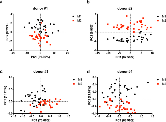
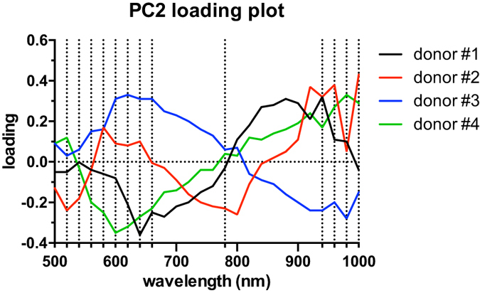
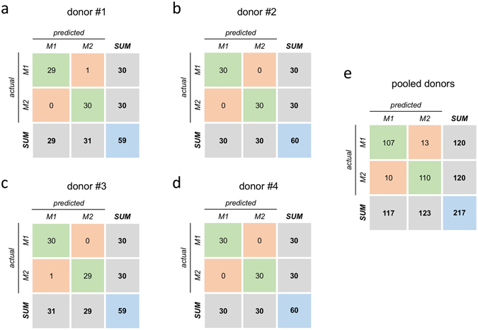
The possibility of detecting and classifying living cells in a label-free and non-invasive manner holds significant theranostic potential. In this work, Hyperspectral Imaging (HSI) has been successfully applied to the analysis of macrophagic polarization, given its central role in several pathological settings, including the regulation of tumour microenvironment. Human monocyte derived macrophages have been investigated using hyperspectral reflectance confocal microscopy, and hyperspectral datasets have been analysed in terms of M1 vs. M2 polarization by Principal Components Analysis (PCA). Following PCA, Linear Discriminant Analysis has been implemented for semi-automatic classification of macrophagic polarization from HSI data. Our results confirm the possibility to perform single-cell-level in vitro classification of M1 vs. M2 macrophages in a non-invasive and label-free manner with a high accuracy (above 98% for cells deriving from the same donor), supporting the idea of applying the technique to the study of complex interacting cellular systems, such in the case of tumour-immunity in vitro models.


Conflict of interest statement

The authors declare that they have no competing interests.

Figures

Figure 1
Figure 1
Epifluorescence microscopy. M1 (a) and M2 (b) MDMs labelled with FITC-phalloidin to stain cytoskeletal actin (in green), and DAPI nuclear counterstain (in blue). Scale bar: 100 μm.
Figure 2
Figure 2
Gene expression profile evaluated by RT-qPCR. Scatter plots of IL-10/IL-12 mRNA ratio (a), ARG1/NOS2 mRNA ratio (b), TNF-α relative mRNA expression (c) and CD206 relative mRNA expression (d) are reported.
Figure 3
Figure 3
Flow cytometry. (ac) Positivity to pan-macrophagic marker CD68 (a), M1 marker CD80 (b) and M2 marker CD206 (c). (d) Mean fluorescence intensity levels for CD206. (e,f) Positivity to M1 marker CD86 (e) and M2 marker CD163 (f). (g) Mean fluorescence intensity levels for CD163.
Figure 4
Figure 4
Hyperspectral microscopy. Representative micrographs for M1 (a) and M2 (b) MDMs obtained as a grey level representation of the reflectance value at 700 nm, as extracted from the hyperspectral datasets. Scale bar: 200 μm.
Figure 5
Figure 5
Principal Component Analysis. PCA score plots of MDM spectra for different donors. Spectra of M1 and M2 MDMs are represented as black circles and red squares, respectively.
Figure 6
Figure 6
PC2 loading plot. Loading plot for the second principal component (PC2), along which the M1/M2 separation occurred. Wavelengths chosen for LDA analysis are marked by dotted lines.
Figure 7
Figure 7
LDA model. Confusion matrices from 10-fold cross validation (each matrix is the summation of 10 confusion matrices from 10 test sets).

References

    1. Davies LC, Jenkins SJ, Allen JE, Taylor PR. Tissue-resident macrophages. Nat. Immunol. 2013;14:986–95. doi: 10.1038/ni.2705. - DOI - PMC - PubMed
    1. Davies LC, Taylor PR. Tissue-resident macrophages: Then and now. Immunology. 2015;144:541–548. doi: 10.1111/imm.12451. - DOI - PMC - PubMed
    1. Allavena P, Sica A, Solinas G, Porta C, Mantovani A. The inflammatory micro-environment in tumor progression: The role of tumor-associated macrophages. Critical Reviews in Oncology/Hematology. 2008;66:1–9. doi: 10.1016/j.critrevonc.2007.07.004. - DOI - PubMed
    1. Chanmee T, Ontong P, Konno K, Itano N. Tumor-associated macrophages as major players in the tumor microenvironment. Cancers (Basel). 2014;6:1670–90. doi: 10.3390/cancers6031670. - DOI - PMC - PubMed
    1. Yang L, Zhang Y. Tumor-associated macrophages: from basic research to clinical application. J. Hematol. Oncol. 2017;10:58. doi: 10.1186/s13045-017-0430-2. - DOI - PMC - PubMed

Publication types