. 2017 Aug 21;7(1):8360.
doi: 10.1038/s41598-017-08641-3.

Efficient generation of bispecific IgG antibodies by split intein mediated protein trans-splicing system

Affiliations

Efficient generation of bispecific IgG antibodies by split intein mediated protein trans-splicing system

Lei Han et al. Sci Rep. .

Abstract

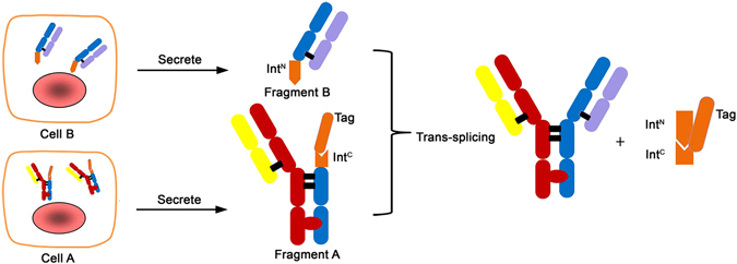
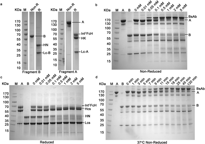
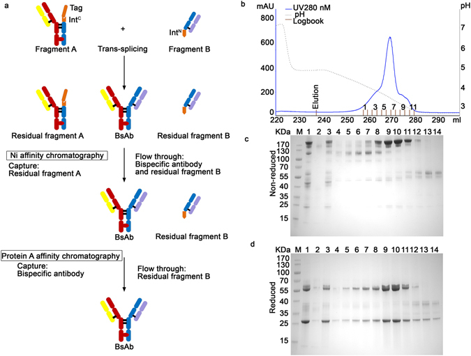
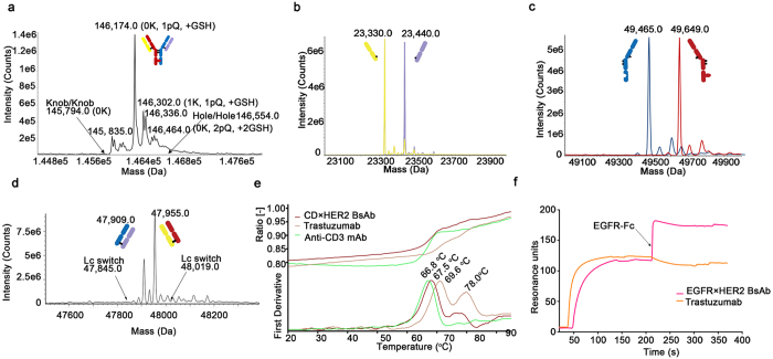
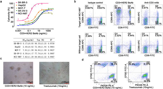
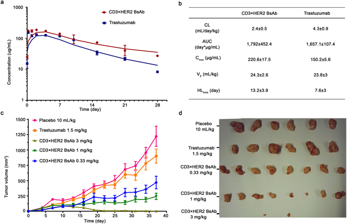
Many methods have been developed to produce bispecific antibodies (BsAbs) for industrial application. However, huge challenges still remain in synthesizing whole length BsAbs, including their assembly, stability, immunogenicity, and pharmacodynamics. Here we present for first time a generic technology platform of generating bispecific IgG antibodies, "Bispecific Antibody by Protein Trans-splicing (BAPTS)". Different from published methods, we assembled two parental antibody fragments in the hinge region by the protein trans-splicing reaction of a split intein to generate BsAbs without heavy/heavy and light/heavy chain mispairing. Utilizing this simple and efficient approach, there have been several BsAbs (CD3×HER2, CD3×EGFR, EGFR×HER2) synthesized to demonstrate its broad applicability. Correctly paired mAb arms were assembled to form BsAbs that were purified through protein A affinity chromatography to demonstrate industrial applicability at large scale. Further, the products were characterized through physical-biochemistry properties and biological activities to confirm expected quality of the products from "BAPTS". More importantly, correct pairing was confirmed by mass spectrum. Proof-of-concept studies with CD3×HER2 BsAb (T-cell recruitment) demonstrated superior bioactivity compared with trastuzumab. The results of undetectable mispairing and high biological activity have indicated that this method has the potential to be utilized to manufacture BsAbs with high efficiency at industrial scale.


Conflict of interest statement

Y.X., H.J., and J.Z. are employees of Jecho 566 Laboratories Inc.

Figures

Figure 1
Figure 1
Schematic of the process for bispecific antibody production using the split intein Npu DnaE trans-splicing activity. Three steps were involved, the first was the expression of the fragment A and B in mammalian cells respectively; the second was trans-splicing of the fragment A and B; and the last was purification of the BsAb. Fragment A and B were harvested after cell culture separately, then after primary purification followed by trans-splicing reaction, the two fragments were ligated to form the BsAb.
Figure 2
Figure 2
“BAPTS” technology mediated generation of the BsAb (CD3 × HER2). (a) SDS-PAGE (4 – 20%) analysis of the fragments A and B under non-reduced and reduced conditions. All lanes were excised from two gels (Supplementary Figure 1a, lanes 4 and 11; Supplementary Figure 1b, lanes 6 and 13) run under the same condition. (b,c) Catalytic trans-splicing reaction between fragments A and B at 37°C in the absence or presence of various concentrations of reducing agent DTT for 2 hours. The expected trans-spliced product BsAb started to appear at 0.01 mM DTT and the reaction reached plateau at 0.5 mM DTT. (d) SDS-PAGE (4 – 20%) of the reaction between fragments A and B performed at 37°C in the presence of 0.5 mM DTT. About 90% of raw material was converted to BsAb within 25 min after the reaction started.
Figure 3
Figure 3
The purification of BsAb (CD3×HER2). (a) Purification workflow. In the first step, unreacted fragment A was removed by capturing on Sepharose affinity column through the His-tag. In the second step, the product was isolated from the rest of reaction mixture by protein A affinity chromatography to remove fragment B. (b) The chromatography of protein A purification of trans-spliced BsAb (CD3×HER2), Using pH gradient elution from pH 5.0 to pH 2.8. (c,d) SDS–PAGE (4 – 20%) analysis of the protein A elutions. Lanes: 1, Sample after the trans-splicing reaction of fragments A (CD3) and B (HER2); 2, Ni column Elution; 3, Ni column flow through; 4 to 14, fractions of protein A elution from 1 to 11 respectively.
Figure 4
Figure 4
Physico-chemical characterization of BsAbs (post-protein A purification). IMS Q-Tof mass spectrometry, Differential scanning fluorimetry and SPR were performed for BsAb (CD3×HER2). (a) Mass spectrometry analysis of the deglycosylated and non-reduced BsAb was performed to demonstrate correct mass for the product without mispairing. Expected mass location for potential knob/knob (K/K), and hole/hole (H/H) homodimers were labeled by the arrows. K, C-terminal lysine presence; pQ, N-terminal pyroglutamic acid cyclization; +GSH, reduced glutathione addition. (b,c) Mass spectrometry analysis of the deglycosylated and reduced sample to confirm the presence of two different light chains and two different heavy chains. (d) Mass spectrometry analysis of the papain digested sample to determine if light chain switching occurred during the BAPTS process. The arrows indicate theoretical masses for the Fab if cognate heavy-light chain pairs had switched. (e) Differential scanning fluorimetry measurement of the BsAb (CD3×HER2) and it’s parental mAbs. (i) Dual antigen binding behavior of the BsAb (EGFR×HER2) measured by SPR. The BsAb was injected over a surface containing immobilized HER2-Fc antigen followed by injection of EGFR-Fc. Trastuzumab was used as a control.
Figure 5
Figure 5
In vitro bioactivity of BsAb (CD3×HER2). (a) Target-dependent T cell mediated cytotoxicity of BsAb (CD3×HER2) was detected using LDH release assay. Effecters human PBMCs, E:T ratio 10:1, time point of 20 hours. The EC50 values were calculated by fitting the dose-response curve with Graphpad Prism software. R2 represents the square of correlation coefficient. (b) T cell activation was detected by staining cells for CD8 and CD69 followed by FACS analysis. Effectors CD3+ T cells, target NCI-N87 cell line, E:T ratio 10:1. The upper panel showed the stimulation of human T cells by 100 ng/mL BsAb (CD3×HER2) or anti-CD3 mAb in the presence of target cells, while the bottom panel was lack of target cells. (c) Photographs of the redirection of T cells to cancer cells by 10 ng/mL BsAb (CD3×HER2). Effecters human PBMCs, target SK-BR-3 cell line, E:T ratio 10:1, time point of 20 hours. (d) FACS analysis of the redirection of CD3+ cells to cancer cells by BsAb (CD3×HER2). Jurkat (CD3+) cells were labeled by PKH26 (PE-A) and NCI-N87 cells were labeled by CFSE (FITC-A) separately. Then the two cells were mixed at equal ratio and treated with 10 ng/mL BsAb (CD3×HER2) or trastuzumab for 30 minutes. Data points in the figure represent the mean of three samples; error bar, SEM.
Figure 6
Figure 6
In vivo PK profile and tumor growth inhibition bioactivity of the BsAb (CD3×HER2). (a) In vivo PK analysis of the BsAb and trastuzumab in Balb/c mice (N = 5). Single i.p. doses of 5 mg/kg BsAb or trastuzumab were injected into Balb/c mice. Test animals’ serum samples were assayed by ELISA. (b) PK parameters of trastuzumab and the BsAb. Data fitted to the non-compartmental analysis model. (c) BsAb inhibited growth of subcutaneously transplanted NCI-N87 cell. NCI-N87 cells were injected together with unstimulated human PBMCs from healthy donors. Mice (N = 8) were treated with 0.33 mg/kg, 1 mg/kg and 3 mg/kg i.p. dose of the BsAb and 1.5 mg/kg dose of Trastuzumab once a week. (d) Photographs of excised tumors at the end of the experiment. Error bar, SEM.

References

    1. Zhu J. Mammalian cell protein expression for biopharmaceutical production. Biotechnology advances. 2012;30:1158–1170. doi: 10.1016/j.biotechadv.2011.08.022. - DOI - PubMed
    1. Bargou R, et al. Tumor regression in cancer patients by very low doses of a T cell-engaging antibody. Science. 2008;321:974–977. doi: 10.1126/science.1158545. - DOI - PubMed
    1. Schaefer G, et al. A two-in-one antibody against HER3 and EGFR has superior inhibitory activity compared with monospecific antibodies. Cancer cell. 2011;20:472–486. doi: 10.1016/j.ccr.2011.09.003. - DOI - PubMed
    1. Huang Y, et al. Engineered Bispecific Antibodies with Exquisite HIV-1-Neutralizing Activity. Cell. 2016;165:1621–1631. doi: 10.1016/j.cell.2016.05.024. - DOI - PMC - PubMed
    1. Wu C, et al. Simultaneous targeting of multiple disease mediators by a dual-variable-domain immunoglobulin. Nat Biotechnol. 2007;25:1290–1297. doi: 10.1038/nbt1345. - DOI - PubMed

Publication types

MeSH terms