. 2017 Aug 21;11(8):e0005809.
doi: 10.1371/journal.pntd.0005809. eCollection 2017 Aug.

Characterization of Monkeypox virus infection in African rope squirrels (Funisciurus sp.)

Affiliations

Characterization of Monkeypox virus infection in African rope squirrels (Funisciurus sp.)

Elizabeth A Falendysz et al. PLoS Negl Trop Dis. .

Abstract

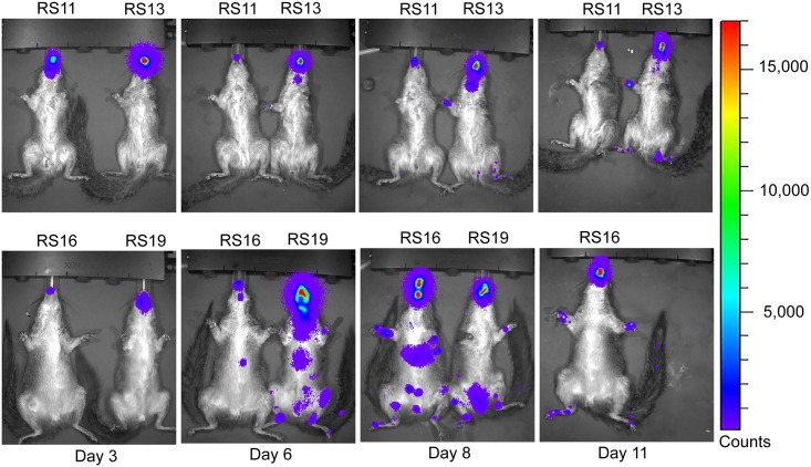
Monkeypox (MPX) is a zoonotic disease endemic in Central and West Africa and is caused by Monkeypox virus (MPXV), the most virulent Orthopoxvirus affecting humans since the eradication of Variola virus (VARV). Many aspects of the MPXV transmission cycle, including the natural host of the virus, remain unknown. African rope squirrels (Funisciurus spp.) are considered potential reservoirs of MPXV, as serosurveillance data in Central Africa has confirmed the circulation of the virus in these rodent species [1,2]. In order to understand the tissue tropism and clinical signs associated with infection with MPXV in these species, wild-caught rope squirrels were experimentally infected via intranasal and intradermal exposure with a recombinant MPXV strain from Central Africa engineered to express the luciferase gene. After infection, we monitored viral replication and shedding via in vivo bioluminescent imaging, viral culture and real time PCR. MPXV infection in African rope squirrels caused mortality and moderate to severe morbidity, with clinical signs including pox lesions in the skin, eyes, mouth and nose, dyspnea, and profuse nasal discharge. Both intranasal and intradermal exposures induced high levels of viremia, fast systemic spread, and long periods of viral shedding. Shedding and luminescence peaked at day 6 post infection and was still detectable after 15 days. Interestingly, one sentinel animal, housed in the same room but in a separate cage, also developed severe MPX disease and was euthanized. This study indicates that MPXV causes significant pathology in African rope squirrels and infected rope squirrels shed large quantities of virus, supporting their role as a potential source of MPXV transmission to humans and other animals in endemic MPX regions.


Conflict of interest statement

The authors have declared that no competing interests exist.

Figures

Fig 1
Fig 1. Progression of disease of rope squirrels infected intradermally with MPXV-Congo/Luc+.
The days post infection (pi) of various clinical signs are shown for the group, above the time scale. The days of luminescence are shown in green. Days of shedding of virus in nasal, ocular, oral and rectal mucosal secretions, as detected by TCID50 assay, are shown below the time scale. Colors coordinate between clinical signs and route of shedding: nasal shedding and respiratory disease is shown in red. Ocular lesions and ocular shedding are shown in yellow. Oral lesions and oral shedding are shown in blue. Rectal shedding, for which no corresponding clinical observations were made, is shown in purple.
Fig 2
Fig 2. Disease progression and shedding of rope squirrels infected intranasally with MPXV-Congo/Luc+.
The days post infection (pi) of various clinical signs are shown for the group, above the time scale. The days of luminescence are shown in green. Days of shedding of virus in nasal, ocular, oral and rectal mucosal secretions, as detected by TCID50 assay, are shown below the time scale. Colors coordinate between clinical signs and route of shedding: nasal shedding and respiratory disease is shown in red. Ocular lesions and ocular shedding are shown in yellow. Oral lesions and oral shedding are shown in blue. Rectal shedding, for which no corresponding clinical observations were made, is shown in purple.
Fig 3
Fig 3. Ventral bioluminescent images of African rope squirrels intradermally infected with MPXV-Congo/Luc+ on days 3, 6, 8, and 11 post infection (pi).
Color scale shows the intensity of luminescence in counts, with purple representing the lowest intensity, green the mid-range and red the highest intensity. Animals were infected on the dorsal scapular area. Luminescence in the oral and nasal areas, which begins on day 3 pi, is the result of replication of the virus distant from the initial site of infection. RS12 died on day 8 pi.
Fig 4
Fig 4. Ventral bioluminescent images of African rope squirrels intradermally infected with MPXV-Congo/Luc+ on days 13, 15, 18, and 20 post infection (pi).
Color scale shows the intensity of luminescence, in counts, with purple representing the lowest intensity, green the mid-range and red the highest intensity. Luminescence, indicative of viral replication, in the oral and nasal areas continued until day 18 pi. Distal limbs and the tail were common sites of replication. These sites often corresponded to lesions on the feet.
Fig 5
Fig 5. Ventral bioluminescent images of African rope squirrels intranasally infected with MPXV-Congo/Luc+ on days 3, 6, 8, and 11 post infection (pi).
Color scale shows the intensity of luminescence, with purple representing the lowest intensity, green the mid-range and red the highest intensity. Detectable luminescence is present in the oral and nasal areas in all animals on day 3 pi, which was the site of inoculation in this group. By day 6 pi viral replication is detectable at distal sites. After day 11 pi RS19 was euthanized.
Fig 6
Fig 6. Ventral bioluminescent images of African rope squirrels intranasally infected with MPXV-Congo/Luc+ on days 13, 15, 18, and 20 post infection (pi).
Color scale shows the intensity of luminescence, with purple representing the lowest intensity, green the mid-range and red the highest intensity. After day 13 pi, RS13 was found dead. RS16 was euthanized after imaging on day 13 pi. RS11 had a large amount of luminescence in the oral and nasal areas on day 13 pi, but decreased throughout the remainder of the days. By day 20 pi, luminescence is not visible in bioluminescent overlay images.
Fig 7
Fig 7. Quantification of luminescence of African rope squirrels infected intradermally (ID) and intranasally (IN) with MPXV-Congo/Luc+.
Both the IN and ID groups showed increasing luminescence from days 0 to 6 post infection (pi), which decreased slightly on day 8 pi, then increased again. The IN group showed peak luminescence on day 11 pi, and the ID group showed peak luminescence on day 13 pi. For both groups luminescence was detected up to day18 pi, and then remained at the background level for the remainder of the study. The sentinel animal did have luminescence above background levels.
Fig 8
Fig 8. Shedding of MPXV in African rope squirrels infected intradermally with MPXV-Congo/Luc+.
Swabs were taken from oral, ocular, nasal and rectal mucosa then tested by TCID50 assay to determine the concentration of virus shed in PFU/mL. Titers increased following infection, peaked between days 8 and 13, and then decreased for all animals except RS17, which showed consistently high titer from ocular swabs until day 25 pi.
Fig 9
Fig 9. Shedding of MPXV in African rope squirrels infected intranasally with MPXV-Congo/Luc+.
Swabs were taken from oral, ocular, nasal and rectal mucosa then tested by TCID50 assay to determine the concentration of virus shed in PFU/mL. Viral shedding increased following infection, peaked between days 8 and 13 pi, and then decreased for all animals. All viral shedding had ceased by day 15 pi in RS11, the lone survivor. All other animals in this group had died by day 13 pi.
Fig 10
Fig 10. Shedding of MPXV by the sentinel African rope squirrel, RS14, following mock intranasal infection.
The animal was housed in its own cage in the same room as the study animals, and was sampled and imaged following the same schedule as the other study animals. Swabs were taken from oral, ocular, nasal and rectal mucosa then tested by TCID50 assay to determine the concentration of virus shed in PFU/mL. Titer for all swabs increased from day 13 pi (oral) or day 15 pi (nasal, ocular, rectal) until day 18 pi, when the animal was euthanized. Oral shedding was the highest, with a final titer greater than 1.3 x 10⁸ PFU/mL on day 18 pi.
Fig 11
Fig 11. Pre-infection neutralizing activity of serum of rope squirrels against MPXV/Congo/Luc+.
Positive controls were serum from a human vaccinated with vaccinia smallpox vaccine (positive control 1) and serum from a Gambian pouched rat (Cricetomys gambianus) that survived MPXV infection (positive control 2). Serum from an uninfected Gambian pouched rat was used as a negative control. RS14 and RS19 had a slightly greater than 50% reduction in viral luminescence at 1:40 dilution. However, this was not considered a protective titer. Neither of these animals survived infection with MPXV.
Fig 12
Fig 12. Endpoint ELISA titers of African rope squirrels infected intradermally (A) and intranasally (B) with MPXV-Congo/Luc+.
Antibodies increased beginning on day 6 post infection (pi) and reached 1:4800 (the highest dilution tested) in the three surviving animals (RS17, RS18, and RS11). RS15 and RS16 had antibody titers of 1:2400 on the day they died, days 22 and 13 pi respectively.

References

    1. Nolen LD, Osadebe L, Katomba J, Likofata J, Mukadi D, et al. (2015) Introduction of Monkeypox into a Community and Household: Risk Factors and Zoonotic Reservoirs in the Democratic Republic of the Congo. American Journal of Tropical Medicine and Hygiene. - PMC - PubMed
    1. Khodakevich L, Szczeniowski M, Manbu ma D, Jezek Z, Marennikova S, et al. (1987) The role of squirrels in sustaining monkeypox virus transmission. Trop Geogr Med 39: 115–122. - PubMed
    1. McCollum AM, Damon IK (2014) Human monkeypox. Clin Infect Dis 58: 260–267. doi: 10.1093/cid/cit703 - DOI - PMC - PubMed
    1. Damon IK (2011) Status of human monkeypox: clinical disease, epidemiology and research. Vaccine 29 Suppl 4: D54–59. - PubMed
    1. Reynolds MG, Carroll DS, Olson VA, Hughes C, Galley J, et al. (2010) A Silent Enzootic of an Orthopoxvirus in Ghana, West Africa: Evidence for Multi-Species Involvement in the Absence of Widespread Human Disease. American Journal of Tropical Medicine and Hygiene 82: 746–754. doi: 10.4269/ajtmh.2010.09-0716 - DOI - PMC - PubMed