. 2017 Aug 21;13(8):e1006974.
doi: 10.1371/journal.pgen.1006974. eCollection 2017 Aug.

mlh3 mutations in baker's yeast alter meiotic recombination outcomes by increasing noncrossover events genome-wide

Affiliations

mlh3 mutations in baker's yeast alter meiotic recombination outcomes by increasing noncrossover events genome-wide

Najla Al-Sweel et al. PLoS Genet. .

Erratum in

Abstract

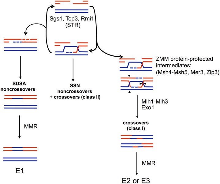
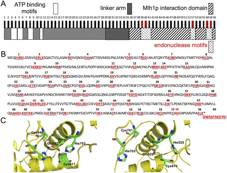
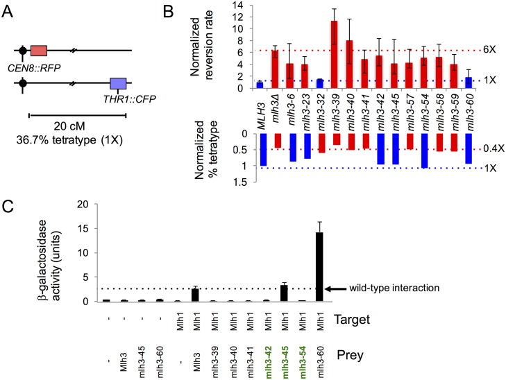
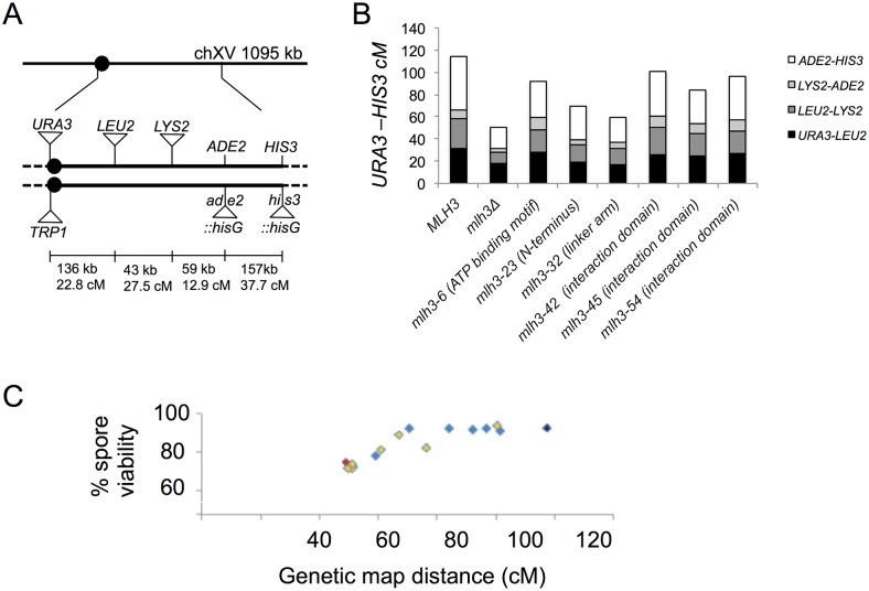
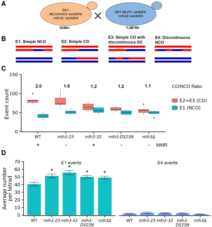
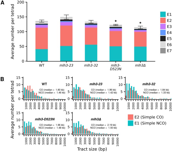
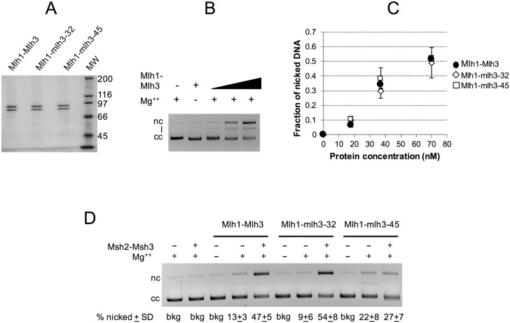
Mlh1-Mlh3 is an endonuclease hypothesized to act in meiosis to resolve double Holliday junctions into crossovers. It also plays a minor role in eukaryotic DNA mismatch repair (MMR). To understand how Mlh1-Mlh3 functions in both meiosis and MMR, we analyzed in baker's yeast 60 new mlh3 alleles. Five alleles specifically disrupted MMR, whereas one (mlh3-32) specifically disrupted meiotic crossing over. Mlh1-mlh3 representatives for each class were purified and characterized. Both Mlh1-mlh3-32 (MMR+, crossover-) and Mlh1-mlh3-45 (MMR-, crossover+) displayed wild-type endonuclease activities in vitro. Msh2-Msh3, an MSH complex that acts with Mlh1-Mlh3 in MMR, stimulated the endonuclease activity of Mlh1-mlh3-32 but not Mlh1-mlh3-45, suggesting that Mlh1-mlh3-45 is defective in MSH interactions. Whole genome recombination maps were constructed for wild-type and MMR+ crossover-, MMR- crossover+, endonuclease defective and null mlh3 mutants in an S288c/YJM789 hybrid background. Compared to wild-type, all of the mlh3 mutants showed increases in the number of noncrossover events, consistent with recombination intermediates being resolved through alternative recombination pathways. Our observations provide a structure-function map for Mlh3 that reveals the importance of protein-protein interactions in regulating Mlh1-Mlh3's enzymatic activity. They also illustrate how defective meiotic components can alter the fate of meiotic recombination intermediates, providing new insights for how meiotic recombination pathways are regulated.


Conflict of interest statement

The authors have declared that no competing interests exist.

Figures

Fig 1
Fig 1. DSB repair pathways in meiosis.
Model adapted from Kaur et al. [30] depicting wild-type meiosis and the central role of the STR complex (Sgs1-Top3-Rmi1 helicase/topoisomerase) in disassembling strand invasion intermediates to facilitate synthesis dependent strand annealing (SDSA) or return of events to the original DSB state to allow capture and protection by the ZMM proteins and dHJ formation for ultimate resolution as class I crossovers by Mlh1-Mlh3 and Exo1. Events that escape STR disassembly form unregulated joint molecules that are resolved by the structure selective nucleases (SSNs-Mus81-Mms4, Yen1, Slx-Slx4) as noncrossovers or class II crossovers. The “E” classification of recombination classes was described in Oke et al. [34]. The majority event classes are presented here and result from MMR of heteroduplex DNA intermediates. E1 events are simple noncrossovers (NCO), E2 are simple crossovers (CO) with or without continuous gene conversion, and E3 are COs with discontinuous gene conversion. A set of definitions for these classes can also be found in Fig 5.
Fig 2
Fig 2. Site directed mutagenesis of MLH3.
A. Functional organization of Mlh3 based on sequence homology and secondary structure prediction [51]. The vertical bars indicate the approximate position of the mlh3 mutations (except mlh3-60) analyzed in this study and described in panel B. mlh3-39, -40, -57, -58, and -59 colored in red are based on highly conserved residues in the endonuclease motifs of Pms1 which were shown in the crystal structure of Mlh1-Pms1 to form a single metal binding site [51] described in panel C. B. Amino acid positions of charged-to-alanine substitutions presented in red on the primary sequence of Saccharomyces cerevisiae Mlh3. Each cluster of underlined residues represents one allele corresponding to the vertical bars in panel A. mlh3-39, -40, -57, -58, and -59 are colored in red as in panel A. mlh3-60 represents the last 11 residues of Pms1 which constitute patch II of the heterodimerization interface of Mlh1-Pms1 [51]. C. Metal binding site of Pms1 (left panel) from [51] comprised of the five highlighted residues (H703, E707, C817, C848, and H850) were found to be highly conserved in Mlh3 (right panel) based on sequence alignment and structural modeling (H525, E529, C670, C701, and H703) and were targeted in the mutagenesis described in this study (alleles represented in red in A and B).
Fig 3
Fig 3. Identification and characterization of mlh3 separation of function alleles.
A. Spore-autonomous fluorescent protein expression was used to quantify crossing over [56]. Shown is the starting parental configuration on chromosome VIII with a map distance of 20 cM separating the red fluorescent protein (RFP) marker and the blue fluorescent protein (CFP) marker. Percent tetratype at this interval in wild-type meiosis is 36.7%. B. MMR (top) and CO (bottom) phenotypes for MLH3 and mlh3 null (mlh3Δ), separation of function, endonuclease, and C-terminal tail (mlh3-60) mutants. Mismatch repair was measured using the lys2-A14 reversion assay [55] and crossing over was measured using the assay depicted in panel A. Bars represent the median reversion rates (error bars based on 95% confidence intervals) and percent tetratype normalized to MLH3 (1X). For mismatch repair (top), bars represent reversion rates of at least 10 independently tested cultures from two independently constructed strains presented here normalized to MLH3 median rate of 1X = 1.43x10⁻⁶ (n = 140). For crossing over (bottom), bars represent percent tetratype of at least 250 tetrads from two independently constructed strains presented here normalized to MLH3 percent tetratype 1X = 36.7% (n = 226; Table 1 and S2 File). Blue and red dotted lines represent MLH3 and mlh3Δ respectively. C. mlh3-42, -54 weaken Mlh1 interaction yet maintain crossover function. Yeast two-hybrid interactions between lexA-Mlh1 (target) and Gal4-Mlh3 (amino acids 481–715; prey) or Gal4-mlh3-39, -40, -41, -42, -45, -54, -60 derivative constructs, as measured in the ONPG assay for β-galactosidase activity. Error bars indicate standard error of mean from at least three independent assays (S2 File). mlh3 separation of function alleles indicated in green font.
Fig 4
Fig 4. Cumulative genetic distance and spore viability of mlh3 separation of function mutants.
A. Distribution of genetic markers on chromosome XV used to determine genetic distances in the EAY1112/EAY2413 background (S1 Table). The solid circle indicates the centromere. The distances between markers are not drawn to scale. The actual physical and genetic distances in the wild-type diploid are given numerically for each interval and for the entire region between CENXV and HIS3 [7]. B. Cumulative genetic distances between URA3 and HIS3 markers from tetrads of MLH3 and indicated mlh3 variants. Each bar is further divided into sectors that correspond to the four genetic intervals that span URA3-HIS3 (S4 Table). C. Spore viabilities are plotted vs. genetic map distances from panel B for MLH3 (dark blue), mlh3Δ (red), and the separation of function mutants (light blue). Yellow diamonds represent data from Sonntag Brown et al. [6] (S2 File).
Fig 5
Fig 5. Genome-wide increase in simple noncrossover events (E1) compared to wild-type in mlh3-23, mlh3-32, mlh3-D523N and mlh3Δ mutants.
A. Generation of S288c/YJM789 isogenic strains with SK1 MLH1, MLH3 and the mlh3-23, mlh3-32, mlh3-D523N and mlh3Δ mutant alleles (Methods). B. Cartoon description of simple NCO (E1; 3:1 tract on one chromatid, not within 5 kb of another CO or NCO), simple CO (E2; CO with or without an associated gene conversion (GC) tract, and not within 5 kb of another CO or NCO), simple CO with discontinuous gene conversion tracts (E3; same definition as for E2, except with one or more gene conversions within 5 kb and on one of the same chromatids as the CO chromatid), and discontinuous NCOs (E4; two or more NCOs consecutively on one chromatid, with 2:2 marker segregation separating them) as presented in Oke et al. [34]. C. Crossover (CO, E2+E3) and noncrossover (NCO, E1) counts per meiosis for wild-type, mlh3-23, mlh3-32, mlh3-D523N, and mlh3Δ. The minimum, first quantile, median, third quantile and maximum count are indicated in the box plot. The ratio of CO to NCO events is presented above the box plots. The proficiency of the mlh3 alleles in mismatch repair is shown as +, MMR proficient, or –, MMR deficient. D. Average number of simple NCO (E1) and NCO with discontinuous tract (E4) events per tetrad (+/- standard error). * p ≤ 0.05 compared to wild-type (Table 3 and S2 File).
Fig 6
Fig 6. Total interhomolog events and distribution of gene conversion tract lengths associated with NCO and CO events in wild-type, mlh3-23, mlh3-32, mlh3-D523N and mlh3Δ mutants.
A. Total average inter-homolog events (IH; S9 Table), and each event type (E1-E7) as a fraction of the total, in MLH3, mlh3-23, mlh3-32, mlh3-D523N, and mlh3Δ mutants. * p ≤ 0.05 compared to wild-type (S2 File). B. Average number of tracts ordered by size for simple CO (E2) and simple NCO (E1) events. Median CO and NCO tract sizes are also presented, and events were assigned as described by Oke et al. [34] (S2 File). The Y axis represents pooled data from all tetrads, normalized by dividing the number of tetrads.
Fig 7
Fig 7. Mlh1-mlh3-32 and Mlh1-mlh3-45 display wild-type endonuclease activities that are differentially stimulated by Msh2-Msh3.
A. SDS-PAGE analysis of purified Mlh1-Mlh3, Mlh1-mlh3-32 and Mlh1-mlh3-45. Coomassie Blue R250-stained 8% Tris-glycine gel. 0.5 μg of each protein is shown. MW = Molecular Weight Standards from top to bottom- 200, 116, 97, 66, 45 kD). B, C. Mlh1-Mlh3, Mlh1-mlh3-32 and Mlh1-mlh3-45 (18, 37, 70 nM) were incubated with 2.2 nM supercoiled pBR322 DNA, and analyzed in agarose gel electrophoresis (C) and the endonuclease activity was quantified (average of 6 independent experiments presented +/-SD) as described in the Methods (S2 File). Ladder: 1 kb DNA ladder (New England BioLabs). Migration of closed circular (cc), nicked (nc) and linear (l) pBR322 DNA is indicated. D. Endonuclease assays were performed as in B., but contained 20 nM of the indicated wild-type or mutant Mlh1-Mlh3 complex and 40 nM Msh2-Msh3 when indicated. Reactions were performed in triplicate, samples were resolved on agarose gels, and the fraction of nicked DNA was quantified, averaged, and the standard deviation between experiments was calculated. The average fraction of supercoiled substrate cleaved is presented +/-S.D. below the gel. (bkg) background, (cc) closed circular DNA, (nc) nicked DNA.
Fig 8
Fig 8. Sgs1 but not sgs1-hd overexpression differentially affects spore viability in mlh3Δ vs. mlh3-32.
Distribution of viable spores in tetrads of MLH3, mlh3Δ, mlh3-23, mlh3-32, and mlh3-D523N strains containing no insert (), pSGS1-2μ, or psgs1-hd-2μ (helicase defective mutant). In all plots, the horizontal axis corresponds to the classes of tetrads with 4, 3, 2, 1 and 0 viable spores, and the vertical axis corresponds to the frequency of each class given in percentage. The overall spore viability (SV) and the total number of spores counted (n) are shown (S2 File). *p<0.05 (χ2 test), comparing spore viability for the indicated strain transformed with a 2μ SGS1 plasmid to that of the same strain transformed with a no insert plasmid.

References

    1. Kunkel TA, Erie DA (2015) Eukaryotic Mismatch Repair in Relation to DNA Replication. Annu Rev Genet 49: 291–313. doi: 10.1146/annurev-genet-112414-054722 - DOI - PMC - PubMed
    1. Flores-Rozas H, Kolodner RD (1998) The Saccharomyces cerevisiae MLH3 gene functions in MSH3-dependent suppression of frameshift mutations. Proc Natl Acad Sci USA 95: 12404–12409. - PMC - PubMed
    1. Harfe BD, Minesinger BK, Jinks-Robertson S (2000) Discrete in vivo roles for the MutL homologs Mlh2p and Mlh3p in the removal of frameshift intermediates in budding yeast. Curr Biol 10: 145–148. - PubMed
    1. Romanova NV, Crouse GF (2013) Different roles of eukaryotic MutS and MutL complexes in repair of small insertion and deletion loops in yeast. PLoS Genet 9: e1003920 doi: 10.1371/journal.pgen.1003920 - DOI - PMC - PubMed
    1. Nishant KT, Plys AJ, Alani E (2008) A mutation in the putative MLH3 endonuclease domain confers a defect in both mismatch repair and meiosis in Saccharomyces cerevisiae. Genetics 179: 747–755. doi: 10.1534/genetics.108.086645 - DOI - PMC - PubMed

MeSH terms