Case Reports
. 2017 Jun 10;12(3):577-584.
doi: 10.1016/j.radcr.2017.04.015. eCollection 2017 Sep.

Persistent sciatic artery found incidentally on hip MRI: report of 4 cases

Affiliations
Case Reports

Persistent sciatic artery found incidentally on hip MRI: report of 4 cases

Ângela Massignan et al. Radiol Case Rep. .

Abstract

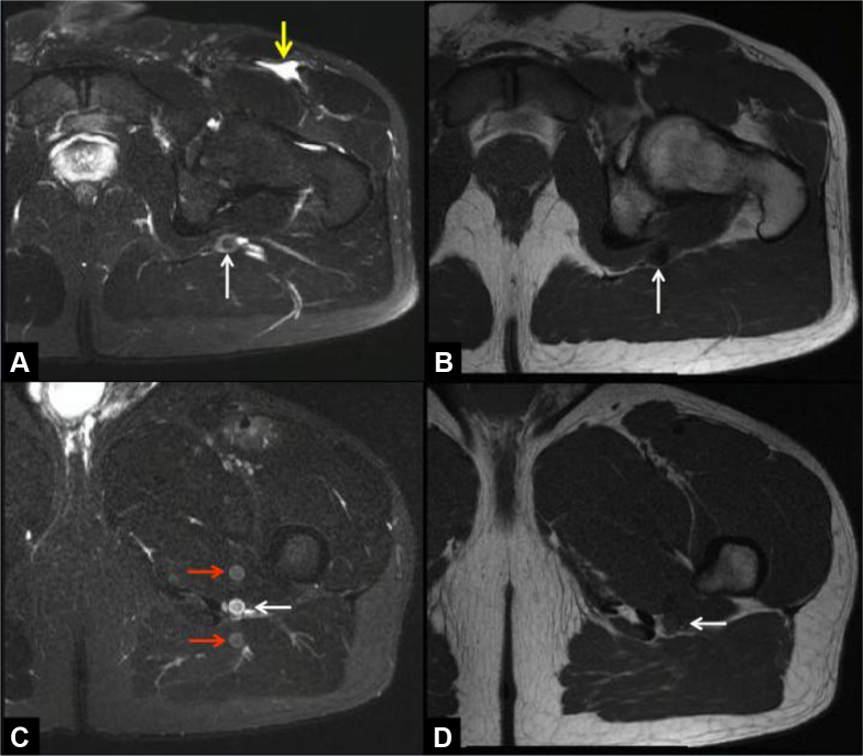
The persistent sciatic artery is a rare anatomical variant, representing the persistence of the sciatic artery in adult life that is responsible for the major blood supply to the lower limb in early embryologic development. Such persistence may be bilateral and can remain asymptomatic for many years. However, aneurysmal degeneration has been described as a complication of the persistent sciatic artery, which may cause critical limb ischemia resulting from thrombosis or embolization of aneurysm thrombus. Digital subtraction angiography, Doppler ultrasound, computed tomography angiography and magnetic resonance angiography are the most frequently used diagnostic tools to detect, classify and determine the presence of complications of a PSA. Early detection of this vascular abnormality on imaging studies can avoid life-threatening complications. We describe 4 patients with PSA that were diagnosed as an incidental finding in magnetic resonance imaging of the hip and demonstrate its characteristic imaging appearance.

Keywords: Computed tomography angiography; Congenital vascular anomaly; Magnetic resonance imaging; Persistent sciatic artery; Vascular anatomical variant.


Figures

Fig. 1
Fig. 1
Case 1. A 31-year-old female with persistent sciatic artery on the right side. (A) and (B) Axial T2-weighted fat saturated MR images demonstrating a persistent sciatic artery at different levels (white arrows). (C) and (D) Coronal T1-weighted (C), coronal T2-weighted fat saturated (D) MR images showing a caliber tubular structure with flow void (white arrows) following the course of the sciatic nerve.
Fig. 2
Fig. 2
Case 1. A 31-year-old female with persistent sciatic artery on the right side. (A) Coronal computed tomography angiography–maximum intensity projections reconstruction demonstrating significant reduction in caliber of the superficial femoral artery, which does not reach the popliteal artery. (B) Coronal CTA volumetric reconstruction demonstrating the PSA originating from the internal iliac artery and continuing as the popliteal artery (arrow). (C) and (D) Coronal CTA volumetric reconstruction with bone structures (posterior and posterior-oblique view) demonstrating the relation of the PSA with the pelvis and the proximal femur and crossing the greater sciatic foramen. This case would be classified as type IIA according to Pillet et al. CTA, computed tomography angiography; PSA, persistent sciatic artery.
Fig. 3
Fig. 3
Case 2. A 28-year-old male. (A) and (B) Axial T2-weighted fat saturated (A), axial T1-weighted (B) MR images at the level of femoral neck demonstrating the PSA (white arrows). (A) Partial tear in the proximal myotendinous junction of the rectus femoris muscle and small hematoma in the intermuscular plane (yellow arrow). (C) and (D) Axial T2-weighted fat saturated (C), axial T1-weighted (D) MR images at the level of the lesser femoral trochanter demonstrating the PSA (white arrow) and pulsation imaging artifacts in the phase encoded direction (red arrows).
Fig. 4
Fig. 4
Case 3. A 40-year-old female with persistent sciatic artery on the right side. (A) and (B) Sagittal and coronal T2-weighted fat saturated MR images demonstrating small partial tear of the proximal iliotibial band surrounded by soft tissue edema (white arrows). (C) and (D) Consecutive pelvic axial T1-weighted fat saturated MR images with gadolinium at the level of the greater femoral trochanter demonstrating the PSA on the right side (red arrow) and normal neurovascular bundle sciatic nerve on the left side (yellow arrow). The common femoral artery (white arrows) and the superficial femoral artery (green arrows) were hypoplastic on the right side.
Fig. 5
Fig. 5
Case 4. A 36-year-old female with persistent sciatic artery on the right side. (A-C) Axial T1-weighted MR image (A), axial T2-weighted fat saturated MR image (B), and axial computed tomography angiography (C) demonstrating the PSA (white arrows) and the hypoplastic superficial femoral artery (yellow arrow).
Fig. 6
Fig. 6
Case 4. A 36-year-old female with persistent sciatic artery on the right side. Coronal computed tomography angiography volumetric reconstruction demonstrating the sciatic artery originating from the right internal iliac artery.
Fig. 7
Fig. 7
Diagram of embryonic and adult vasculature of the lower limbs. The vessels painted in yellow represent the embryonic vasculature that regresses with development of the embryo (sciatic or axial artery). The vessels painted in green constitute the embryonic vasculature that normally persists and form part of the adult vasculature (popliteal artery). The vessels painted in red represent the adult vasculature.

References

    1. Paraskevas G., Papaziogas B., Gigis J., Mylonas A., Gigis P. The persistence of the sciatic artery. Folia Morphol. 2004;63(4):515–518. - PubMed
    1. Van Hooft I.M., Zeebregts C.J., van Sterkenburg S.M., de Vries W.R., Reijnen M.M. The persistent sciatic artery. Eur J Vasc Endovasc Surg. 2009;37(5):585–591. - PubMed
    1. Brantley S.K., Rigdon E.E., Raju S. Persistent sciatic artery: embryology, pathology, and treatment. J Vasc Surg. 1993;18(2):242–248. - PubMed
    1. Santaolalla V., Bernabe M.H., Hipola Ulecia J.M., De Loyola Agundez Gomez I., Hoyos Y.G., Otero F.J.M. Persistent sciatic artery. Ann Vasc Surg. 2010;24(5):691.e7–691.e10. - PubMed
    1. Maldini G., Teruya T.H., Kamida C., Eklof B. Combined percutaneous endovascular and open surgical approach in the treatment of a persistent sciatic artery aneurysm presenting with acute limb-threatening ischemia–a case report and review of the literature. Vasc Endovascular Surg. 2002;36(5):403–408. - PubMed

Publication types