

Tendon injuries: Basic science and new repair proposals
- PMID: 28828182
- PMCID: PMC5549180
- DOI: 10.1302/2058-5241.2.160075
Tendon injuries: Basic science and new repair proposals
Abstract
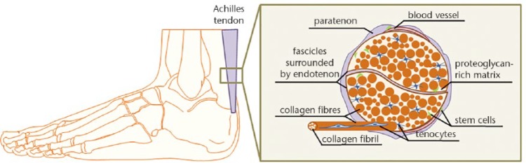
Tendons connect muscles to bones, ensuring joint movement. With advanced age, tendons become more prone to degeneration followed by injuries. Tendon repair often requires lengthy periods of rehabilitation, especially in elderly patients. Existing medical and surgical treatments often fail to regain full tendon function.The development of novel treatment methods has been hampered due to limited understanding of basic tendon biology. Recently, it was discovered that tendons, similar to other mesenchymal tissues, contain tendon stem/progenitor cells (TSPCs) which possess the common stem cell properties.The current strategies for enhancing tendon repair consist mainly of applying stem cells, growth factors, natural and artificial biomaterials alone or in combination. In this review, we summarise the basic biology of tendon tissues and provide an update on the latest repair proposals for tendon tears. Cite this article: EFORT Open Rev 2017;2:332-342. DOI: 10.1302/2058-5241.2.160075.
Keywords: biomaterials; cell-based therapy; growth factors; mesenchymal stem cells; tendon repair; tendon stem/progenitor cells.
Conflict of interest statement
ICMJE Conflict of interest statement: None declared.
Figures
References
-
- Sharma P, Maffulli N. Tendinopathy and tendon injury: the future. Disabil Rehabil 2008;30:1733-1745. - PubMed
-
- Jósza L, Lehto MU, Järvinen M, et al. A comparative study of methods for demonstration and quantification of capillaries in skeletal muscle. Acta Histochemica 1993;94:89-96. - PubMed
-
- Magnusson SP, Kjaer M. Region-specific differences in Achilles tendon cross-sectional area in runners and non-runners. Eur J Appl Physiol 2003;90:549-553. - PubMed
-
- James R, Kesturu G, Balian G, Chhabra AB. Tendon: biology, biomechanics, repair, growth factors, and evolving treatment options. J Hand Surg Am 2008;33:102-112. - PubMed
Publication types
LinkOut - more resources
Full Text Sources
Other Literature Sources
