

Cortical thickness and subcortical structure volume abnormalities in patients with major depression with and without anxious symptoms
- PMID: 28828215
- PMCID: PMC5561315
- DOI: 10.1002/brb3.754
Cortical thickness and subcortical structure volume abnormalities in patients with major depression with and without anxious symptoms
Abstract
Background: Anxious depression is one of the common subtypes of major depressive disorder (MDD). Clinically, patients with anxious depression exhibit more severe depressive symptoms than patients with nonanxious depression. The aim of the present study was to explore the common and differing cortical and subcortical structural changes between patients with anxious and nonanxious depression.
Methods: Patients were placed into one of three groups: the anxious depression group (MDD patients with high levels of anxiety symptoms, n = 23), the nonanxious depression group (n = 22), and healthy controls (n = 43) that were matched for age, sex, and education level. All participants underwent T1-weighted MRI. The Freesurfer, which uses a set of automated sequences to analyze the abnormal changes of cortical thickness, cortical and subcortical structures, was used to process the T1 images.
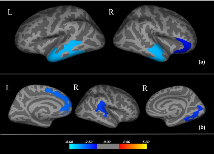
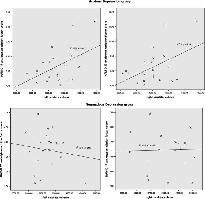
Results: Compared to controls, MDD patients showed thinner cortical thickness in the left inferior temporal, the right superior temporal, and the right parsorbitalis, and a smaller volume of the left hippocampus. Compared to nonanxious depression, anxious depressive patients showed a cortical thinning of the left superior frontal and right superior temporal, as well as the right lingual, and significantly increased subcortical volume of the bilateral caudate nuclei. Correlation analysis showed that the volumes of the bilateral caudate nuclei were directly proportional to the anxiety/somatization factor score.
Conclusions: These findings suggest that smaller hippocampal volume and atrophic prefrontal and temporal cortices might be a common pattern of cortical and subcortical alterations in patients with depression and/or anxiety. However, the change in the caudate nucleus volume may be indicative of anxious depression and may potentially be used to distinguish anxious from nonanxious depression.
Keywords: anxiety; biological markers; brain imaging; depression; mood disorders.
Figures
Similar articles
-
Gray Matter Abnormalities in Non-comorbid Medication-naive Patients with Major Depressive Disorder or Social Anxiety Disorder.EBioMedicine. 2017 Jul;21:228-235. doi: 10.1016/j.ebiom.2017.06.013. Epub 2017 Jun 15. EBioMedicine. 2017. PMID: 28633986 Free PMC article.
-
Altered patterns of association between cortical thickness and subcortical volume in patients with first episode major depressive disorder: A structural MRI study.Psychiatry Res Neuroimaging. 2017 Feb 28;260:16-22. doi: 10.1016/j.pscychresns.2016.12.001. Epub 2016 Dec 1. Psychiatry Res Neuroimaging. 2017. PMID: 28012422
-
Cortical thickness, cortical and subcortical volume, and white matter integrity in patients with their first episode of major depression.J Affect Disord. 2014 Feb;155:42-8. doi: 10.1016/j.jad.2013.10.021. Epub 2013 Oct 21. J Affect Disord. 2014. PMID: 24210630
-
The impact of cortical and subcortical volumes on major depression risk: A genetic study.J Affect Disord. 2025 Apr 1;374:356-364. doi: 10.1016/j.jad.2025.01.069. Epub 2025 Jan 15. J Affect Disord. 2025. PMID: 39824313
-
Neurobiology of anxious depression: a review.Depress Anxiety. 2013 Apr;30(4):374-85. doi: 10.1002/da.22095. Epub 2013 Mar 11. Depress Anxiety. 2013. PMID: 23495126 Free PMC article. Review.
Cited by
-
Anxious depression as a clinically relevant subtype of pediatric major depressive disorder.J Neural Transm (Vienna). 2019 Sep;126(9):1217-1230. doi: 10.1007/s00702-019-02069-x. Epub 2019 Aug 27. J Neural Transm (Vienna). 2019. PMID: 31456039 Clinical Trial.
-
Identifying tripartite relationship among cortical thickness, neuroticism, and mood and anxiety disorders.Sci Rep. 2024 Apr 11;14(1):8449. doi: 10.1038/s41598-024-59108-1. Sci Rep. 2024. PMID: 38600283 Free PMC article.
-
A translational exploration of the effects of WNT2 variants on altered cortical structures in autism spectrum disorder.J Psychiatry Neurosci. 2021 Dec 3;46(6):E647-E658. doi: 10.1503/jpn.210022. Print 2021 Nov-Dec. J Psychiatry Neurosci. 2021. PMID: 34862305 Free PMC article.
-
Hypoechogenicity of brainstem raphe correlates with depression in migraine patients.J Headache Pain. 2019 May 15;20(1):53. doi: 10.1186/s10194-019-1011-2. J Headache Pain. 2019. PMID: 31092190 Free PMC article.
-
Identification of structural brain alterations in adolescents with depressive symptomatology.Brain Res Bull. 2023 Sep;201:110723. doi: 10.1016/j.brainresbull.2023.110723. Epub 2023 Aug 1. Brain Res Bull. 2023. PMID: 37536609 Free PMC article.
References
-
- Bell‐McGinty, S. , Butters, M. A. , Meltzer, C. C. , Greer, P. J. , Reynolds, C. F. 3rd. , & Becker, J. T . (2002). Brain morphometric abnormalities in geriatric depression: Long‐term neurobiological effects of illness duration. American Journal of Psychiatry, 159(8), 1424–1427. - PubMed
-
- Bracht, T. , Horn, H. , Strik, W. , Federspiel, A. , Schnell, S. , Hofle, O. , … Walther, S . (2014). White matter microstructure alterations of the medial forebrain bundle in melancholic depression. Journal of Affective Disorders, 155, 186–193. - PubMed
-
- Brzezicka, A. (2013). Integrative deficits in depression and in negative mood states as a result of fronto‐parietal network dysfunctions. Acta Neurobiologiae Experimentalis (Wars), 73(3), 313–325. - PubMed
-
- Buckner, R. L. , Head, D. , Parker, J. , Fotenos, A. F. , Marcus, D. , Morris, J. C. , & Snyder, A. Z. (2004). A unified approach for morphometric and functional data analysis in young, old, and demented adults using automated atlas‐based head size normalization: Reliability and validation against manual measurement of total intracranial volume. NeuroImage, 23(2), 724–738. - PubMed
MeSH terms
Grants and funding
LinkOut - more resources
Full Text Sources
Other Literature Sources
Medical
