

Management of Penetrating Skull Base Injury: A Single Institutional Experience and Review of the Literature
- PMID: 28828384
- PMCID: PMC5554568
- DOI: 10.1155/2017/2838167
Management of Penetrating Skull Base Injury: A Single Institutional Experience and Review of the Literature
Abstract
Background: Penetrating skull base injury (PSBI) is uncommon among head injuries, presenting unique diagnostic and therapeutic challenges. Although many cases of PSBIs have been reported, comprehensive understanding of its initial diagnosis, management, and outcome is still unavailable.
Materials and methods: A retrospective review was performed for patients treated in neurosurgical department of Changzheng Hospital for PSBIs. Presurgical three-dimensional (3D) Slicer-assisted reconstructions were conducted for each patient. Then we reviewed previous literature about all the published cases of PSBIs worldwide and discussed their common features.
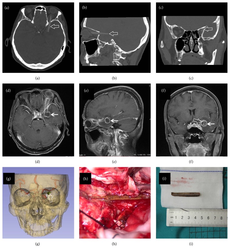
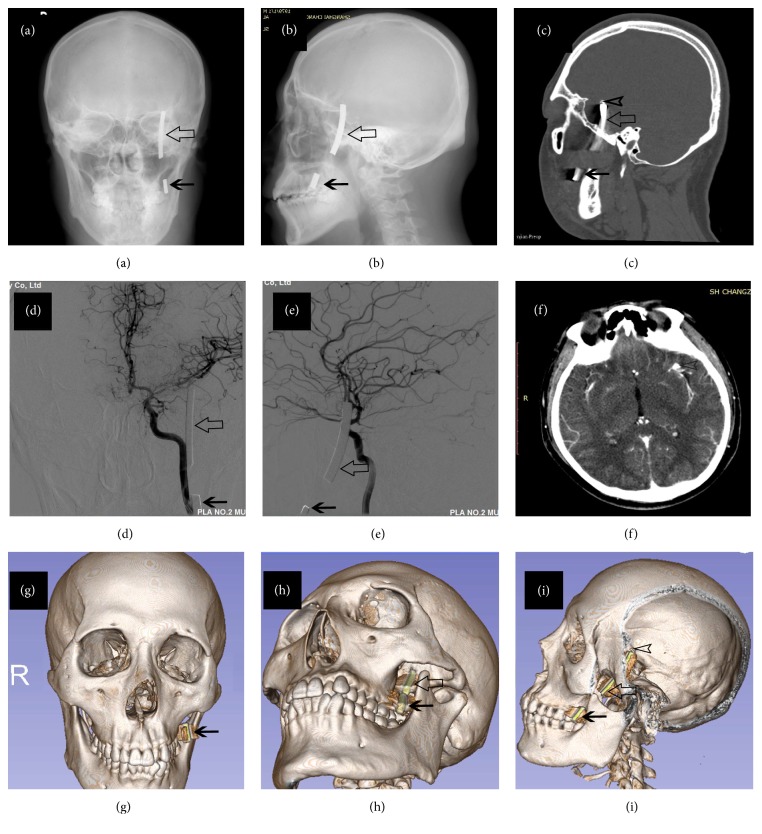
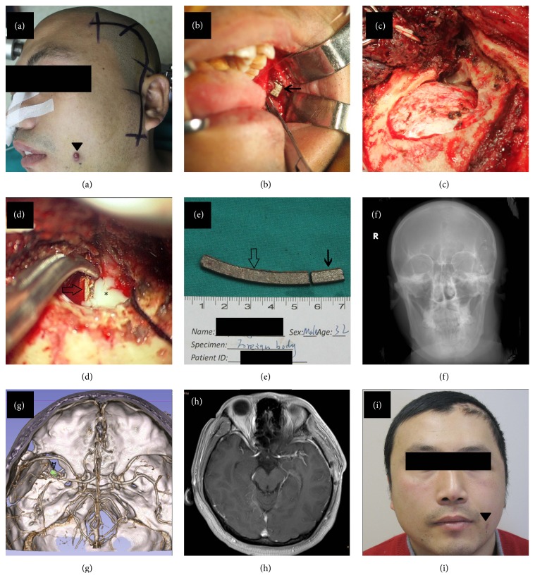
Results: A total of 5 patients suffering PSBIs were identified. Penetrating points as well as the surrounding neurovascular structures were clearly visualized, assisting in the presurgical planning of optimal surgical approach and avoiding unexpected vascular injury. Four patients underwent craniotomy with foreign bodies removed successfully and 1 patient received conservative treatment. All of them presented good outcomes after proper management.
Conclusion: Careful physical examination and radiological evaluation are essential before operation, and angiography is recommended for those with suspected vascular injuries. 3D modeling with 3D Slicer is practicable and reliable, facilitating the diagnosis and presurgical planning. Treatment decision should be made upon the comprehensive evaluation of patient's clinicoradiological features and characteristics of foreign bodies.
Figures
Similar articles
-
Computed Tomography Angiography is the Definitive Vascular Imaging Modality for Penetrating Neck Injury: A South African Experience.Scand J Surg. 2018 Mar;107(1):23-30. doi: 10.1177/1457496917731187. Epub 2017 Sep 27. Scand J Surg. 2018. PMID: 28950788
-
Transoral penetrating craniocerebral injury by a bamboo chopstick in a child.J Clin Neurosci. 2013 May;20(5):746-8. doi: 10.1016/j.jocn.2012.03.053. Epub 2013 Feb 26. J Clin Neurosci. 2013. PMID: 23453158
-
[Penetrating head injury from harpoon. Case report].Neurocirugia (Astur). 2002 Oct;13(5):397-400. doi: 10.1016/s1130-1473(02)70596-4. Neurocirugia (Astur). 2002. PMID: 12444413 Spanish.
-
A systematic review of penetrating extracranial vertebral artery injuries.J Vasc Surg. 2020 Jun;71(6):2161-2169. doi: 10.1016/j.jvs.2019.10.084. Epub 2020 Jan 2. J Vasc Surg. 2020. PMID: 31902594
-
Industrial nail gun injury to the anterior skull base: a case report and review of the literature.J Trauma. 2008 Mar;64(3):E29-32. doi: 10.1097/01.ta.0000197145.19876.38. J Trauma. 2008. PMID: 18332791 Review. No abstract available.
Cited by
-
Intracranial penetrating injury by clothes fork in an infant: case report and review of the literature.Childs Nerv Syst. 2023 Jan;39(1):47-55. doi: 10.1007/s00381-022-05706-1. Epub 2022 Oct 22. Childs Nerv Syst. 2023. PMID: 36273084 Review.
-
Complex orbitocranial penetrating injury-managed through neuroplastic surgery - a rare case report and review of literature.Ann Med Surg (Lond). 2024 Dec 19;87(2):924-928. doi: 10.1097/MS9.0000000000002963. eCollection 2025 Feb. Ann Med Surg (Lond). 2024. PMID: 40110324 Free PMC article.
-
Transorbital nonmissile penetrating brain injury: Report of two cases.World J Clin Cases. 2020 Jan 26;8(2):471-478. doi: 10.12998/wjcc.v8.i2.471. World J Clin Cases. 2020. PMID: 32047800 Free PMC article.
-
Transorbital-penetrating intracranial injury due to a homemade metal arrow: A case report.Ann Med Surg (Lond). 2020 Jul 28;57:183-189. doi: 10.1016/j.amsu.2020.07.049. eCollection 2020 Sep. Ann Med Surg (Lond). 2020. PMID: 32774851 Free PMC article.
-
Clinical application of 3D Slicer combined with Sina/MosoCam multimodal system in preoperative planning of brain lesions surgery.Sci Rep. 2022 Nov 10;12(1):19258. doi: 10.1038/s41598-022-22549-7. Sci Rep. 2022. PMID: 36357434 Free PMC article.
References
-
- Gennarelli T. A., Champion H. R., Sacco W. J., Copes W. S., Alves W. M. Mortality of patients with head injury and extracranial injury treated in trauma centers. Journal of Trauma. 1989;29(9):1193–1201. - PubMed
-
- Maruya J., et al. Brain abscess following transorbital penetrating injury due to bamboo fragments—case report. Neurol Med Chir (Tokyo) 2002;42(3):143–146. - PubMed
Publication types
MeSH terms
LinkOut - more resources
Full Text Sources
Other Literature Sources
Medical
