. 2017 Jul 24:6:171-182.
doi: 10.1016/j.omtm.2017.07.003. eCollection 2017 Sep 15.

Thermal Stability as a Determinant of AAV Serotype Identity

Affiliations

Thermal Stability as a Determinant of AAV Serotype Identity

Antonette Bennett et al. Mol Ther Methods Clin Dev. .

Abstract

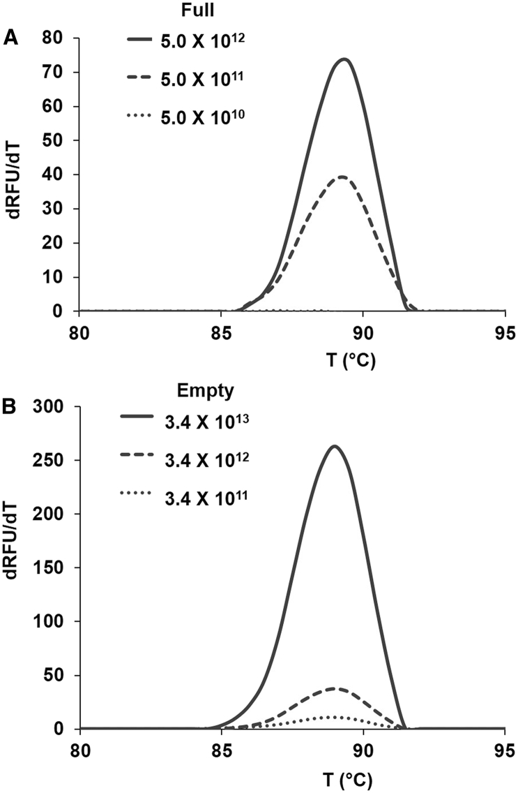
Currently, there are over 150 ongoing clinical trials utilizing adeno-associated viruses (AAVs) to target various genetic diseases, including hemophilia (AAV2 and AAV8), congenital heart failure (AAV1 and AAV6), cystic fibrosis (AAV2), rheumatoid arthritis (AAV2), and Batten disease (AAVrh.10). Prior to patient administration, AAV vectors must have their serotype, concentration, purity, and stability confirmed. Here, we report the application of differential scanning fluorimetry (DSF) as a good manufacturing practice (GMP) capable of determining the melting temperature (Tm) for AAV serotype identification. This is a simple, rapid, cost effective, and robust method utilizing small amounts of purified AAV capsids (∼25 μL of ∼1011 particles). AAV1-9 and AAVrh.10 exhibit specific Tms in buffer formulations commonly used in clinical trials. Notably, AAV2 and AAV3, which are the least stable, have varied Tms, whereas AAV5, the most stable, has a narrow Tm range in the different buffers, respectively. Vector stability was dictated by VP3 only, specifically, the ratio of basic/acidic amino acids, and was independent of VP1 and VP2 content or the genome packaged. Furthermore, stability of recombinant AAVs differing by a single basic or acidic amino acid residue are distinguishable. Hence, AAV DSF profiles can serve as a robust method for serotype identification of clinical vectors.

Keywords: AAV; AAV buffer formulations; AAV capsid stability; AAV serotype identification; AAV vector buffers; adeno-associated virus; differential scanning fluorimetry; parvovirus stability; viral vectors.


Figures

Figure 1
Figure 1
Sample Evaluation and Stability of Full rAAV1–rAAV9-gfp and rAAVrh.10-gfp, Packaging the GFP Transgene, Vectors in PBS (A) Coomassie blue stained SDS-PAGE (left) and negative-stained EM (right) of rAAV1–rAAV9 and rAAVrh.10 as indicated above each panel. Scale bar, 100 nm, is shown in the AAVrh.10 EM image. (B) Thermal profile (shown as normalized relative fluorescence units [RFUs]) versus temperature (T [°C]) of rAAV1–rAAV9 and rAAVrh.10 obtained by DSF analysis. A representative profile is shown for each serotype. See also Table 1. Each profile is colored according to the serotype, as shown on the right-hand side.
Figure 2
Figure 2
Comparative Thermal Profiles of Full AAV1–AAV9-gfp and AAVrh.10-gfp Capsids in Commonly Used AAV Formulation and Storage Buffers and UB (A–J) Comparison of (A) AAV1, (B) AAV2, (C) AAV3, (D) AAV4, (E) AAV5, (F) AAV6, (G) AAV7, (H) AAV8, (I) AAV9, and (J) AAVrh.10 thermal profiles (shown as in Figure 1) in different buffers. Each buffer profile is colored according to the series legend in (K). (K) Discrete dots representing the distribution of Tms (°C) for each AAV serotype in the different buffers, as indicated to the right-hand side.
Figure 3
Figure 3
Comparative Analysis of the Tm of Full, Packaging GFP Transgene, and Empty Capsids in the Different AAV Formulation and Storage Buffers Plots show Tm (°C) (y axis) versus buffer (x axis) for two selected rAAV serotypes from the buffer stability groupings. Group I was AAVs with Tms varying by ∼15°C–20°C, AAV2 and AAV3 (top); group II was AAVs with Tms varying by ∼3°C–6°C, AAV8 and AAVrh.10 (middle); and group III was AAVs with Tms varying by <2.0°C, AAV5 and AAV9. The Tm of the full rAAV capsids is shown in a black line, and the empty capsids are shown in a gray line.
Figure 4
Figure 4
Concentration Survey of AAV5 (A) Full rAAV5-luc capsids at 5.0 × 1010–5.0 × 1012 vg/mL. (B) Empty rAAV5 capsids at 3.4 × 1011–3.4 × 1013 particles/mL. The inverted measured rate of change of fluorescence with time, dRFU/dT, is shown plotted in the y axis against T (°C) on the x axis.
Figure 5
Figure 5
Sample Evaluation and Stability of WT and Mutant AAV2 VLPs (A) Negative-stained EM (top) and silver-stained SDS-PAGE of AAV2-VP123 (WT), AAV2-VP13, AAV2-VP23, and AAV2-VP3 VLPs (bottom). Top: white scale bar, 50 nm. Bottom: the position of VP1, VP2, and VP3 is indicated with arrows. (B) Thermal profiles for AAV2-VP123 (WT), AAV2-VP13, AAV2-VP23, and AAV2-VP3 (different shades of blue), and rAAV5 (gray) VLPs. The AAV2 sample profiles are superposable and have identical Tms of 67.5°C. (C) Thermal profiles (normalized RFU v T [°C] of full capsids of rAAV1-luc [purple], rAAV1-K531-luc [purple broken], rAAV6-luc [pink], and AAV1-E531-luc [pink broken]). The stabilizing/destabilizing effect of E531K is evident.
Figure 6
Figure 6
AAV Capsid Tm Is pI Dependent A comparative analysis of the Tm of rAAV1–rAAV9 and AAVrh.10 in PBS and UB is plotted against the calculated capsid VP3 pI.
Figure 7
Figure 7
Transduction Efficiency of rAAV Diluted into Different Buffers The normalized relative fluorescence (RFU) in HEK293 cells is shown for rAAV1-luc, rAAV2-luc, rAAV5-luc, and rAAV8-luc post incubation in the different buffers (as indicated on the right-hand side) above a plot of the Tm (left-hand side) and TE (right-hand side) against the same buffers. A similar trend in stability and TE is only observed for AAV8.

References

    1. Gao G., Vandenberghe L.H., Alvira M.R., Lu Y., Calcedo R., Zhou X., Wilson J.M. Clades of adeno-associated viruses are widely disseminated in human tissues. J. Virol. 2004;78:6381–6388. - PMC - PubMed
    1. Rose J.A., Maizel J.V., Jr., Inman J.K., Shatkin A.J. Structural proteins of adenovirus-associated viruses. J. Virol. 1971;8:766–770. - PMC - PubMed
    1. Snijder J., van de Waterbeemd M., Damoc E., Denisov E., Grinfeld D., Bennett A., Agbandje-McKenna M., Makarov A., Heck A.J. Defining the stoichiometry and cargo load of viral and bacterial nanoparticles by Orbitrap mass spectrometry. J. Am. Chem. Soc. 2014;136:7295–7299. - PMC - PubMed
    1. Girod A., Wobus C.E., Zádori Z., Ried M., Leike K., Tijssen P., Kleinschmidt J.A., Hallek M. The VP1 capsid protein of adeno-associated virus type 2 is carrying a phospholipase A2 domain required for virus infectivity. J. Gen. Virol. 2002;83:973–978. - PubMed
    1. Zádori Z., Szelei J., Lacoste M.C., Li Y., Gariépy S., Raymond P., Allaire M., Nabi I.R., Tijssen P. A viral phospholipase A2 is required for parvovirus infectivity. Dev. Cell. 2001;1:291–302. - PubMed