. 2017 Aug 18;4(4):ENEURO.0089-17.2017.
doi: 10.1523/ENEURO.0089-17.2017. eCollection 2017 Jul-Aug.

Poly (ADP-Ribose) Polymerase-1 (PARP-1) Induction by Cocaine Is Post-Transcriptionally Regulated by miR-125b

Affiliations

Poly (ADP-Ribose) Polymerase-1 (PARP-1) Induction by Cocaine Is Post-Transcriptionally Regulated by miR-125b

Sabyasachi Dash et al. eNeuro. .

Erratum in

Abstract

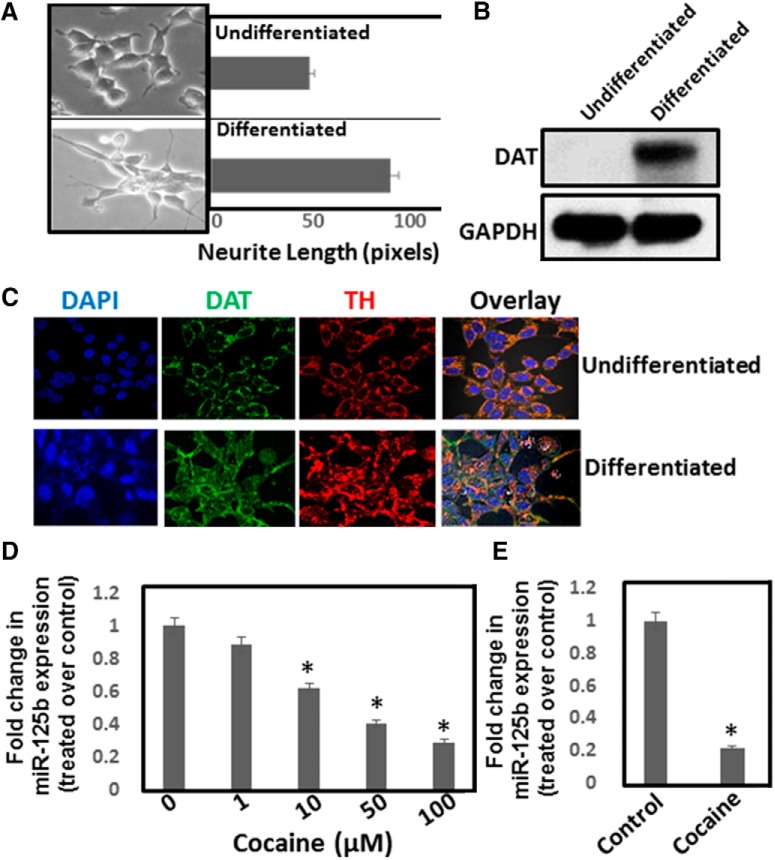
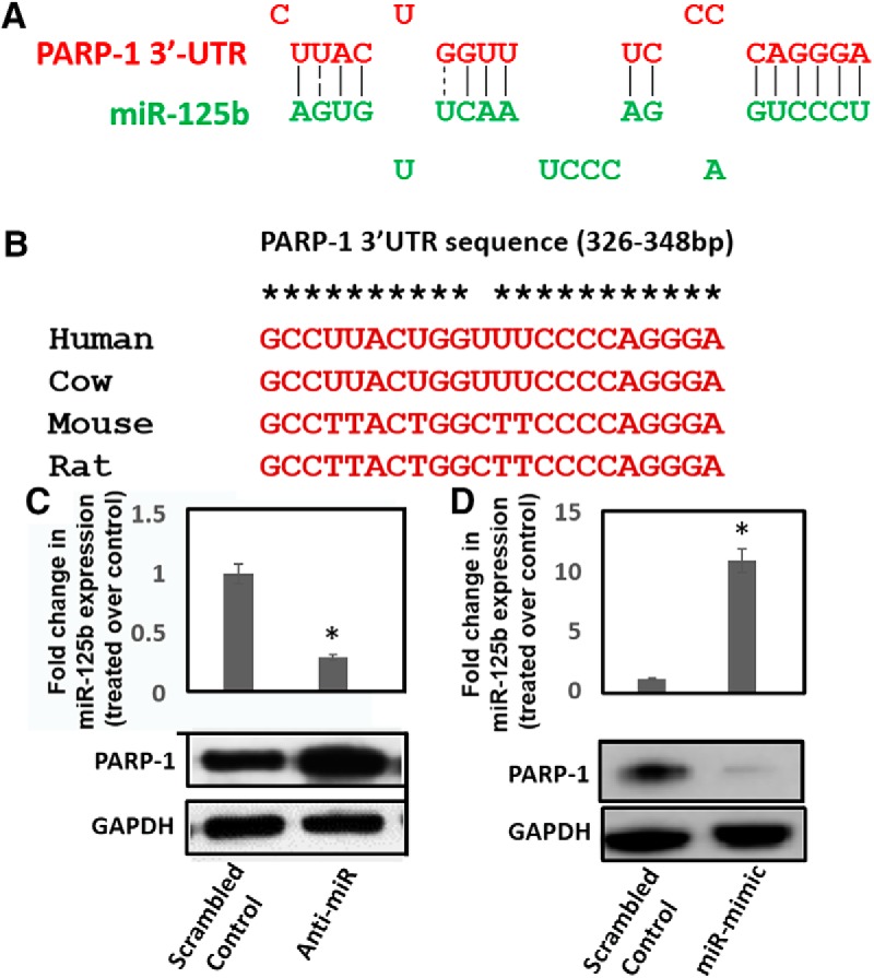
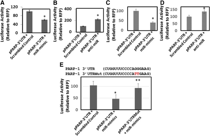
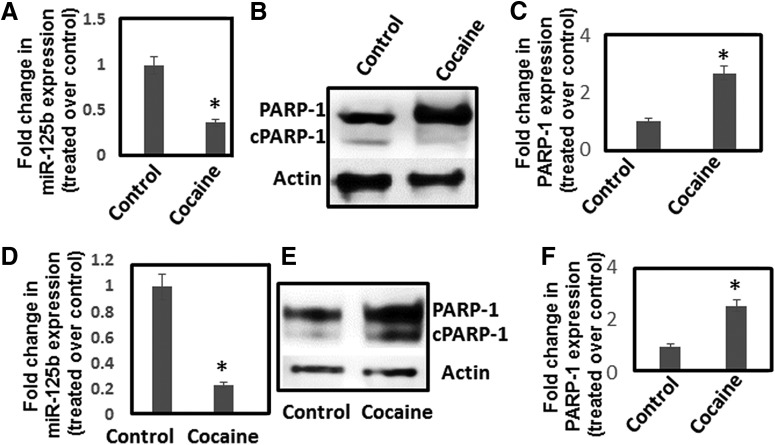
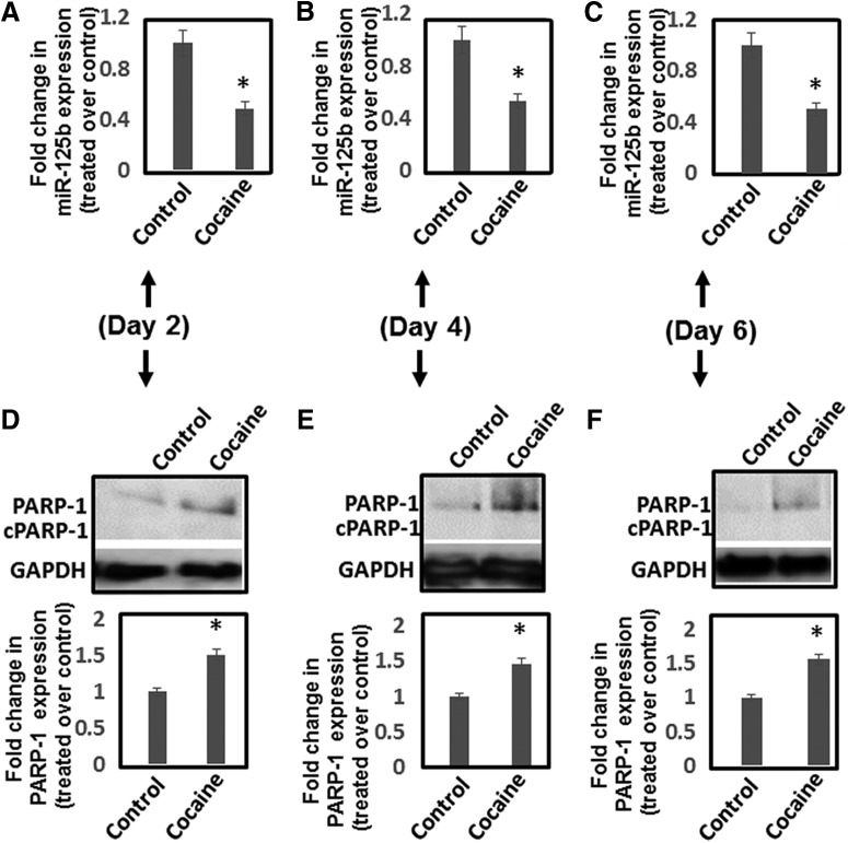
Cocaine exposure alters gene expression in the brain via methylation and acetylation of histones along with methylation of DNA. Recently, poly (ADP-ribose) polymerase-1 (PARP-1) catalyzed PARylation has been reported as an important regulator of cocaine-mediated gene expression. In this study, we report that the cellular microRNA "miR-125b" plays a key role for cocaine-induced PARP-1 expression. Acute and chronic cocaine exposure resulted in the downregulation of miR-125b concurrent with upregulation of PARP-1 in dopaminergic neuronal cells and nucleus accumbens (NAc) of mice but not in the medial prefrontal cortex (PFC) or ventral tegmental area (VTA). In silico analysis predicted a binding site of miR-125b in a conserved 3'-untranslated region (3'UTR) of the PARP-1 mRNA. Knockdown and overexpression studies showed that miR-125b levels negatively correlate with PARP-1 protein expression. Luciferase reporter assay using a vector containing the 3'UTR of PARP-1 mRNA confirmed regulation of PARP-1 by miR-125b. Specific nucleotide mutations within the binding site abrogated miR-125b's regulatory effect on PARP-1 3'UTR. Finally, we established that downregulation of miR-125b and concurrent upregulation of PARP-1 is dependent on binding of cocaine to the dopamine transporter (DAT). Collectively, these results identify miR-125b as a post-transcriptional regulator of PARP-1 expression and establish a novel mechanism underlying the molecular effects of cocaine action.

Keywords: PARP-1; PARylation; cocaine; miRNA; post-transcriptional regulation.


Figures

Figure 1.
Figure 1.
Cocaine exposure downregulates miR-125b expression. SH-SY5Y cells were cultured overnight and then differentiated for 3–5 d. A, Dopaminergic phenotype of these cells was monitored via neurite length measurement by phase-contrast microscopy. B, Western blot analysis of DAT expression in differentiated SH-SY5Y cells. C, DAT and TH expression in differentiated SH-SY5Y cells were detected by confocal microscope. Data are representative of three independent experiments. D, Differentiated SH-SY5Y cells were treated with varying concentrations of cocaine. Cells were harvested and miRNA expression was analyzed by qPCR. miR-125b expression was normalized to the 5S rRNA levels. Relative expression of miR-125b in cocaine-treated cells was compared to the untreated control cells and are plotted as fold change. E, Mice were injected (i.p.) with a single dose of cocaine (20 mg/kg weight of mice) or saline and animals were sacrificed. NAc tissues were used for miRNA isolations and were subjected to qPCR. miR-125b expression was normalized to the 5s rRNA levels and relative expression of miR-125b in the NAc of cocaine-treated mice (n = 6) are expressed as fold changes to the NAc of saline-treated mice (n = 6). *p < 0.05 for the comparison of cocaine-treated samples versus untreated controls.
Figure 2.
Figure 2.
Cocaine exposure induces PARP-1 protein expression without increasing mRNA levels. A, Differentiated SH-SY5Y cells were exposed to different concentrations of cocaine overnight and apoptosis was measured by flow cytometry using AV- as an early apoptosis marker and PI- as the late apoptotic marker. After cocaine treatment, cells were harvested and stained with FITC-conjugated AV/PI and analyzed by flow cytometry. Representative dot blot data showing percentage of cells stained positive for AV, PI, and AV + PI. B, Differentiated SH-SY5Y cells were treated with varying concentrations of cocaine. Cells were harvested, and cell lysates were electrophoresed on denaturing acrylamide gels and transferred onto nitrocellulose membrane by electroblotting. Western blot analyses were performed using appropriate antibody and ECL kit. Representative data of PARP-1 expression in untreated and cocaine-treated cells. C, Densitometry of PARP-1 expression from three independent experiments. D, Mice were injected (i.p.) with a single dose of cocaine (20 mg/kg weight of mice, i.p.) or saline. Animals were sacrificed and NAc was isolated. Lysates prepared from the NAc tissues were subjected to western blot analysis. (E) Relative expression of PARP-1 in the NAc of cocaine-treated mice (n = 6) compared to the NAc of saline-treated mice (n = 6). *p < 0.05 for the comparison of cocaine-treated samples versus untreated controls. (F) Differentiated SH-SY5Y cells were treated with varying concentrations of cocaine overnight. Cells were harvested and total RNA was isolated. PARP-1 mRNA expression was measured by qPCR and was normalized to the levels of GAPDH mRNA. PARP-1 mRNA in cocaine-treated cells were expressed as fold changes over the untreated control cells based on ΔΔCt values. G, Mice were injected (i.p.) with a single dose of cocaine (20 mg/kg weight of mice) or saline. Animals were sacrificed and NAc was isolated. Total RNA was isolated from the NAc tissues and were subjected to qPCR. PARP-1 mRNA expression was normalized to the GAPDH mRNA levels and fold changes in PARP-1 mRNA in the NAc of cocaine-treated mice (n = 6) over the NAc of saline-treated mice (n = 6) were calculated based on ΔΔCt values.
Figure 3.
Figure 3.
miR-125b expression negatively correlates with PARP-1 protein levels. A, In silico analysis predicts putative binding site of miR-125b in the 3’UTR of PARP-1 mRNA. B, Sequence alignment of miR-125b binding site in the 3’UTR of PARP-1 mRNA among different mammalian species. C, upper panel, miR-125b knockdown. Chemically synthesized anti-miR-125b oligonucleotides were transfected into differentiated SH-SY5Y cells and reduction of miR-125b expression was measured 24 h post-transfection by qPCR. C, lower panel, PARP-1 expression in miR-125b knocked-down cells was measured in the cellular lysates by immunoblot. D, upper panel, miR-125b overexpression. Chemically synthesized miR-125b mimic oligonucleotides were transfected into differentiated SH-SY5Y cells and miR-125b expression was measured 24 h post-transfection by qPCR. D, lower panel, PARP-1 expression in miR-125b overexpressing cells was measured in the cellular lysates by immunoblot. Data in C, D are representative of three independent experiments. *p < 0.05 for the comparison of anti-miR-125b or miR-125b mimic samples versus scrambled controls.
Figure 4.
Figure 4.
miR-125b targets 3’UTR of PARP-1 mRNA. We used a luciferase reporter vector, pPARP-3’UTR, containing the 3’UTR of PARP-1 mRNA cloned downstream of the luciferase stop codon. The pPARP-3’UTR was transfected into HEK-293T cells or differentiated SH-SY5Y cells in the presence or absence of miR-125b mimics or anti-miR-125b. A, Luciferase activity of cellular lysates prepared from HEK-293T cells cotransfected with (pPARP-3’UTR) and scrambled control or miR-125b mimics. B, Luciferase activity in cellular lysates prepared from HEK-293T cells cotransfected with (pPARP-3’UTR) and scrambled control or anti-miR-125b. C, Luciferase activity in cellular lysates prepared from differentiated SH-SY5Y cells cotransfected with (pPARP-3’UTR) and scrambled control or miR-125b mimics. D, Luciferase activity in cellular lysates prepared from differentiated SH-SY5Y cells cotransfected with (pPARP-3’UTR) and scrambled control or anti-miR-125b. E, Site-directed mutagenesis of miR-125b binding site within the PARP-1 3’UTR. Upper panel, PARP 3’UTRMut represents mutation of two critical residues GG > TT. Lower panel, Luciferase activity in cellular lysates prepared from differentiated SH-SY5Y cells transfected with pPARP 3’UTR and pPARP 3’UTRMut in the presence or absence of miR-125b mimics. *p < 0.05 for the comparison of anti-miR-125b or miR-125b mimic samples versus scrambled controls. E, **p < 0.05 for the comparison of miR-125b mimic samples of pPARP-3’UTR versus pPARP-3’UTRMut.
Figure 5.
Figure 5.
Cocaine-induced downregulation of miR-125b and concurrent upregulation of PARP-1 is dependent on DAT. A, Detection of DAT in the HEK-293T-DATNull and HEK-293T-DAT cells by immunoblot. Fold change in miR-125b expression in (B) HEK-293T-DAT cells and (C) HEK-293T-DAT-Null cells on exposure to varying concentration of cocaine relative to untreated cells as measured by qPCR. D, Fold change in miR-125b expression in HEK-293T-DAT cells pretreated with nomifensine. E, G, Differentiated SH-SY5Y cells with or without nomifensine pretreatment were treated with cocaine. E, miR-125b expression was measured in these cells by qPCR. F, PARP-1 expression was measured in the cell lysates by Western blotting. G, Densitometry of PARP-1 expression from three independent experiments. H, Differentiated SH-SY5Y cells were transfected with miR-125b mimics or scrambled controls and then treated with cocaine (50 μM) overnight. PARP-1 expression was measured in the lysates of these cells by immunoblot. Lower panel shows representative data, whereas upper panel shows fold change in PARP-1 expression from three independent experiments. *p < 0.05 for the comparison of cocaine-treated samples versus untreated controls. D, E, G, **p < 0.05 for the comparison of cocaine-treated samples versus nomifensine + cocaine-treated samples. H, **p < 0.05 for the comparison of cocaine-treated samples versus miR-mimic + cocaine-treated samples.
Figure 6.
Figure 6.
Chronic cocaine exposure reduces miR-125b expression and upregulates PARP-1 expression. A–C, Differentiated SH-SY5Y cells were treated daily with varying concentrations of cocaine for 7 d. Cells were harvested, and miR-125b expression was measured by qPCR (A), whereas PARP-1 expression was detected by Western blotting (B, C). A, miR-125b expression was normalized to 5S rRNA levels and plotted as fold changes in miR-125b expression in cocaine-treated cells relative to untreated control. B, Representative immunoblot of PARP-1 expression in control and cocaine-treated cells. C, Relative PARP-1 expression in cocaine-treated cells and untreated cells as quantified by densitometry from three independent experiments. D-F, Mice were injected (i.p.) with cocaine (20 mg/kg weight of mice) or saline for 7 d. Thereafter, animals were sacrificed and NAc was isolated. RNA and lysates from the NAc tissues were subjected to qPCR and Western blotting, respectively. D, miR-125b expression was normalized to the 5s rRNA levels and relative expression of miR-125b in the NAc of cocaine-treated mice (n = 6) compared to the NAc of saline-treated mice (n = 6) are expressed as fold changes. E, Representative immunoblot showingPARP-1 expression in the NAc of cocaine-treated mice and saline-treated control mice. F, Relative expression of PARP-1 in the NAc of cocaine-treated mice (n = 6) compared to the NAc of saline-treated control mice (n = 6) based on densitometry analysis. *p < 0.05 for the comparison of cocaine-treated samples versus untreated controls.
Figure 7.
Figure 7.
Time-dependent effects of cocaine exposure on miR-125b and PARP-1 expression in NAc. Mice were injected (i.p.) with cocaine (20 mg/kg weight of mice) or saline for 2, 4, and 6 d. Thereafter, animals were sacrificed, and NAc regions were isolated. RNA and lysates from the NAc tissues were subjected to qPCR and Western blotting for measuring miR-125b and PARP-1 expression, respectively. A–C, Fold changes in miR-125b expression in cocaine-treated (n = 6) and saline-treated control (n = 6) animals on (A) day 2, (B) day 4, and (C) day 6. D–F, PARP-1 protein expression in the NAc tissues was analyzed by immunoblot and subjected to quantification by densitometry. Upper panels show representative data of PARP-1, whereas lower panels show densitometry of PARP-1 expression from cocaine-treated mice (n = 6) and saline-treated control animals (n = 6). Fold changes in PARP-1 expression in cocaine-treated mice and saline-treated controls as measured by densitometry of immunoblots of samples on (D) day 2, (E) day 4, and (F) day 6. *p < 0.05 for the comparison of cocaine-treated samples versus untreated controls.
Figure 8.
Figure 8.
Effects of cocaine exposure on miR-125b and PARP-1 expression in PFC and VTA. Mice were injected (i.p.) with cocaine (20 mg/kg weight of mice) or saline under acute and chronic conditions. Thereafter, animals were sacrificed and PFC (A–C) and VTA (D–F) were isolated. RNA and lysates from the tissues were subjected to qPCR and Western blotting for measuring miR-125b and PARP-1 expression, respectively. Fold changes in miR-125b expression in (A) the PFC and (D) the VTA of cocaine-treated animals and saline-treated control animals were calculated. PARP-1 expression in (B) the PFC and (E) the VTA of cocaine-treated mice and saline-treated controls as measured by immunoblot. Fold changes in PARP-1 protein expression in (C) the PFC and (F) the VTA of cocaine-treated mice (n = 6) compared to saline-treated control mice (n = 6). *p < 0.05 for the comparison of cocaine-treated samples versus untreated controls.

References

    1. Anderson EM, Self DW (2017) It's only a matter of time: longevity of cocaine-induced changes in dendritic spine density in the nucleus accumbens. Curr Opin Behav Sci 13:117–123. 10.1016/j.cobeha.2016.11.013 - DOI - PMC - PubMed
    1. Barr TL, Conley YP (2007) Poly(ADP-ribose) polymerase-1 and its clinical applications in brain injury. J Neurosci Nurs 39:278–284. - PubMed
    1. Bartel DP (2004) MicroRNAs: genomics, biogenesis, mechanism, and function. Cell 116:281–297. - PubMed
    1. Bartel DP (2009) MicroRNAs: target recognition and regulatory functions. Cell 136:215–233. 10.1016/j.cell.2009.01.002 - DOI - PMC - PubMed
    1. Beuming T, Kniazeff J, Bergmann ML, Shi L, Gracia L, Raniszewska K, Newman AH, Javitch JA, Weinstein H, Gether U, Loland CJ (2008) The binding sites for cocaine and dopamine in the dopamine transporter overlap. Nat Neurosci 11:780–789. 10.1038/nn.2146 - DOI - PMC - PubMed

Publication types

MeSH terms

LinkOut - more resources