Review
. 2017 Aug 21;27(16):R816-R824.
doi: 10.1016/j.cub.2017.06.012.

How Kinetochore Architecture Shapes the Mechanisms of Its Function

Affiliations
Review

How Kinetochore Architecture Shapes the Mechanisms of Its Function

Ajit P Joglekar et al. Curr Biol. .

Abstract

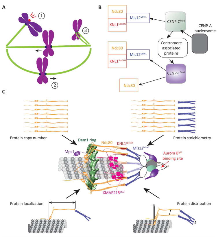
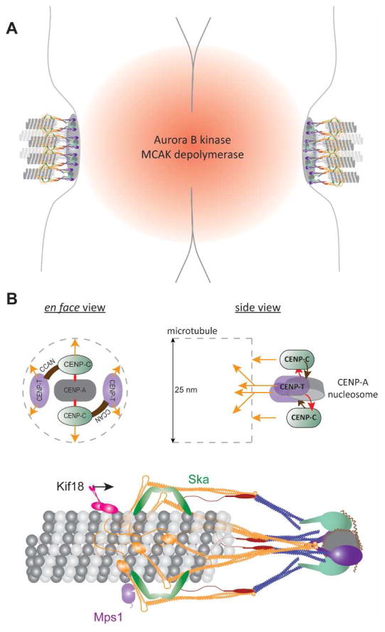
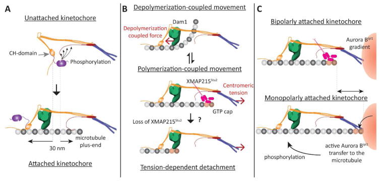
The eukaryotic kinetochore is a sophisticated multi-protein machine that segregates chromosomes during cell division. To ensure accurate chromosome segregation, it performs three major functions using disparate molecular mechanisms. It operates a mechanosensitive signaling cascade known as the spindle assembly checkpoint (SAC) to detect and signal the lack of attachment to spindle microtubules, and delay anaphase onset in response. In addition, after attaching to spindle microtubules, the kinetochore generates the force necessary to move chromosomes. Finally, if the two sister kinetochores on a chromosome are both attached to microtubules emanating from the same spindle pole, they activate another mechanosensitive mechanism to correct the monopolar attachments. All three of these functions maintain genome stability during cell division. The outlines of the biochemical activities responsible for these functions are now available. How the kinetochore integrates the underlying molecular mechanisms is still being elucidated. In this Review, we discuss how the nanoscale protein organization in the kinetochore, which we refer to as kinetochore 'architecture', organizes its biochemical activities to facilitate the realization and integration of emergent mechanisms underlying its three major functions. For this discussion, we will use the relatively simple budding yeast kinetochore as a model, and extrapolate insights gained from this model to elucidate functional roles of the architecture of the much more complex human kinetochore.


Figures

Figure 1
Figure 1. The function and protein architecture of the kinetochore
A Cartoon of a mitotic spindle displaying the three main kinetochore functions: (1) Activation of the Spindle Assembly Checkpoint, (2) generation of bidirectional chromosome movement that is coupled to microtubule polymerization and depolymerization, and (3) correction of monopolar attachment of sister kinetochores. B (left to right = microtubule plus-end to centromere) The conserved, dual pathways (solid arrows – direct interaction, dashed arrow – indirect interaction) that assemble the KMN network, which forms the interface of the kinetochore with the microtubule plus-end. C Reconstruction of the protein architecture of the budding yeast kinetochore using fluorescence microscopy measurements and protein structures [, , , , , , –50, 73]. Centromere-associated proteins are represented by white, oblong shapes.
Figure 2
Figure 2. Protein architecture of the human kinetochore
A Cartoon of the organization of sister kinetochores on a human chromosome. The inter-centromeric localization of Aurora BIpl1 and MCAK is highlighted. B Top: Schematic displays a hypothetical spatial manifestation of the biochemical pathways of kinetochore assembly. Orange arrows indicate pathways of Ndc80 recruitment; grey dashed lines represent the microtubule. Note that CENP-TCnn1 recruits two Ndc80 molecules. Bottom: Cartoon of a hypothetical local architecture of the kinetochore-microtubule attachment in humans.
Figure 3
Figure 3. Proposed architecture-function relationships for the yeast kinetochore (1-D representations of the kinetochore shown)
A Role of kinetochore architecture in SAC inactivation: Separation of the CH-domains of Ndc80 and the phosphodomain of KNL1Spc105 by end-on attachment (highlighted by dashed lines) disrupts the phosphorylation of KNL1Spc105 by the Mps1 kinase (magenta) bound to the CH-domain. B Proposed roles of the architecture of microtubule-binding proteins in generating bidirectional movement. Top: When the plus-end is depolymerizing, the Dam1 ring (green) mechanically opposes the curling of tubulin protofilaments, and experiences a pushing force (red arrow). Middle panel: We propose that XMAP215Stu2 localizes to the kinetochore by recognizing the GTP-tubulin cap on the polymerizing plus-end. Its microtubule-destabilizing activity reverts the plus-end back to the depolymerizing state. Bottom panel: In the absence of XMAP215 activity, centromeric tension can slide the kinetochore off the growing plus-end. C Potential roles for kinetochore architecture in correcting monopolar attachment: The position of the plus-end may be significantly different in kinetochores with bipolar and monopolar attachment (highlighted by the arrow, top and bottom panels respectively). The proximity of the lattice to the centromere may facilitate the transport of hyper-activated Aurora BIpl1 kinase to its targets – microtubule-binding kinetochore proteins.

References

    1. Picco A, Mund M, Ries J, Nedelec F, Kaksonen M. Visualizing the functional architecture of the endocytic machinery. Elife. 2015:4. - PMC - PubMed
    1. Alber F, Dokudovskaya S, Veenhoff LM, Zhang W, Kipper J, Devos D, Suprapto A, Karni-Schmidt O, Williams R, Chait BT, et al. The molecular architecture of the nuclear pore complex. Nature. 2007;450:695–701. - PubMed
    1. Musacchio A, Desai A. A Molecular View of Kinetochore Assembly and Function. Biology. 2017:6. - PMC - PubMed
    1. Nezi L, Musacchio A. Sister chromatid tension and the spindle assembly checkpoint. Current Opinion in Cell Biology. 2009;21:785–795. - PubMed
    1. Winey M, Mamay CL, O’Toole ET, Mastronarde DN, Giddings TH, Jr, McDonald KL, McIntosh JR. Three-dimensional ultrastructural analysis of the Saccharomyces cerevisiae mitotic spindle. J Cell Biol. 1995;129:1601–1615. - PMC - PubMed

MeSH terms

Substances

LinkOut - more resources