

Sciatic neuromuscular variants on MR neurography: frequency study and interobserver performance
- PMID: 28830192
- PMCID: PMC5963375
- DOI: 10.1259/bjr.20170116
Sciatic neuromuscular variants on MR neurography: frequency study and interobserver performance
Abstract
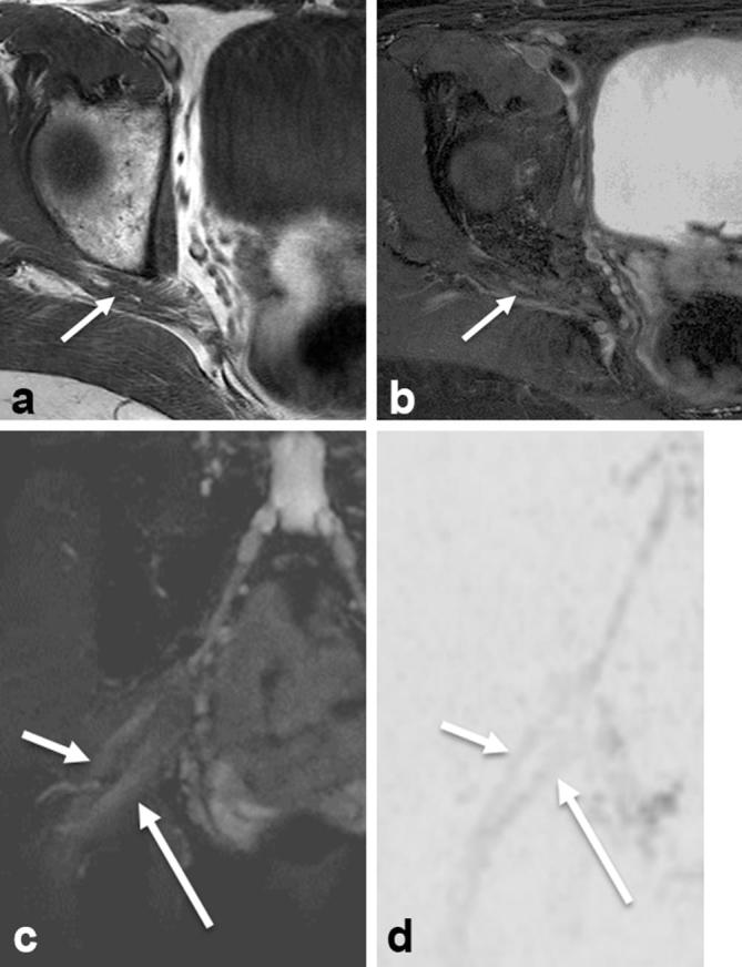
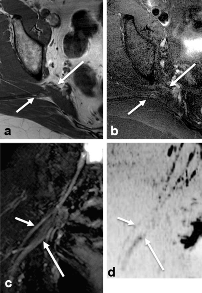
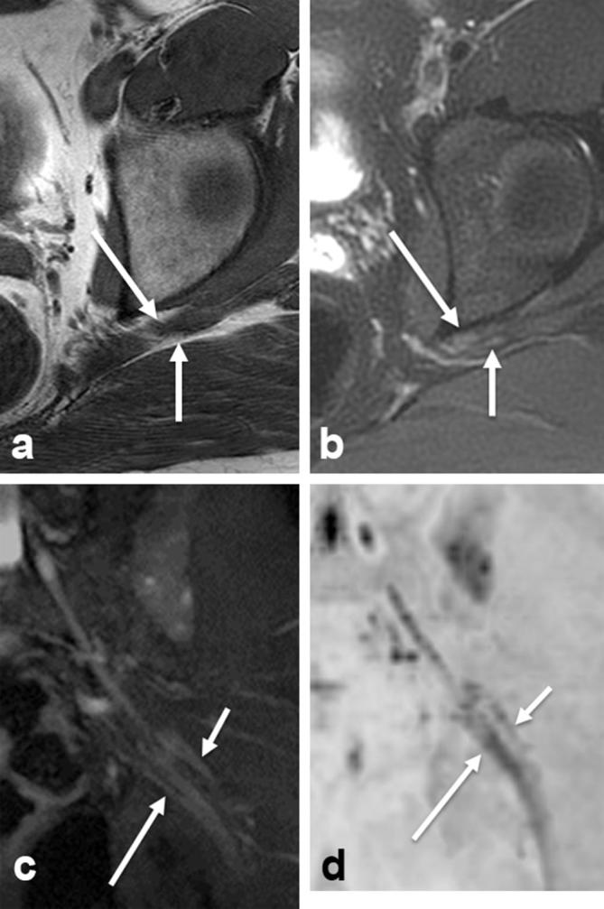
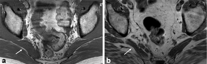
Objective: To evaluate the frequency of sciatic neuromuscular variants on MR neurography and determine the interobserver variability.
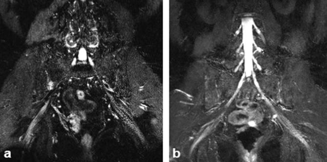
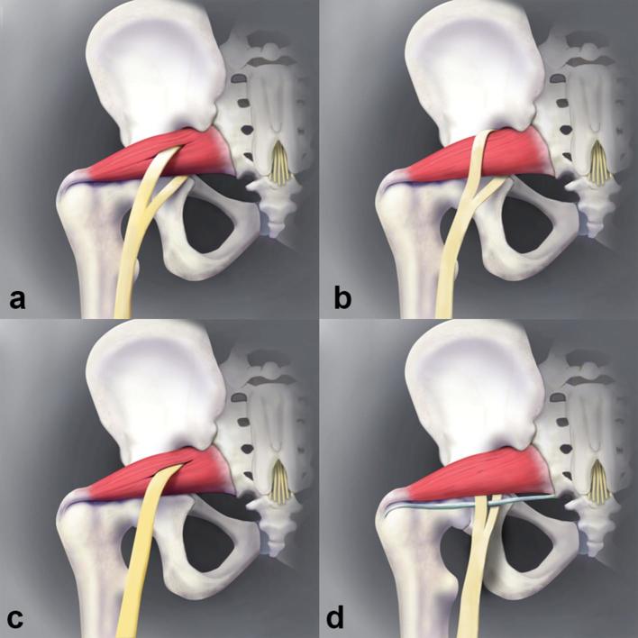
Methods: A retrospective evaluation of 137 consecutive lumbosacral plexus magnetic resonance neurography examinations was performed. All examinations were performed using nerve selective 3D imaging and independently reviewed by two readers for the presence of sciatic neuromuscular variants and piriformis muscle asymmetry. Inter- and intraobserver performance were evaluated.
Results: There were a total of 44/268 (16.4%) extremities with sciatic neuromuscular variants. The interobserver performance in the identification of sciatic nerve variants was excellent (kappa values from 0.8-0.9). There was a total of 45/134 (33.6%) patients with piriformis muscle asymmetry. Of these, 7/134 (5.2%) had piriformis muscle atrophy and 38/134 (28.4%) had piriformis muscle hypertrophy. The interobserver performance in the identification of piriformis muscle atrophy and hypertrophy was moderate to good (kappa values from 0.39-0.61). The intraobserver performance revealed kappa values of 0.735 and 0.821 on right and left, respectively.
Conclusion: Sciatic neuromuscular variants and piriformis muscle asymmetry are frequent on lumbosacral plexus MRN with moderate to excellent interobserver performance. Advances in knowledge: Sciatic neuromuscular variants and piriformis asymmetry on MR neurography are frequent and the prevalence is similar to cumulative prevalence from available scientific series. Interobserver performance for identification of sciatic neuromuscular variants is excellent, and moderate-good for piriformis muscle asymmetry.
Figures
References
-
- Bardeen CR, Elting AW. A statistical study of the variations in the formation and position of the lumbosacral plexus in man. Anatomical Record 1901; 19: 209–32.
-
- Trotter M. The relation of the sciatic nerve to the piriformis muscle in American whites and negroes. Anat Rec . 1932; 52: 321–3. DOI: https://doi.org/10.1002/ar.1090520404 - DOI
-
- Ming-Tzu P'an. The relation of the sciatic nerve to the piriformis muscle in the Chinese. Am J Phys Anthropol 1941; 28: 375–80. DOI: https://doi.org/10.1002/ajpa.1330280403 - DOI
-
- Misra B. The relations of the sciatic nerve to the piriformis in Indian cadavers. J Anat Soc India 1954; 3: 28–33.
MeSH terms
LinkOut - more resources
Full Text Sources
Other Literature Sources
Medical
