

A robotic C-arm cone beam CT system for image-guided proton therapy: design and performance
- PMID: 28830239
- PMCID: PMC5963391
- DOI: 10.1259/bjr.20170266
A robotic C-arm cone beam CT system for image-guided proton therapy: design and performance
Abstract
Objective: A ceiling-mounted robotic C-arm cone beam CT (CBCT) system was developed for use with a 190° proton gantry system and a 6-degree-of-freedom robotic patient positioner. We report on the mechanical design, system accuracy, image quality, image guidance accuracy, imaging dose, workflow, safety and collision-avoidance.
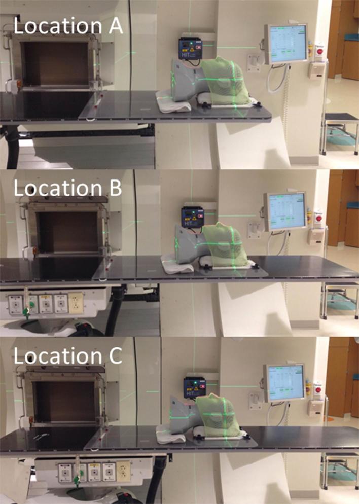
Methods: The robotic CBCT system couples a rotating C-ring to the C-arm concentrically with a kV X-ray tube and a flat-panel imager mounted to the C-ring. CBCT images are acquired with flex correction and maximally 360° rotation for a 53 cm field of view. The system was designed for clinical use with three imaging locations. Anthropomorphic phantoms were imaged to evaluate the image guidance accuracy.
Results: The position accuracy and repeatability of the robotic C-arm was high (<0.5 mm), as measured with a high-accuracy laser tracker. The isocentric accuracy of the C-ring rotation was within 0.7 mm. The coincidence of CBCT imaging and radiation isocentre was better than 1 mm. The average image guidance accuracy was within 1 mm and 1° for the anthropomorphic phantoms tested. Daily volumetric imaging for proton patient positioning was specified for routine clinical practice.
Conclusion: Our novel gantry-independent robotic CBCT system provides high-accuracy volumetric image guidance for proton therapy. Advances in knowledge: Ceiling-mounted robotic CBCT provides a viable option than CT on-rails for partial gantry and fixed-beam proton systems with the added advantage of acquiring images at the treatment isocentre.
Figures


References
-
- Uematsu M, Fukui T, Shioda A, Tokumitsu H, Takai K, Kojima T, et al. A dual computed tomography linear accelerator unit for stereotactic radiation therapy: a new approach without cranially fixated stereotactic frames. Int J Radiat Oncol Biol Phys 1996; 35: 587–92. DOI: https://doi.org/10.1016/S0360-3016(96)80022-8 - DOI - PubMed
-
- Mosleh-Shirazi MA, Evans PM, Swindell W, Webb S, Partridge M. A cone-beam megavoltage CT scanner for treatment verification in conformal radiotherapy. Radiother Oncol 1998; 48: 319–28. DOI: https://doi.org/10.1016/S0167-8140(98)00042-5 - DOI - PubMed
-
- Mackie TR, Kapatoes J, Ruchala K, Lu W, Wu C, Olivera G, et al. Image guidance for precise conformal radiotherapy. Int J Radiat Oncol Biol Phys 2003; 56: 89–105. DOI: https://doi.org/10.1016/S0360-3016(03)00090-7 - DOI - PubMed
-
- Jaffray DA, Siewerdsen JH, Wong JW, Martinez AA. Flat-panel cone-beam computed tomography for image-guided radiation therapy. Int J Radiat Oncol Biol Phys 2002; 53: 1337–49. DOI: https://doi.org/10.1016/S0360-3016(02)02884-5 - DOI - PubMed
-
- Mori S, Zenklusen S, Knopf AC. Current status and future prospects of multi-dimensional image-guided particle therapy. Radiol Phys Technol 2013; 6: 249–72. DOI: https://doi.org/10.1007/s12194-013-0199-0 - DOI - PubMed
MeSH terms
Substances
LinkOut - more resources
Full Text Sources
Other Literature Sources
