. 2017 Aug 22;12(1):60.
doi: 10.1186/s13024-017-0201-0.

High-density lipoproteins suppress Aβ-induced PBMC adhesion to human endothelial cells in bioengineered vessels and in monoculture

Affiliations

High-density lipoproteins suppress Aβ-induced PBMC adhesion to human endothelial cells in bioengineered vessels and in monoculture

Jérôme Robert et al. Mol Neurodegener. .

Abstract

Background: Alzheimer's Disease (AD), characterized by accumulation of beta-amyloid (Aβ) plaques in the brain, can be caused by age-related failures to clear Aβ from the brain through pathways that involve the cerebrovasculature. Vascular risk factors are known to increase AD risk, but less is known about potential protective factors. We hypothesize that high-density lipoproteins (HDL) may protect against AD, as HDL have vasoprotective properties that are well described for peripheral vessels. Epidemiological studies suggest that HDL is associated with reduced AD risk, and animal model studies support a beneficial role for HDL in selectively reducing cerebrovascular amyloid deposition and neuroinflammation. However, the mechanism by which HDL may protect the cerebrovascular endothelium in the context of AD is not understood.

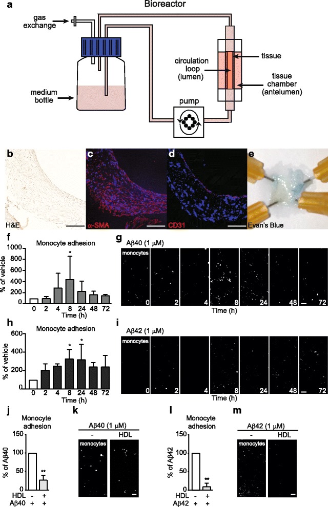
Methods: We used peripheral blood mononuclear cell adhesion assays in both a highly novel three dimensional (3D) biomimetic model of the human vasculature composed of primary human endothelial cells (EC) and smooth muscle cells cultured under flow conditions, as well as in monolayer cultures of ECs, to study how HDL protects ECs from the detrimental effects of Aβ.

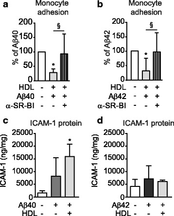
Results: Following Aβ addition to the abluminal (brain) side of the vessel, we demonstrate that HDL circulated within the lumen attenuates monocyte adhesion to ECs in this biofidelic vascular model. The mechanism by which HDL suppresses Aβ-mediated monocyte adhesion to ECs was investigated using monotypic EC cultures. We show that HDL reduces Aβ-induced PBMC adhesion to ECs independent of nitric oxide (NO) production, miR-233 and changes in adhesion molecule expression. Rather, HDL acts through scavenger receptor (SR)-BI to block Aβ uptake into ECs and, in cell-free assays, can maintain Aβ in a soluble state. We confirm the role of SR-BI in our bioengineered human vessel.

Conclusion: Our results define a novel activity of HDL that suppresses Aβ-mediated monocyte adhesion to the cerebrovascular endothelium.

Keywords: Alzheimer’s disease; Beta-amyloid; Endothelial cells; Engineered vessel; High-density lipoprotein.


Conflict of interest statement

Springer Nature remains neutral with regard to jurisdictional claims in published maps and institutional affiliations.

Figures

Fig. 1
Fig. 1
Aβ induces monocyte adhesion in engineered vessels, which is suppressed by HDL. a Schematic representation of bioengineered tissue. b Histological structure of engineered tissue using hematoxalin-eosin staining to reveal a dense tissue formation composed of cells and extracellular matrix in engineered vessels. c α-smooth muscle actin (α-SMA) confirmed the smooth muscle phenotype of cells in the inner layers and d CD31 confirmed an endothelial monolayer. Scale bar represents 200 μm. e Evans blue staining confirmed a tight endothelium. f-g 1 μM of Aβ40 or h-i Aβ42 monomers were injected within the tissue chamber (abluminal). Fluorescently labeled human monocytes (THP1, white) were circulated in the lumen of the engineered vessels. Tissues were counterstained with DAPI (blue) and analyzed using confocal microscopy over time. j-m 200 μg/mL of HDL were circulated through the lumen of the grafts for 2 h prior injection of 1 μM of j-k Aβ40 or l-m Aβ42 monomers within the tissue chamber (abluminal) for 8 h prior to circulating fluorescent THP1 in the lumen. Graphs represent means ± SD of adhered monocytes relative to Aβ treated tissues from at least 3 individual grafts. *p < 0.05, **p < 0.01
Fig. 2
Fig. 2
Aβ40 and Aβ42 induce PBMC adhesion to ECs, which is suppressed by HDL. HUVEC (a-b) or hCMEC/D3 (c-d) were primed with 100 μg/mL of HDL and stimulated with 0.1 μM a, c Aβ40 (light grey) or b, dAβ42 (dark grey) monomers for 3 h. Fluorescently labelled PBMC were allowed to adhere to stimulated cells for 3 h followed by washing, fixation, imaging, and counting. hCMEC/D3 were primed with either 100 μg/mL of HDL or 50 μg/mL or 100 μg/mL lipid-free human apoA-I for 2 h followed by stimulation with 0.1 μM e Aβ40 (light grey) or f Aβ42 (dark grey) monomers for 3 h. Cells were washed, imaged and counted as above. Graphs represent mean ± SD of adhered PBMC relative to vehicle control from at least 3 independent trials where * p < 0.05, **p < 0.01, ***p < 0.001 versus vehicle, § p < 0.05, §§ p < 0.01 versus Aβ
Fig. 3
Fig. 3
HDL delays beta-sheet formation and attenuates Aβ-induced PBMC adherence independent of Aβ structure. a Representative graph ± SD of technical triplicates of 3 individual experiments where 10 μM Aβ40 or Aβ42 with or without 10 mg/mLl HDL were incubated with 20 μM of Thio-T in 150 mM NaCl and 5 μM of HEPES (pH 7.4) for 12 h at 37 °C. Formation of β-amyloid pleated sheets was monitored every 5 min at excitation 440 nm and emission 490 nm. b-c Aβ structures were confirmed using dot blot with antibodies against oligomers (A11) or fibrils (OC) and electron microscopy (EM). d-e hCMEC/D3 were stimulated for 3 h with 0.1 μM monomeric (m-Aβ, solid bar), oligomeric (o-Aβ, striped bar) or fibrillar-aggregated (f-A Aβ, cross-hatched bar) d Aβ40 or e Aβ42 in the presence or absence of HDL. f-g hCMEC/D3 were pre-incubated for 2 h with 100 μg/mL HDL prior to Aβ addition (pre, solid bar), co-incubated with 100 μg/mL HDL and Aβ (co, striped bar), or post-incubated by adding 100 μg/mL HDL 1 h following Aβ stimulation (post, double striped bar). Graphs represent means ± SD of adhered PBMC relative to vehicle treated cells from at least 5 independent trials where. *p < 0.05, **p < 0.01, ***p < 0.001 * p < 0.05, **p < 0.01, ***p < 0.001 versus vehicle, § p < 0.05, §§ p < 0.01, §§§p < 0.001 versus Aβ
Fig. 4
Fig. 4
HDL suppression of Aβ-induced inflammation is independent of eNOS and S1P. a Intracellular NO production was measured by treating hCMEC/D3 with 100 μg/mL HDL in the presence of 1 μM DAF-2 for 6 h. Fluorescence was measured at 485 nm. b Phosphorylation of eNOS was measured by treating hCMEC/D3 with 100 μg/mL HDL for 15 min before immunoblotting for phosphorylated eNOS (p-eNOS) or total eNOS. Representative immunoblots are shown in (c). hCMEC/D3 were pretreated for 1 h with the eNOS inhibitor L-NAME (d-f) or the S1P1 and S1P3 inhibitor VPC23019 (g-i) followed by 100 μg/mL HDL for 2 h. Cells were then stimulated with 0.1 μM Aβ40 (d,g) Aβ42 monomers, (e,h) or 1 ng/mL of TNF-α (f,i) for 3 h before testing PBMC adherence. Graphs represent means ± SD of adhered PBMC relative to vehicle treated cells for at least 5 independent trials. *p < 0.05, **p < 0.01, ***p < 0.001 * p < 0.05, **p < 0.01, ***p < 0.001 versus vehicle, § p < 0.05, §§ p < 0.01, §§§p < 0.001 versus Aβ or TNF-α
Fig. 5
Fig. 5
HDL reduces Aβ association, binding and uptake to hCMEC/D3 whereas blocking Aβ binding or uptake reduces PBMC adhesion to hCMEC/D3. a-d hCMEC/D3 were pre-treated with 100 μg/mL of HDL and 0.1 μM a,c Aβ40 or b,d Aβ42 monomers as described in Fig. 2 at either 37 °C (association a,b) or at 4 °C (binding c,d). Cells were lysed in RIPA buffer and Aβ were measured using commercial ELISA. e hCMEC/D3 were pre-treated with HDL (1 mg/mL) for 2 h before stimulating with 1 mM of fluorescently labelled Aβ40 or Aβ42 monomers. Scale bar represents 10 μm. f-n hCMEC/D3 were pre-treated with (f-h) RAGE blocking antibody, i-k RAP or l-n heparin or heparinase III 60 min before stimulation with Aβ40 or Aβ42 monomers or TNF-α for 3 h. PBMC adhesion assays were conducted as described in Fig. 2. Graphs represent means ± SD relative to vehicle treated cells for at least 3 independent trials. *p < 0.05, **p < 0.01 versus vehicle
Fig. 6
Fig. 6
HDL suppression of Aβ-induced inflammation requires SR-BI. hCMEC/D3 were pre-treated for 1 h with a-c SR-BI or d-f CD36 blocking antibodies or g-i BLT1 followed by 100 μg/mL HDL for 2 h. Cells were then stimulated with 0.1 μM monomeric a,d,g Aβ40 or b,e,h Aβ42 or (c,f,i) 1 ng/mL of TNF-α for 3 h before evaluating PBMC adherence. j hCMEC/D3 were pre-treated for 1 h with SR-BI blocking antibody followed by HDL (1 mg/mL) for 2 h before stimulating with 1 mM of fluorescently labelled Aβ40 or Aβ42 monomers. Scale bar represents 10 μm. Graphs represent means ± SD of adhered PBMC relative to vehicle treated cells from at least 4 independent trials. *p < 0.05, **p < 0.01, ***p < 0.001 * p < 0.05, **p < 0.01, ***p < 0.001 versus vehicle, § p < 0.05 versus Aβ or TNF-α.
Fig. 7
Fig. 7
HDL suppresses Aβ-induced monocyte adhesion in engineered vessels via SR-BI. SR-BI blocking antibody was circulated through the lumen of engineered vessels prepared using HUVEC for 1 h prior treatment with 200 μg/mL of HDL for 2 h followed by injection of 1 μM of monomeric a Aβ40 or b Aβ42 within the tissue chamber (abluminal) for 8 h prior to injecting circulating fluorescent THP1 into the lumen. c-d ICAM-1 protein was measured in tissue lysates prepared in RIPA buffer by commercial ELISA. Graphs represent means ± SD of adhered monocytes relative to Aβ treated tissues from at least 4 individual grafts. * and # p < 0.05

References

    1. Alzheimer’s Association 2015 Alzheimer’s disease facts and figures. Alzheimer’s & dementia : the journal of the Alzheimer’s Association. 2015;11(3):332–384. doi: 10.1016/j.jalz.2015.02.003. - DOI - PubMed
    1. Perl DP. Neuropathology of Alzheimer’s disease. The Mount Sinai journal of medicine, New York [Internet]. 2010;77(1):32–42. - PMC - PubMed
    1. Debette S. Vascular risk factors and cognitive disorders. Rev Neurol. 2013;169(10):757–764. doi: 10.1016/j.neurol.2013.07.022. - DOI - PubMed
    1. Justin BN, Turek M, Hakim AM. Heart disease as a risk factor for dementia. Clin Epidemiol. 2013;5:135–145. - PMC - PubMed
    1. Craft S. The role of metabolic disorders in Alzheimer disease and vascular dementia: two roads converged. Arch Neurol. 2009;66(3):300–305. doi: 10.1001/archneurol.2009.27. - DOI - PMC - PubMed

LinkOut - more resources