. 2017 Dec;298(Pt A):1-12.
doi: 10.1016/j.expneurol.2017.08.010. Epub 2017 Aug 19.

Contribution of amygdala CRF neurons to chronic pain

Affiliations

Contribution of amygdala CRF neurons to chronic pain

Matthew Andreoli et al. Exp Neurol. 2017 Dec.

Abstract

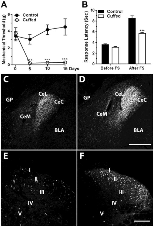
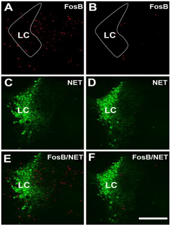
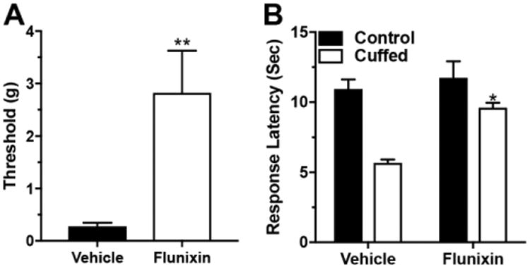
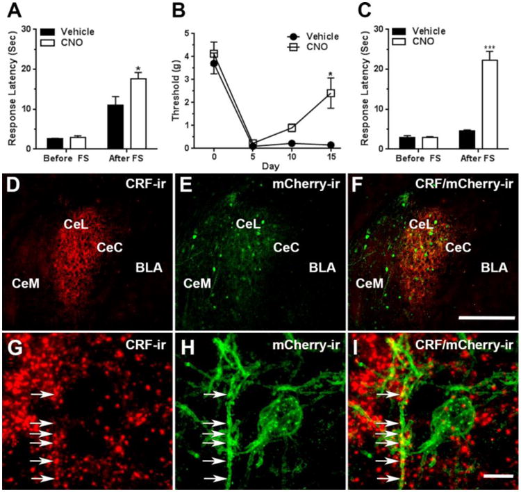
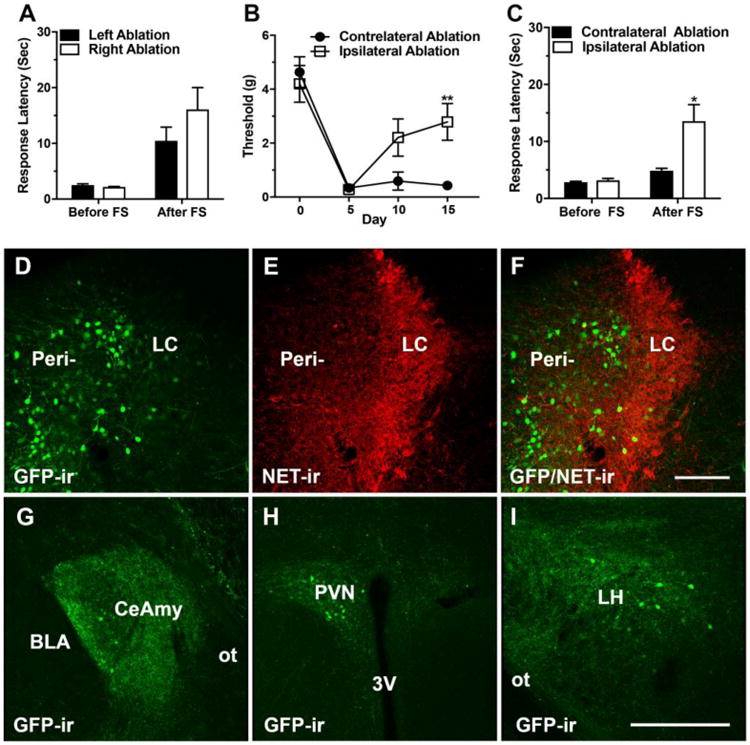
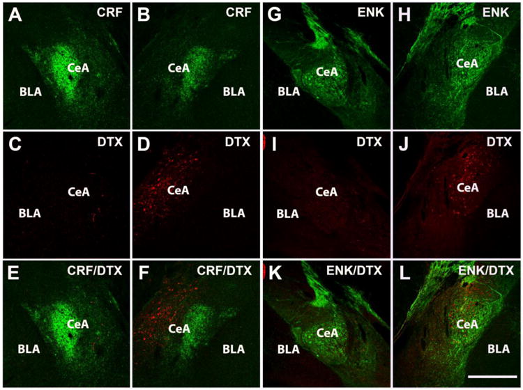
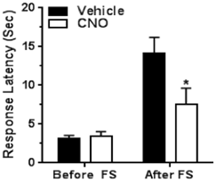
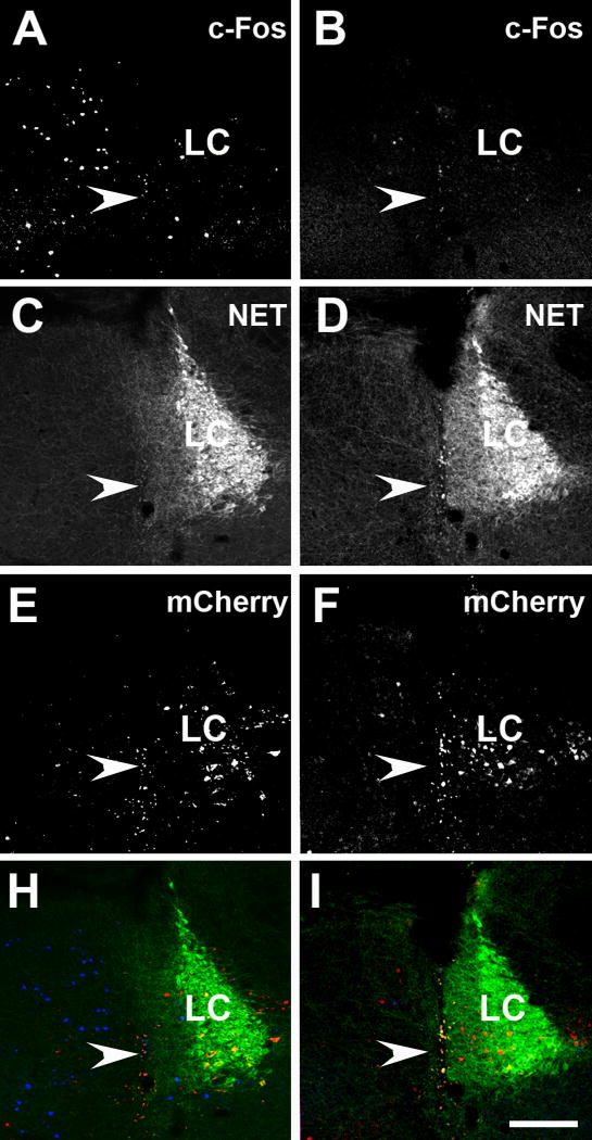
We investigated the role of amygdala corticotropin-releasing factor (CRF) neurons in the perturbations of descending pain inhibition caused by neuropathic pain. Forced swim increased the tail-flick response latency in uninjured mice, a phenomenon known as stress-induced analgesia (SIA) but did not change the tail-flick response latency in mice with neuropathic pain caused by sciatic nerve constriction. Neuropathic pain also increased the expression of CRF in the central amygdala (CeAmy) and ΔFosB in the dorsal horn of the spinal cord. Next, we injected the CeAmy of CRF-cre mice with cre activated AAV-DREADD (Designer Receptors Exclusively Activated by Designer Drugs) vectors. Activation of CRF neurons by DREADD/Gq did not affect the impaired SIA but inhibition of CRF neurons by DREADD/Gi restored SIA and decreased allodynia in mice with neuropathic pain. The possible downstream circuitry involved in the regulation of SIA was investigated by combined injections of retrograde cre-virus (CAV2-cre) into the locus ceruleus (LC) and cre activated AAV-diphtheria toxin (AAV-FLEX-DTX) virus into the CeAmy. The viral injections were followed by a sciatic nerve constriction ipsilateral or contralateral to the injections. Ablation of amygdala projections to the LC on the side of injury but not on the opposite side, completely restored SIA, decreased allodynia and decreased ΔFosB expression in the spinal cord in mice with neuropathic pain. The possible lateralization of SIA impairment to the side of injury was confirmed by an experiment in which unilateral inhibition of the LC decreased SIA even in uninjured mice. The current view in the field of pain research attributes the process of pain chronification to abnormal functioning of descending pain inhibition. Our results demonstrate that the continuous activity of CRF neurons brought about by persistent pain leads to impaired SIA, which is a symptom of dysregulation of descending pain inhibition. Therefore, an over-activation of amygdala CRF neurons is very likely an important contributing factor for pain chronification.

Keywords: Central amygdala; Chronic pain; Corticotropin-releasing factor; Descending pain inhibition; Neuropathic pain; Norepinephrine; Stress-induced analgesia.


Figures

Figure 1
Figure 1. Sciatic nerve constriction impaired SIA, increased CRF-ir in the central amygdala and ΔFosB-ir in the dorsal horn of the spinal cord
Panel A: Insertion of cuffs on the sciatic nerve decreased the mechanical paw withdrawal thresholds when compared to pre-surgical day 0 or when compared to the control group from day 5 to the end of the experiment at day 15. Two-Way ANOVA, *** - significant for interaction (time × cuff), F 3,27 = 12.4, P < 0.001 and for nerve-cuffed versus control at day 5, 10 and 15, Repeated Measures Two-Way ANOVA, F 1,9 = 41.2, P < 0.001. Panel B: Non-stressed, nerve-cuffed mice showed a normal response to thermal stimulus in a tail-flick test, but they were not able to generate SIA to the level of the control group after five minutes of forced swim. Two-Way ANOVA, *** - significant for interaction (pain × stress), F1,84 = 16.5, P < 0.001. Panels C and D: CRF-ir in the CeAmy was increased in nerve-cuffed mice (D) when compared to the controls (C); T-test, t16 = 7.2, P < 0.001. Panels E and F: Sciatic nerve constriction also increased ΔFosB-ir on the cuffed side of the dorsal horn of the spinal cord (F) when compared to the controls (E); T-test, t16 = 2.6, P < 0.05. Abbreviations: Roman numerals from I to V label the laminae of the dorsal horn. BLA -basolateral amygdala, CeC - centrocentral nucleus, CeM - cenrtromedial nucleus, CeL-centrolateral nucleus, FS – forced swim, GP - globus pallidus. Scale bar = 200 μm in D and 100 μm in F.
Figure 2
Figure 2. Sciatic nerve constriction for fifteen days decreased expression of ΔFosB-ir in LC and perinuclear zone
Panels A and B show ΔFosB-ir, C and D show NET-ir, and E and F show double ΔFosB/NET-ir of control (A, C and E) and nerve-cuffed mice (B, D and F) 15 days after insertion of the cuffs. The white contours in A and B outline LC proper. There were fewer ΔFosB labeled cells in the LC of the nerve-cuffed mice compared to the control mice; T-test, t16 = 3.3, P < 0.01. Abbreviation: LC – locus ceruleus. Scale bar = 200 μm.
Figure 3
Figure 3. Providing adequate analgesia restored SIA in mice with neuropathic pain
Panel A: Subcutaneous injection of the NSAID flunixin increased the mechanical paw withdrawal thresholds of nerve-cuffed mice toward a pre-surgical level, T-test, t16 = 3.5, P < 0.01. Panel B: Nerve-cuffed mice were able to generate SIA when pre-treated with analgesic. Two-Way ANOVA, * - significant for interaction (pain × drug), F1,44 = 4.7, P < 0.05.
Figure 4
Figure 4. Activation of CRF neurons in the CeAmy increased SIA in control mice, whereas the inhibition of amygdala CRF neurons for the full duration of the pain period relieved the mechanical allodynia and restored SIA in mice with neuropathic pain
Panel A: Activation of CRF neurons expressing AAV-DREADD/Gq by a single injection of CNO did not change the tail-flick response latency of unstressed mice, but increased the response latency after a forced swim, Two-Way ANOVA, * - significant for interaction (stress × treatment), F1, 30 = 5.3, P < 0.05. Panel B: The mice expressing the inhibitory AAV-DREADD/Gi in CeAmy CRF neurons and treated with CNO in their drinking water developed mechanical allodynia after cuff placement, but their mechanical paw withdrawal threshold increased by day 15 of the CNO treatment, Two-Way Repeated Measures ANOVA, * - significant for interaction (treatment × subjects), F3,21 = 3.2, P < 0.05. Panel C: While the inhibition of CRF neurons for the entire pain period did not change tail-flick response latency of unstressed mice, it increased the tail-flick response latency after a forced swim, Two-Way ANOVA, *** - significant for interaction (stress × treatment), F1, 40 = 85.6, P < 0.001. Panels D to F: Expression of AAV-DREADD/Gi viral constructs in the CeAmy and their colocalization with CRF-ir after fifteen days of CNO treatment. Panel D: A dense network of CRF-ir fibers that primarily occupy CeL, but are also visible in neighboring CeC and CeM subdivisions of CeAmy. Panel E: The expression of AAV-DREADD/Gi is confined to CeAmy, where the colocalization of DREADD/Gi with CRF-ir is indicated by a yellow tone in panel F. Panels G to I: The arrows in these high magnification images show the colocalization of CRF-ir positive fibers with mCherry-ir, the fluorescent reporter of AAV-DREADD/Gi. Abbreviations: BLA - basolateral amygdala, CeC - centrocentral nucleus, CeM -cenrtromedial nucleus, CeL- centrolateral nucleus, FS – forced swim. Scale bar = 200 μm in D to F and scale bar = 10 μm in G to I.
Figure 5
Figure 5. Ablation of CeAmy projections to the LC did not affect SIA in uninjured mice, but ablation of CeAmy projections to the LC on the side of sciatic nerve constriction decreased the mechanical allodynia and recovered SIA
Panel A: SIA in uninjured mice, remains unaffected by ablation of the left or the right CeAmy projections to the LC. Two-Way ANOVA, non-significant for interaction (stress × ablation side), P > 0.05. Panel B: Mice with ablation of CeAmy projections to the LC ipsilateral to the nerve cuff developed mechanical allodynia, but their thresholds increased by postsurgical day 15, Two-Way Repeated Measures ANOVA, ** - significant for interaction (ablation side × subjects), F3,24 = 4.1, P < 0.01. Panel C: SIA was restored in nerve-cuffed mice after ablation of the CeAmy projections to the LC ipsilateral to the nerve cuff, Two-Way ANOVA, * - significant for interaction (stress × side), F1, 24 = 6.5, P < 0.05. Panels D to F show the injection of rAAV-CAG-eGFP-F2A-Cre (D) into the medial dendritic peri-LC zone (E and F). Panels G to I show the retrograde labeling of the CeAmy (G), PVN (H) and LH, (I) all of which are brain areas that unilaterally project to LC. Abbreviations: 3V – third ventricle, BLA - basolateral amygdala, CeAmy – central amygdala, FS – forced swim, GFP – green fluorescent protein, LH – lateral hypothalamus, LC – locus ceruleus, NET – norepinephrine extracellular transporter, ot – optic tract, PVN – paraventricular nucleus, Peri – peri-locus ceruleus zone. Scale bar = 100 μm in D to F and scale bar = 200 μm in G to I.
Figure 6
Figure 6. Injection of AAV-FLEX/DTX into the CeAmy and CAV2-cre into the LC decreased expression of CRF-ir in the CeAmy. The ablation of CRF neurons in CeAmy on the side of injury decreased the expression of ΔFosB-ir in the dorsal horn of the spinal cord in mice with sciatic nerve constriction
Panel A and B: CRF-ir expression in the contralateral CeAmy (A) and ipsilateral CeAmy (B) of nerve-cuffed mice; T-test, t14 = 4.3, P < 0.001. Panel C and D: ΔFosB-ir in the dorsal horn of the spinal cord after 15 days of neuropathic pain in mice with ablation contralateral to the nerve cuff (C) and ipsilateral to the nerve cuff (D), T-test, t27 = 3.9, P < 0.001. Abbreviations: BLA - basolateral amygdala, CeC - centrocentral nucleus, CeM -cenrtromedial nucleus and CeL- centrolateral nucleus. Roman numerals from I to V indicate spinal cord laminae. Scale bar = 200 μm in A and B and scale bar = 100 μm in C and D.
Figure 7
Figure 7. While the ablation of neuronal projections from the CeAmy to the LC decreased CRF-ir expression, it did not affect the expression of ENK-ir
Panels A to F: Expression of CRF-ir and AAV-DTX-mCherry in CeAmy of non-injected side (A, C and E) and injected side (B, D and F). Panels G to L: Expression of ENK-ir and DTX-mCherry-ir in the CeAmy of non-injected side (G, I and K) and injected side (H, J and L). mCherry is expressed only by non-active neurons or neurons without cre recombinase, which was delivered retrogradely to CeAmy by CAV2 injection into the LC. Whereas CRF-ir decreased in the injected side when compared to the non-injected side (see figure 6 for statistical analysis), the expression of ENK-ir was similar in the CeAmy on both sides, T-test, t14 = 0.4, P >0.04. Abbreviations: BLA - basolateral amygdala, CeAmy – central amygdala, Scale bar = 200 μm.
Figure 8
Figure 8. Inhibition of the left LC impairs SIA in uninjured mice
The baseline tail -flick response latency was not affected by the inhibition of LC neurons of mice without nerve cuffs and under non-stressful conditions. However, the inhibition of LC neurons significantly decreased the tail-flick response latency after a forced swim, Two-Way ANOVA, * - significant for interaction (stress × treatment), F1,28 = 5.1, P < 0.05. Abbreviation: FS – forced swim.
Figure 9
Figure 9. Activation of DREADD/Gi receptors in TH-cre mice injected into the LC area with AAV-DREADD/Gi decreased the expression of c-Fos-ir in the LC and perinuclear zone
Panels A and B show c-Fos-ir, panels C and D show NET-ir, panels E and F show mCherry-ir and panels H and I show triple labeling of c-Fos (blue)/NET (green)/mCherry-ir (red) in vehicle-only treated (A, C, E and H) and CNO treated (B, D, F and I) mice. CNO treated group showed significantly lower c-Fos counts, T-test, t14 = 2.6, P < 0.05. The arrowheads point to cannula tracks left after the viral injections. Abbreviation: LC – locus ceruleus. Scale bar = 100 μm.

References

    1. Lewis JW, Cannon JT, Liebeskind JC. Opioid and nonopioid mechanisms of stress analgesia. Science. 1980;208:623–625. - PubMed
    1. Long CC, Sadler KE, Kolber BJ. Hormonal and molecular effects of restraint stress on formalin-induced pain-like behavior in male and female mice. Physiol Behav. 2016;165:278–285. - PMC - PubMed
    1. Benedetti F, Arduino C, Amanzio M. Somatotopic activation of opioid systems by target-directed expectations of analgesia. J Neurosci. 1999;19:3639–3648. - PMC - PubMed
    1. Mayer DJ, Price DD. Central nervous system mechanisms of analgesia. Pain. 1976;2:379–404. - PubMed
    1. Liebeskind JC, Mayer DJ. Somatosensory evoked responses in the mesencephalic central gray matter of the rat. Brain Res. 1971;27:133–151. - PubMed

Publication types

Substances