Case Reports
. 2017 Aug 22;7(1):9146.
doi: 10.1038/s41598-017-09629-9.

Adult-Onset Vitelliform Macular Dystrophy caused by BEST1 p.Ile38Ser Mutation is a Mild Form of Best Vitelliform Macular Dystrophy

Affiliations
Case Reports

Adult-Onset Vitelliform Macular Dystrophy caused by BEST1 p.Ile38Ser Mutation is a Mild Form of Best Vitelliform Macular Dystrophy

Ikhyun Jun et al. Sci Rep. .

Abstract

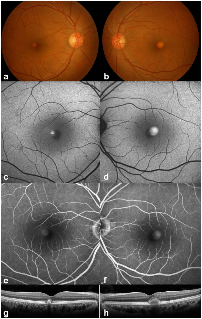
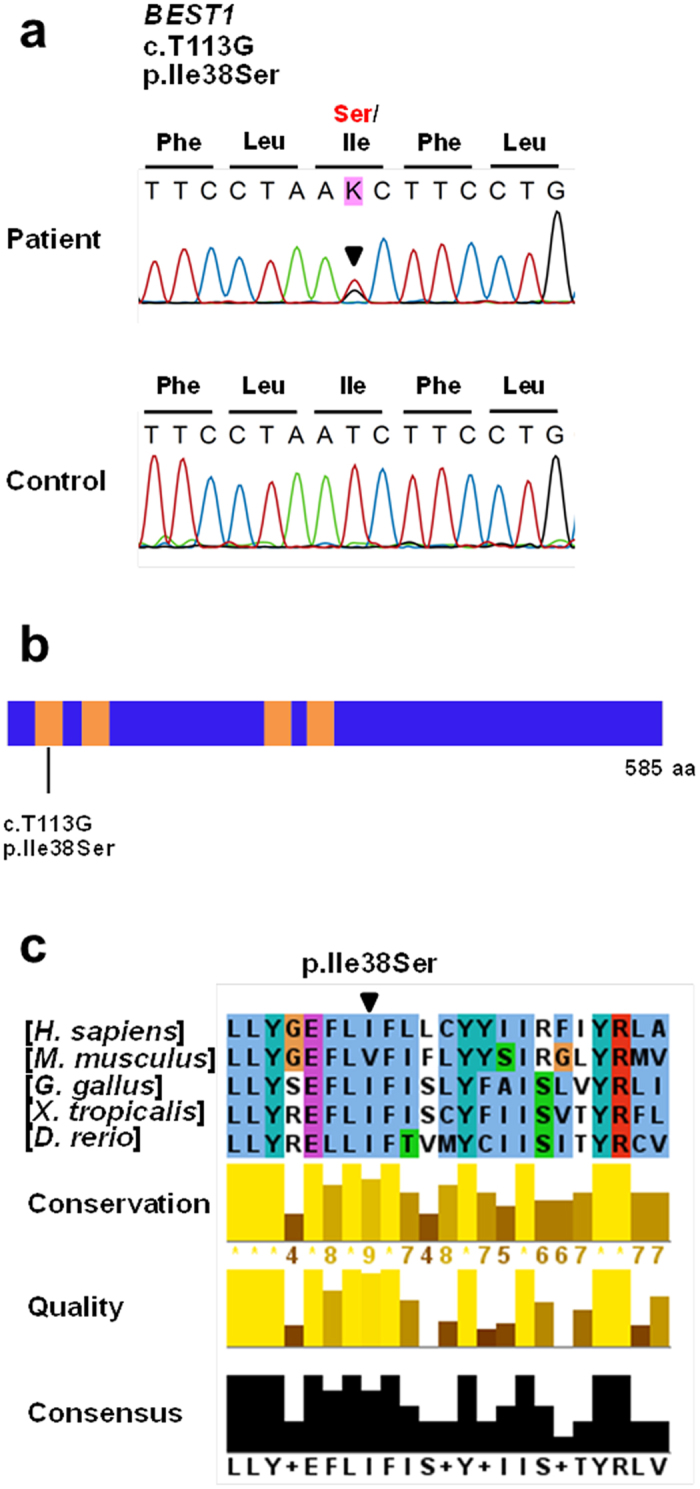
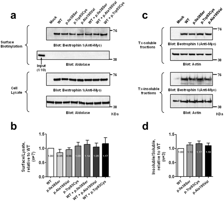
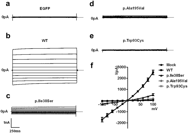
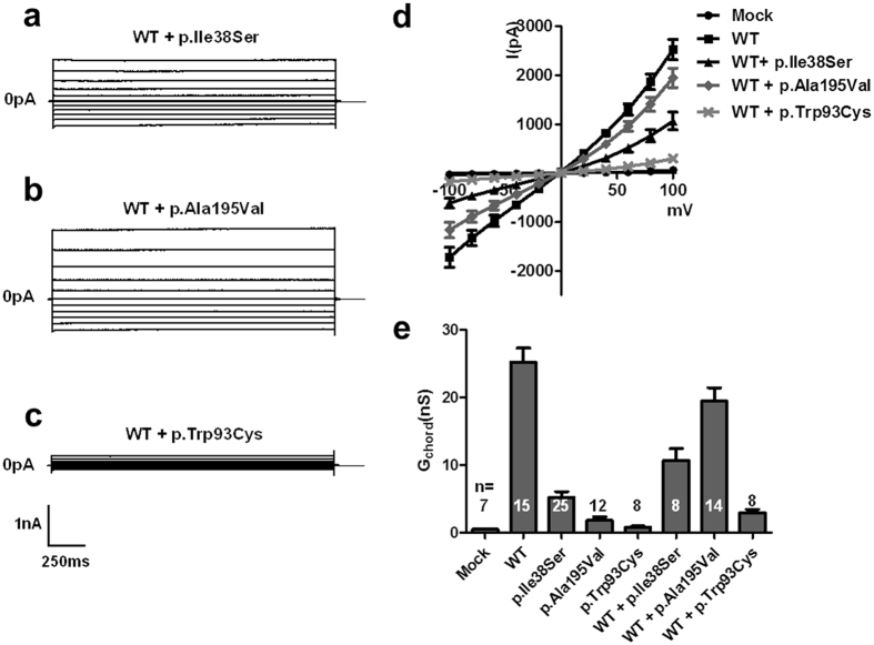
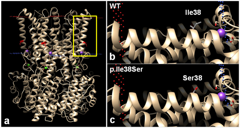
Adult-onset vitelliform macular dystrophy (AVMD) is a common and benign macular degeneration which can be caused by BEST1 mutation. Here, we investigated the clinical characteristics associated with a newly identified BEST1 mutation, p.Ile38Ser and confirmed the associated physiological functional defects. The 51-year-old patient presented bilateral small subretinal yellow deposits. Consistent with AVMD, the corresponding lesions showed hyperautofluorescence, late staining in fluorescein angiography, and subretinal hyper-reflective materials in spectral-domain optical coherence tomography. Genetic analysis demonstrated that the patient presented with a heterozygous p.Ile38Ser BEST1 mutation. Surface biotinylation and patch clamp experiments were performed in transfected HEK293T cells. Although, the identified BEST1 mutant maintains normal membrane expression, p.Ile38Ser mutant showed significantly smaller currents than wild type (WT). However, it showed larger currents than other BEST1 mutants, p.Trp93Cys, causing autosomal dominant best vitelliform macular dystrophy (BVMD), and p.Ala195Val, causing autosomal recessive bestrophinopathy (ARB). The cells expressing both WT and each BEST1 mutant showed that the functional defect of p.Ile38ser was milder than that of p.Trp93Cys, whereas combination of p.Ala195Val with WT showed good current. We identified and described the phenotype and in vitro functions of a novel BEST1 mutation causing AVMD. AVMD induced by p.Ile38Ser BEST1 mutation is a mild form of BVMD.


Conflict of interest statement

E.K.K. is the medical advisory board member of the Avellino LAB USA. Other authors have no conflict of interest to declare.

Figures

Figure 1
Figure 1
Clinical features of the patient. (a,b) Color fundus photographs showing bilateral, round subretinal yellowish deposits (vitelliform) in the posterior pole. (c,d) Blue light autofluorescence imaging showed intense hyper-autofluorescence at the corresponding yellowish deposit lesions. (e,f) Corresponding lesions showed diffuse hyperfluorescence in the late-phase images of fluorescein angiography. (g,h) Horizontal spectral-domain optical coherence tomography images through fovea showed well demarcated subretinal hyper-reflective material in the affected lesion.
Figure 2
Figure 2
Genetic analysis of the patient identified a novel BEST1 mutation, p.Ile38Ser. (a) Sequencing traces of the mutation and wild type control. Altered nucleotide and amino-acid changes are indicated above the sequence traces. (b) Domain structure of BEST1. The transmembrane (TM) domains are depicted by orange colored bars. The BEST1 mutation is located in the TM1 domain. (c) Partial protein alignment of BEST1 TM1 domain showed evolutionary conservation of the identified missense changes.
Figure 3
Figure 3
BEST1 p.Ile38Ser mutation does not interfere with membrane expression. HEK293T cells were transfected with plasmids expressing wild type and mutant hBEST1. (a) Surface biotinylation assays of hBEST1 were performed. (b) Quantitation of multiple experiments shows that the membrane expression was not different among groups by ANOVA (P = 0.589). Fold changes relative to WT are provided. (c) Detergent solubility assay of wild type and mutant hBEST1. (d) Quantification of multiple experiments shows that the detergent solubility was not different among the groups by ANOVA (P = 0.300). Fold changes relative to WT are provided. Western blots were cropped to show specific bands only. For uncropped blots see Supplementary Fig. S2.
Figure 4
Figure 4
p.Ile38Ser BEST1 mutant shows smaller currents compared to wild type (WT) BEST1 and bigger currents compared to p.Ala195Val or p.Trp93Cys mutants. (a–e) Representative traces of HEK293T cells transfected with EGFP alone (a), WT (b), p.Ile38Ser (c), p.Ala195Val (d), or p.Trp93Cys (e) BEST1. Voltage was stepped from a holding potential of 0 mV to between −100 and +100 mV in 20 mV steps. Step duration was 2000 ms. (f) Current-voltage relationship of mock, WT, and mutant BEST1. Data are presented as mean ± SEM.
Figure 5
Figure 5
p.Ile38Ser mutation induces mild deterioration of protein function. (a–c) Representative traces of HEK293T cells transiently transfected with wild type (WT) BEST1 plus either p.Ile38Ser (a), p.Ala195Val (b), or p.Trp93Cys (c). (d) Current-voltage relationship of mock, WT, and co-transfection of WT and mutant BEST1. (e) The mean outward chord conductance (Gchord) in transfected HEK293T cells, calculated over 0 mV to +100 mV. Co-expression of WT and p.Ile38Ser BEST1 results into approximately one-third of the currents of WT BEST1, while p.Trp93Cys generated very small currents. The p.Ala195Val mutant, an autosomal recessive type, elicited large currents when co-expressed with WT BEST1. Data are presented as mean ± SEM. (Conductance values are provided in Supplementary Table S2. WT vs. WT + p.Ile38Ser, p < 0.01; WT + p.Ile38Ser vs. WT + p.Ala195Val, p < 0.001; WT + p.Ile38Ser vs. WT + p.Trp93Cys, p < 0.01 by ANOVA followed by Newman-Keuls multiple comparison test, details are provided in Supplementary Table S2).
Figure 6
Figure 6
Predicted computational tertiary structure of wild-type (WT) and p.Ile38Ser hBEST1 proteins. 4RDQ crystal structure was used as template. (a) Homopentameric structure of WT BEST1 full structure. Red dots represent outer membrane border and blue dots represent intracellular membrane border. Isoleucine 38 position, which is located in the transmembrane (TM) domain 1, is highlighted in red. The yellow box region was magnified in (b) and (c). (b) The side chain of Ile38 in the TM1 domain was described, which consisted of a hydrophobic carbon chain (gray). The side chain protrudes toward the lipid membrane. (c) The side chain of Ser38 presents hydrophilic oxygen residue (red), which may generate a repulsive force with the hydrophobic lipid membrane.

References

    1. Petrukhin K, et al. Identification of the gene responsible for Best macular dystrophy. Nat Genet. 1998;19:241–247. doi: 10.1038/915. - DOI - PubMed
    1. Johnson AA, et al. Bestrophin 1 and retinal disease. Prog Retin Eye Res. 2017 - PMC - PubMed
    1. Boon CJ, et al. The spectrum of ocular phenotypes caused by mutations in the BEST1 gene. Prog Retin Eye Res. 2009;28:187–205. doi: 10.1016/j.preteyeres.2009.04.002. - DOI - PubMed
    1. Marmorstein AD, et al. Bestrophin, the product of the Best vitelliform macular dystrophy gene (VMD2), localizes to the basolateral plasma membrane of the retinal pigment epithelium. Proc Natl Acad Sci USA. 2000;97:12758–12763. doi: 10.1073/pnas.220402097. - DOI - PMC - PubMed
    1. Kane Dickson V, Pedi L, Long SB. Structure and insights into the function of a Ca(2+)-activated Cl(−) channel. Nature. 2014;516:213–218. doi: 10.1038/nature13913. - DOI - PMC - PubMed

Publication types

LinkOut - more resources