. 2017:2017:4387081.
doi: 10.1155/2017/4387081. Epub 2017 Jul 31.

Expression and Clinical Significance of ILF2 in Gastric Cancer

Affiliations

Expression and Clinical Significance of ILF2 in Gastric Cancer

Zi-Huan Yin et al. Dis Markers. 2017.

Abstract

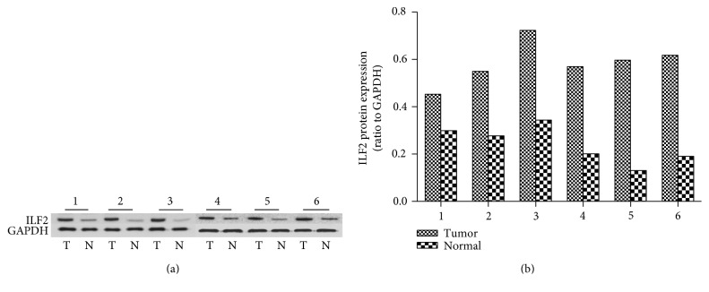
The aim of this study is to investigate the expression levels and clinical significance of ILF2 in gastric cancer. The mRNA and protein expression levels of ILF2 were, respectively, examined by quantitative real-time PCR (qRT-PCR) and Western blot from 21 paired fresh frozen GC tissues and corresponding normal gastric tissues. In order to analyze the expression pattern of ILF2 in GC, 60 paired paraffin-embedded GC slides and corresponding normal gastric slides were detected by immunohistochemistry (IHC) assay. The correlation between ILF2 protein expression levels and clinicopathological parameters, overall survival (OS), disease-free survival (DFS), and clinical prognosis were analyzed by statistical methods. Significantly higher levels of ILF2 were detected in GC tissues compared with normal controls at both mRNA and protein level. High expression of ILF2 was tightly correlated with depth of invasion, lymph node metastasis, pathological stage, and histological differentiation. Log-rank test showed that high expression of ILF2 was positively associated with poor clinical prognosis. Multivariate analysis identified that ILF2 was an independent prognostic factor for OS and DFS. Our findings suggest that ILF2 may be a valuable biomarker and a novel potential prognosis predictor for GC patients.


Figures

Figure 1
Figure 1
The expression levels of ILF2 mRNA in 21 paired samples by qRT-PCR. Scatter plots of the relative expression of ILF2 between GC tissues (tumor) and normal controls (normal) to GAPDH, GAPDH as an endogenous control (p = 0.031, paired t-test, two-tailed) (a). Bar plots of the relative expression of ILF2 in GC tissues compared with adjacent normal tissues; each patient was presented as the log2 ratio of tumor tissues/normal tissues (b).
Figure 2
Figure 2
The expression levels of ILF2 protein in six representative paired clinical samples by Western blot analysis. The levels of ILF2 protein were higher in GC tissues (T) compared with adjacent normal tissues (N), GAPDH as an endogenous control (a). Relative quantification results of the intensity of ILF2 bands of GC tissues (tumor) and normal controls (normal) to GAPDH were quantified by gray analysis (b).
Figure 3
Figure 3
Correlation between ILF2 mRNA and ILF2 protein expression levels in 21 GC tissues was analyzed by Pearson correlation analysis (r = 0.753, p < 0.001).
Figure 4
Figure 4
Representative microphotographs for ILF2 protein expression by IHC staining in GC tissues and corresponding normal gastric tissues. ILF2 protein in GC tissues in strong staining (sepia) (a), moderate staining (claybank) (b), weak staining (light yellow) (c), no staining (d), and negative controls (e). Weak staining (light yellow) (f) and barely stained (g) adjacent normal gastric tissues. The comparison of ILF2 immunostaining score between GC tissues (n = 60) and normal gastric tissues (n = 60); the results are presented as mean ± SEM (∗∗∗p < 0.001) Wilcoxon matched-pairs signed-ranks test (h) (original magnification: ×400 in (a)–(g)).
Figure 5
Figure 5
Kaplan-Meier survival analysis and log-rank test for OS and DFS of GC patients. The OS of GC patients with ILF2 high expression and low expression (a). The DFS of GC patients with ILF2 high expression and low expression (b).

Similar articles

Cited by

References

    1. Torre L. A., Bray F., Siegel R. L., Ferlay J., Lortet-Tieulent J., Jemal A. Global cancer statistics, 2012. CA: A Cancer Journal for Clinicians. 2015;65(2):87–108. doi: 10.3322/caac.21262. - DOI - PubMed
    1. Chen W., Zheng R., Baade P. D., et al. Cancer statistics in China, 2015. CA: A Cancer Journal for Clinicians. 2016;66(2):115–132. doi: 10.3322/caac.21338. - DOI - PubMed
    1. Chen W., Zheng R., Zeng H., Zhang S. The incidence and mortality of major cancers in China, 2012. Chinese Journal of Cancer. 2016;35(1):p. 73. doi: 10.1186/s40880-016-0137-8. - DOI - PMC - PubMed
    1. Smyth E. C., Verheij M., Allum W., et al. Gastric cancer: ESMO clinical practice guidelines for diagnosis, treatment and follow-up. Annals of Oncology. 2016;27(Supplement 5):38–49. doi: 10.1093/annonc/mdw350. - DOI - PubMed
    1. Lee J. H., Nam B. H., Ryu K. W., et al. Comparison of outcomes after laparoscopy-assisted and open total gastrectomy for early gastric cancer. The British Journal of Surgery. 2015;102(12):1500–1505. doi: 10.1002/bjs.9902. - DOI - PubMed

LinkOut - more resources