. 2017:2017:2786427.
doi: 10.1155/2017/2786427. Epub 2017 Aug 2.

Intrathecal Resiniferatoxin Modulates TRPV1 in DRG Neurons and Reduces TNF-Induced Pain-Related Behavior

Affiliations

Intrathecal Resiniferatoxin Modulates TRPV1 in DRG Neurons and Reduces TNF-Induced Pain-Related Behavior

M Leo et al. Mediators Inflamm. 2017.

Abstract

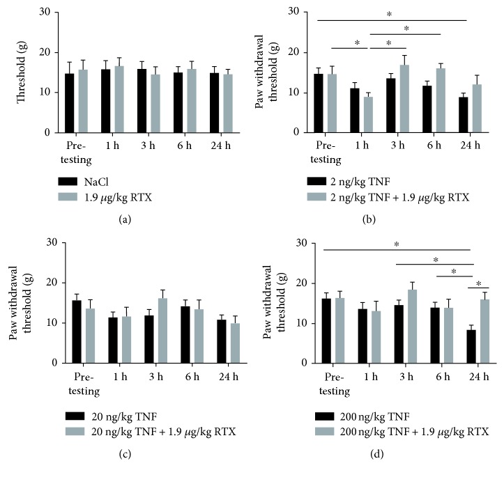
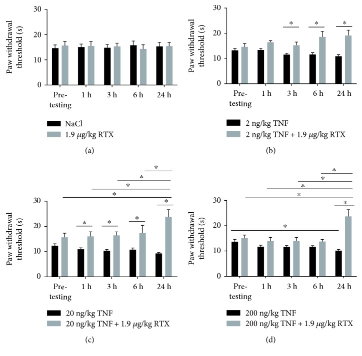
Transient receptor potential vanilloid-1 (TRPV1) is a nonselective cation channel, predominantly expressed in sensory neurons. TRPV1 is known to play an important role in the pathogenesis of inflammatory and neuropathic pain states. Previous studies suggest interactions between tumor necrosis factor- (TNF-) alpha and TRPV1, resulting in a modulation of ion channel function and protein expression in sensory neurons. We examined the effect of intrathecal administration of the ultrapotent TRPV1 agonist resiniferatoxin (RTX) on TNF-induced pain-associated behavior of rats using von Frey and hot plate behavioral testing. Intrathecal injection of TNF induces mechanical allodynia (2 and 20 ng/kg) and thermal hyperalgesia (200 ng) 24 h after administration. The additional intrathecal administration of RTX (1.9 μg/kg) alleviates TNF-induced mechanical allodynia and thermal hyperalgesia 24 h after injection. In addition, TNF increases the TRPV1 protein level and number of TRPV1-expressing neurons. Both effects could be abolished by the administration of RTX. These results suggest that the involvement of TRPV1 in TNF-induced pain offers new TRPV1-based experimental therapeutic approaches and demonstrates the analgesic potential of RTX in inflammatory pain diseases.


Figures

Figure 1
Figure 1
Influence of intrathecal administration of TNF and TNF + RTX on mechanical allodynia. (a) Administration of NaCl or RTX did not affect paw withdrawal thresholds, compared to control conditions (p > 0.05). (b) While 2 ng TNF does not influence paw withdrawal threshold 1, 3, or 6 h after administration, withdrawal threshold was reduced after 24 h (p < 0.05). Additional administration of RTX (1.9 mg/kg) did not abolish the TNF-induced reduction of withdrawal threshold 1 h, 3 h, 6 h, or 24 h after injection (p > 0.05). (c) Administration of TNF (20 ng) or the combination of TNF + RTX did not change paw withdrawal threshold (p > 0.05). (d) In contrast, administration of 200 ng TNF led to a reduction of paw withdrawal threshold after 24 h (p < 0.05). Additional administration of RTX abolished the effect of TNF after 24 h (p < 0.05).
Figure 2
Figure 2
Influence of intrathecal administration of TNF and TNF + RTX on thermal hyperalgesia. (a) Administration of NaCl or RTX did not affect paw withdrawal thresholds, compared to control conditions (p > 0.05). (b) Two nanograms of TNF did not lead to changes in paw withdrawal thresholds 1 h, 3 h, 6 h, or 24 h after administration (p > 0.05). Additional administration of RTX (1.9 mg/kg) resulted in changes in withdrawal threshold over the experimental time. When TNF + RTX were administered, a significant increase of withdrawal threshold was observed after 3 (p < 0.05), 6 (p < 0.05), and 24 h (p < 0.05), compared to TNF administration alone. (c) Administration of TNF did not change paw withdrawal thresholds over the experimental time, compared to pretesting (p > 0.05). When RTX was additionally administered, paw withdrawal threshold was significantly increased (p < 0.05). When TNF + RTX was compared to TNF administration alone, an increase of withdrawal threshold was observed after 1 h (p < 0.05), 3 h (p < 0.05), 6 h (p < 0.05), and 24 h (p < 0.05). (d) Administration of 200 ng TNF resulted in a reduction of paw withdrawal threshold after 24 h (p < 0.05), while no changes were observed after 1 h, 3 h, or 6 h (p > 0.05). Additional RTX led to significant increase of paw withdrawal threshold 24 h after administration (p < 0.05). Additional RTX abolished the TNF-induced reduction of paw withdrawal threshold 24 h after administration (p < 0.05).
Figure 3
Figure 3
Influence of intrathecal administration of TNF and TNF + RTX on the protein level of TRPV1 and the number of TRPV1-expressing DRG neurons. (a) Immunohistochemical staining of DRG slices 24 h after administration of either 200 ng/kg TNF, 1.9 μg/kg RTX, or 200 ng/kg TNF + 1.9 μg/kg RTX. DRG neurons were stained against NeuN (red), as a neuronal marker and TRPV1 (green). Quantification of TRPV1-derived signals revealed significant increase in TRPV1 protein level after TNF administration, (p < 0.001), while administration RTX decreased the TRPV1 protein level significantly (p < 0.001), compared to control conditions. When TNF and TRPV1 were administrated together, a significant change to sole TNF administration was observed. Combined administration of TNF and RTX abolished the TNF-mediated increase of the TRPV1 protein level (p < 0.001). No significant change was observed when compared to untreated control (p > 0.05). In addition to the protein level of TRPV1, the number of TRPV1-expressing neurons within the DRG was significantly increased after TNF administration, compared to control conditions (p < 0.05). RTX administration significantly decreased the number of TRPV1-expressing neurons (p < 0.05). Combined application of TNF + RTX led to a significant reduction of TRPV1-positive neurons, compared to TNF administration (p < 0.01). When compared to control conditions, no significant alteration in the number of TRPV1-expressing neurons was observed (p > 0.05). (b) Western blot analysis showed an increase of TRPV1 protein level 24 h after TNF administration (p < 0.05), while additional administration of RTX reduced this effect (p < 0.05).

Similar articles

Cited by

References

    1. Ohtori S., Takahashi K., Moriya H., Myers R. R. TNF-alpha and TNF-alpha receptor type 1 upregulation in glia and neurons after peripheral nerve injury: studies in murine DRG and spinal cord. Spine. 2004;29:1082–1088. - PubMed
    1. Clark A. K., Old E. A., Malcangio M. Neuropathic pain and cytokines: current perspectives. Journal of Pain Research. 2013;6:803–814. doi: 10.2147/JPR.S53660. - DOI - PMC - PubMed
    1. Murphy G. M., Jr., Lee Y. L., Jia X. C., et al. Tumor necrosis factor-alpha and basic fibroblast growth factor decrease glial fibrillary acidic protein and its encoding mRNA in astrocyte cultures and glioblastoma cells. Journal of Neurochemistry. 1995;65:2716–2724. doi: 10.1046/j.1471-4159.1995.65062716.x. - DOI - PubMed
    1. Schafers M., Lee D. H., Brors D., Yaksh T. L., Sorkin L. S. Increased sensitivity of injured and adjacent uninjured rat primary sensory neurons to exogenous tumor necrosis factor-alpha after spinal nerve ligation. The Journal of Neuroscience : The Official Journal of the Society for Neuroscience. 2003;23:3028–3038. - PMC - PubMed
    1. Schafers M., Sorkin L. S., Geis C., Shubayev V. I. Spinal nerve ligation induces transient upregulation of tumor necrosis factor receptors 1 and 2 in injured and adjacent uninjured dorsal root ganglia in the rat. Neuroscience Letters. 2003;347:179–182. - PubMed