

Changes in macular parameters in different types of amblyopia: optical coherence tomography study
- PMID: 28831240
- PMCID: PMC5552145
- DOI: 10.2147/OPTH.S143223
Changes in macular parameters in different types of amblyopia: optical coherence tomography study
Abstract
Purpose: The purposes of this study were to investigate the changes in macular parameters (thickness, volume) and peripapillary retinal nerve fiber layer (RNFL) thickness (RNFLT) in different cases of amblyopia versus the normal fellow eyes using optical coherence tomography (OCT) and to estimate the relationship of OCT changes with various defined patients' parameters.
Design: This is a prospective, observational, cross-sectional case series.
Methods: The method involved measuring the peripapillary RNFLT, macular thickness, and macular volume via spectral domain (OCT) in different types of amblyopia and comparing with the other fellow eyes. This study was conducted at the Mansoura Ophthalmic Center.
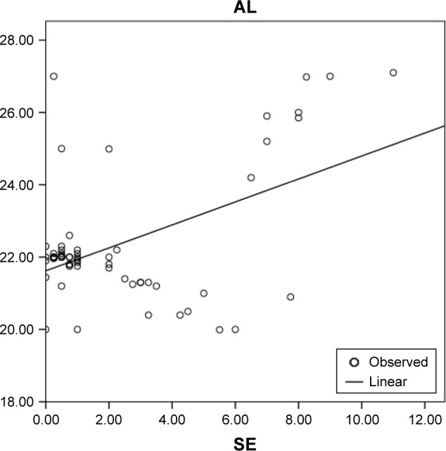
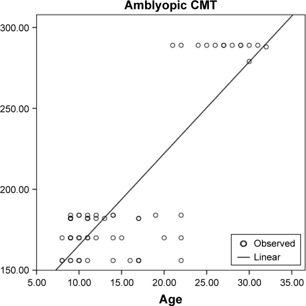
Results: A total of 64 patients with different types of amblyopia were included. The mean central macular thickness (CMT) was 196.2±50.03 µm in the amblyopic eyes versus 167±12.76 µm in the fellow eyes (P=0.000), the mean average macular thickness was 265.80±12.77 µm in the amblyopic eyes versus 259.10±3.09 µm in the fellow eyes (P=0.000), the mean macular volume was 7.59±0.32 mm3 in the amblyopic eyes versus 7.34±0.071 mm3 in the fellow eyes (P=0.002), and the mean global RNFLT was 97.00±11.60 µm in the amblyopic eyes versus 78.50±13.05 µm in the fellow eyes (P=0.029). There was a discrepancy between the different amblyopic types. Age and the axial length were the only independent variables that statistically significantly correlated with the CMT.
Conclusion: The unilateral amblyopic eyes were prone to have a higher CMT and thicker global RNFL compared to those of the sound fellow eyes. Retinal variations between different types of the amblyopia differ from one type to another. The age could be considered as a predictor of the disease worsening and treatment prognosis. Further studies are required to emphasize these results.
Keywords: amblyopia; anisometropia; optical coherence tomography; strabismus.
Conflict of interest statement
Disclosure The authors report no conflicts of interest in this work.
Figures


References
-
- Von Norden GK, Crawford ML. The lateral geniculate nucleus in human strabismic amblyopia. Invest Ophthalmol Vis Sci. 1992;33(9):2729–2732. - PubMed
-
- Asper L, Crewther D, Crewthei SG. Strabismic amblyopia. Part 2. Neural processing. Clin Exp Optom. 2000;83(4):200–211. - PubMed
-
- Hess RF. Amblyopia: site unseen. Clin Exp Optom. 2001;84(6):321–336. - PubMed
LinkOut - more resources
Full Text Sources
Other Literature Sources
