PDGF-metronidazole-encapsulated nanofibrous functional layers on collagen membrane promote alveolar ridge regeneration
- PMID: 28831251
- PMCID: PMC5548280
- DOI: 10.2147/IJN.S137342
PDGF-metronidazole-encapsulated nanofibrous functional layers on collagen membrane promote alveolar ridge regeneration
Abstract
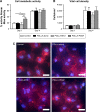
This study aimed to develop a functionally graded membrane (FGM) to prevent infection and promote tissue regeneration. Poly(l-lactide-co-d,l-lactide) encapsulating platelet-derived growth factor (PDLLA-PDGF) or metronidazole (PDLLA-MTZ) was electrospun to form a nanofibrous layer on the inner or outer surface of a clinically available collagen membrane, respectively. The membrane was characterized for the morphology, molecule release profile, in vitro and in vivo biocompatibility, and preclinical efficiency for alveolar ridge regeneration. The PDLLA-MTZ and PDLLA-PDGF nanofibers were 800-900 nm in diameter, and the thicknesses of the functional layers were 20-30 μm, with sustained molecule release over 28 days. All of the membranes tested were compatible with cell survival in vitro and showed good tissue integration with minimal fibrous capsule formation or inflammation. Cell proliferation was especially prominent on the PDLLA-PDGF layer in vivo. On the alveolar ridge, all FGMs reduced wound dehiscence compared with the control collagen membrane, and the FGM with PDLLA-PDGF promoted osteogenesis significantly. In conclusion, the FGMs with PDLLA-PDGF and PDLLA-MTZ showed high biocompatibility and facilitated wound healing compared with conventional membrane, and the FGM with PDLLA-PDGF enhanced alveolar ridge regeneration in vivo. The design represents a beneficial modification, which may be easily adapted for future clinical use.
Keywords: alveolar process; animal models; metronidazole; platelet-derived growth factor; tissue engineering.
Conflict of interest statement
Disclosure The authors report no conflicts of interest in this work.
Figures


Similar articles
-
Functionally graded membrane deposited with PDLLA nanofibers encapsulating doxycycline and enamel matrix derivatives-loaded chitosan nanospheres for alveolar ridge regeneration.Int J Biol Macromol. 2022 Apr 1;203:333-341. doi: 10.1016/j.ijbiomac.2022.01.147. Epub 2022 Jan 28. Int J Biol Macromol. 2022. PMID: 35093432
-
Sequential platelet-derived growth factor-simvastatin release promotes dentoalveolar regeneration.Tissue Eng Part A. 2014 Jan;20(1-2):356-64. doi: 10.1089/ten.TEA.2012.0687. Epub 2013 Oct 17. Tissue Eng Part A. 2014. PMID: 23980713 Free PMC article.
-
Combination of a biomolecule-aided biphasic cryogel scaffold with a barrier membrane adhering PDGF-encapsulated nanofibers to promote periodontal regeneration.J Periodontal Res. 2020 Aug;55(4):529-538. doi: 10.1111/jre.12740. Epub 2020 Feb 24. J Periodontal Res. 2020. PMID: 32096217
-
Evaluation of emulsion electrospun polycaprolactone/hyaluronan/epidermal growth factor nanofibrous scaffolds for wound healing.J Biomater Appl. 2016 Jan;30(6):686-98. doi: 10.1177/0885328215586907. Epub 2015 May 26. J Biomater Appl. 2016. PMID: 26012354
-
Promises of Functionally Graded Material in Bone Regeneration: Current Trends, Properties, and Challenges.ACS Biomater Sci Eng. 2022 Mar 14;8(3):1001-1027. doi: 10.1021/acsbiomaterials.1c01416. Epub 2022 Feb 24. ACS Biomater Sci Eng. 2022. PMID: 35201746 Review.
Cited by
-
Antibiotic-Loaded Polymeric Barrier Membranes for Guided Bone/Tissue Regeneration: A Mini-Review.Polymers (Basel). 2022 Feb 21;14(4):840. doi: 10.3390/polym14040840. Polymers (Basel). 2022. PMID: 35215754 Free PMC article. Review.
-
Nanomaterials for Periodontal Tissue Regeneration: Progress, Challenges and Future Perspectives.J Funct Biomater. 2023 May 24;14(6):290. doi: 10.3390/jfb14060290. J Funct Biomater. 2023. PMID: 37367254 Free PMC article. Review.
-
A Bibliometric Analysis of Electrospun Nanofibers for Dentistry.J Funct Biomater. 2022 Jul 9;13(3):90. doi: 10.3390/jfb13030090. J Funct Biomater. 2022. PMID: 35893458 Free PMC article. Review.
-
Effect of metronidazole on placental and fetal development in albino rats.Anim Reprod. 2019 Nov 18;16(4):810-818. doi: 10.21451/1984-3143-AR2018-0149. Anim Reprod. 2019. PMID: 32368258 Free PMC article.
-
Near-infrared light responsive gold nanoparticles coating endows polyetheretherketone with enhanced osseointegration and antibacterial properties.Mater Today Bio. 2024 Feb 1;25:100982. doi: 10.1016/j.mtbio.2024.100982. eCollection 2024 Apr. Mater Today Bio. 2024. PMID: 38371468 Free PMC article.
References
-
- Haffajee AD, Socransky SS. Microbial etiological agents of destructive periodontal diseases. Periodontol 2000. 1994;5:78–111. - PubMed
-
- Albandar JM, Brunelle JA, Kingman A. Destructive periodontal disease in adults 30 years of age and older in the United States, 1988–1994. J Periodontol. 1999;70(1):13–29. - PubMed
-
- Lai H, Su CW, Yen AM, et al. A prediction model for periodontal disease: modelling and validation from a national survey of 4061 Taiwanese adults. J Clin Periodontol. 2015;42(5):413–421. - PubMed
-
- Bottino MC, Thomas V, Schmidt G, et al. Recent advances in the development of GTR/GBR membranes for periodontal regeneration–a materials perspective. Dent Mater. 2012;28(7):703–721. - PubMed
-
- Corinaldesi G, Lizio G, Badiali G, Morselli-Labate AM, Marchetti C. Treatment of intrabony defects after impacted mandibular third molar removal with bioabsorbable and non-resorbable membranes. J Periodontol. 2011;82(10):1404–1413. - PubMed
MeSH terms
Substances
LinkOut - more resources
Full Text Sources
Other Literature Sources

