

Capnocytophaga canimorsus sepsis in a methotrexate-treated patient with rheumatoid arthritis
- PMID: 28831382
- PMCID: PMC5554928
- DOI: 10.1016/j.idcr.2017.08.002
Capnocytophaga canimorsus sepsis in a methotrexate-treated patient with rheumatoid arthritis
Abstract
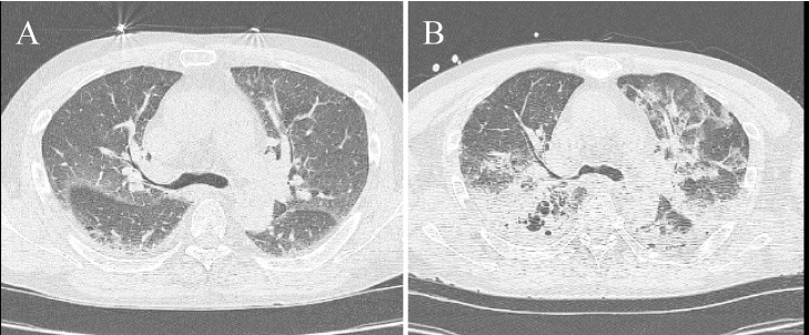
Capnocytophaga canimorsus is a gram-negative rod that can be transmitted primarily by dog bites. This life-threatening organism commonly causes sepsis in patients with splenectomy or alcoholism. A 53-year-old rheumatoid arthritis male treated with methotrexate (MTX) for 5 years was admitted for a 4-day history of fever and dyspnea. He had been bitten on a finger by the family dog 4 days before onset. Laboratory tests revealed pancytopenia, acute renal failure, and evidence of disseminated intravascular coagulation, and he subsequently developed acute respiratory distress syndrome. Furthermore, blood cultures grew gram-negative bacilli and despite intensive treatment, he died 5 days after admission. Later, C. canimorsus was identified from his culture samples using a species-specific polymerase chain reaction. C. canimorsus infections should be considered in the differential diagnosis of sepsis for immunocompromised hosts following animal bites.
Keywords: Capnocytophaga canimorsus; Iatrogenic immunocompromised hosts; Methotrexate; Rheumatoid arthritis; Sepsis.
Figures


Similar articles
-
A Bite So Bad: Septic Shock Due to Capnocytophaga Canimorsus Following a Dog Bite.Cureus. 2021 Apr 24;13(4):e14668. doi: 10.7759/cureus.14668. Cureus. 2021. PMID: 34055517 Free PMC article.
-
[Capnocytophaga canimorsus sepsis after a dog bite].Ugeskr Laeger. 2008 May 26;170(22):1941. Ugeskr Laeger. 2008. PMID: 18513481 Danish.
-
Rapid diagnosis of Capnocytophaga canimorsus septic shock in an immunocompetent individual using real-time Nanopore sequencing: a case report.BMC Infect Dis. 2019 Jul 24;19(1):660. doi: 10.1186/s12879-019-4173-2. BMC Infect Dis. 2019. PMID: 31340776 Free PMC article.
-
Fatal septicemia with Capnocytophaga canimorsus in a compromised host. A case report with review of the literature.Acta Clin Belg. 1991;46(6):364-70. doi: 10.1080/17843286.1991.11718192. Acta Clin Belg. 1991. PMID: 1665939 Review.
-
[Dog bite in a splenectomized patient].Rev Med Liege. 2002 Jan;57(1):40-4. Rev Med Liege. 2002. PMID: 11899497 Review. French.
Cited by
-
The Great pretender: the first case of septic shock due to Capnocytophaga canimorsus in Sardinia. A Case report and review of the literature.J Public Health Res. 2022 Nov 26;11(4):22799036221133234. doi: 10.1177/22799036221133234. eCollection 2022 Oct. J Public Health Res. 2022. PMID: 36451937 Free PMC article.
-
A classic case of Capnocytophaga induced septic shock with multi-organ failure after a dog bite in an asplenic patient.IDCases. 2023 May 23;32:e01808. doi: 10.1016/j.idcr.2023.e01808. eCollection 2023. IDCases. 2023. PMID: 37273844 Free PMC article.
-
Capnocytophaga gingivalis Bacteremia After Upper Gastrointestinal Bleeding in Immunocompromised Patient.J Investig Med High Impact Case Rep. 2021 Jan-Dec;9:23247096211020672. doi: 10.1177/23247096211020672. J Investig Med High Impact Case Rep. 2021. PMID: 34041953 Free PMC article.
References
-
- Gaastra W., Lipman L.J. Capnocytophaga canimorsus. Vet Microbiol. 2010;140:339–346. - PubMed
-
- Butler T. Capnocytophaga canimorsus: an emerging cause of sepsis, meningitis, and post-splenectomy infection after dog bites. Eur J Clin Microbiol Infect Dis. 2015;34:1271–1280. - PubMed
-
- Hästbacka J., Hynninen M., Kolho E. Capnocytophaga canimorsus bacteremia: clinical features and outcomes from a Helsinki ICU cohort. Acta Anaesthesiol Scand. 2016;60:1437–1443. - PubMed
-
- Pers C., Gahrn-Hansen B., Frederiksen W. Capnocytophaga canimorsus septicemia in Denmark, 1982–1995: review of 39 cases. Clin Infect Dis. 1996;23:71–75. - PubMed
-
- Lion C., Escande F., Buridin J.C. Capnocytophaga canimorsus infections in human: review of the literature and cases report. Eur J Epidemiol. 1996;12:521–533. - PubMed
Publication types
LinkOut - more resources
Full Text Sources
Other Literature Sources
