Review
. 2017 Mar 3;3(3):170-175.
doi: 10.1016/j.ijwd.2017.01.003. eCollection 2017 Sep.

Analogs of human genetic skin disease in domesticated animals

Affiliations
Review

Analogs of human genetic skin disease in domesticated animals

Justin Finch et al. Int J Womens Dermatol. .

Abstract

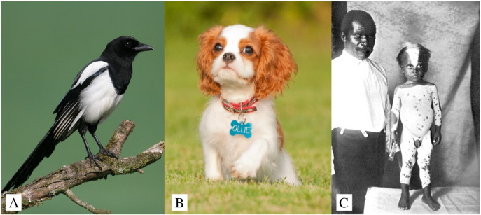
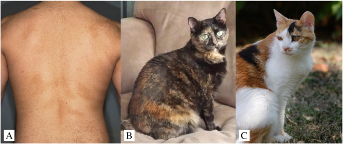
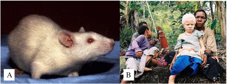
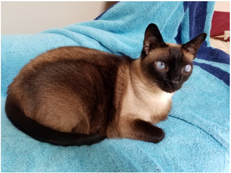
Genetic skin diseases encompass a vast, complex, and ever expanding field. Recognition of the features of these diseases is important to ascertain a correct diagnosis, initiate treatment, consider genetic counseling, and refer patients to specialists when the disease may impact other areas. Because genodermatoses may present with a vast array of features, it can be bewildering to memorize them. This manuscript will explain and depict some genetic skin diseases that occur in both humans and domestic animals and offer a connection and memorization aid for physicians. In addition, we will explore how animal diseases serve as a model to uncover the mechanisms of human disease. The genetic skin diseases we will review are pigmentary mosaicism, piebaldism, albinism, Griscelli syndrome, ectodermal dysplasias, Waardenburg syndrome, and mucinosis in both humans and domesticated animals.


Figures

Fig. 1
Fig. 1
Piebaldism. (A) A magpie that exhibits midline depigmentation (Available from: https://upload.wikimedia.org/wikipedia/commons/5/50/01-Magpie.jpg User: diginatur [own work], CC BY-SA 3.0). (B) Cavalier King Charles Spaniel dog that demonstrates axial depigmentation (Justin Finch, MD). (C) Human with piebaldism who lives in El Cayo, British Honduras (Available from: https://commons.wikimedia.org/wiki/File:ALBINISM;_Human_Piebald_living_in_El_Cayo_Wellcome_L0032353.jpg Credit: Wellcome Library, London. Originally published in Karl Pearson and C. H. Usher. London: 1913. Plate BB).
Fig. 2
Fig. 2
Variations in pigmentary mosaicism. (A) Pigmentary mosaicism in a human (Justin Finch, MD). (B) Pigmentary mosaicism in a tortoise shell cat (Stephanie Abrams, DVM). (C) Pigmentary mosaicism in a calico cat (photo courtesy of Tatiana Chessa; under CC-BY SA 3.0 license).
Fig. 3
Fig. 3
Albinism. (A) Pet rat with albinism (Available from: http://dir.niehs.nih.gov/dirlep/Webpages/clinpath.html, public domain). (B) An albino girl from Papua New Guinea (Available from: https://commons.wikimedia.org/wiki/File:Albinistic_girl_papua_ new_guinea.jpg, public domain).
Fig. 4
Fig. 4
Siamese cat (Stephanie Abrams, DVM).
Fig. 5
Fig. 5
Foal with Lavender Foal Syndrome demonstrating pigmentary dilution and opisthotonos, one cardinal neurological sign of the disorder (PLoS Genet 2010;6, CC-BY 3.0).
Fig. 6
Fig. 6
Phenotypic variation of Keratin 71 mutations in humans and cats. (A) Autosomal dominant wooly hair and hypotrichosis that is caused by KRT 71. Note the sparse eyebrow and wooly scalp hair (photo courtesy of Simomura Yukata, MD). (B) Pure breed Sphinx cat. This phenotype is also caused by a KRT71 mutation. (photo: by User: Artourp [Own work], CC BY-SA 3.0; Available from: https://upload.wikimedia.org/wikipedia/commons/9/95/Shabbat.jpg). (C) Selkirk Rex. The wooly hair is caused by a KRT71 mutation. (photo: Nickolas Titkov [own work], CC BY-SA 3.0; Available from: https://www.flickr.com/photos/titkov/5297138877/).
Fig. 7
Fig. 7
Pigmentary alteration in Waardenburg syndrome. (A) Deaf Frame Overo horse displayed at a show (photo: by Sandysphotos2009; licensed under CC BY 2.0). (B) Deaf blaze-color ferret. (image courtesy of John Porter and Erika Matulich, PhD, PCM). (C) Waardenburg syndrome in two siblings (image courtesy of Chirsophe Hsu, MD).
Fig. 8
Fig. 8
(A) Leonine facies due to follicular mucinosis (photo: Peter Heald, MD). (B) Shar-Pei dog with facial features that result from hereditary mucinosis (Available from: https://www.flickr.com/photos/48685334@N00/388814720; User: whartonds; licensed under CC BY-SA 2.0).

Similar articles

Cited by

References

    1. Brooks S.A., Gabreski N., Miller D., Brisbin A., Brown H.E., Streeter C. Whole genome SNP association in the horse: identification of a deletion in myosin Va responsible for lavender foal syndrome. PLoS Genet. 2010;6:e1000909. - PMC - PubMed
    1. Cvejic D., Steinberg T.A., Kent M.S., Fischer A. Unilateral and bilateral congenital sensorineural deafness in client-owned pure-breed white cats. J Vet Intern Med. 2009;23:392–395. - PubMed
    1. Drögemüller C., Karlsson E.K., Hytönen M.K., Perloski M., Dolf G., Sainio K. A mutation in hairless dogs implicates FOXI3 in ectodermal development. Science. 2008;321:1462. - PubMed
    1. Faletra F., Berti I., Tommasini A., Pecile V., Cleva L., Alberini E. Phylloid pattern of hypomelanosis closely related to chromosomal abnormalities in the 13q detected by SNP array analysis. Dermatology. 2012;225:294–297. - PubMed
    1. Finch J.J., Payette M. Genoderms Made Ludicrously Easy. Journal of Drugs in Dermatology. 2017 New York, NY, 2017 (in press)